- Physical Examination
- Surgical Examination
- Ophthalmology
- Clinical Skills
- Orthopedics
- Surgery Videos
- Laparoscopy
- Pediatrics
- Funny Videos
- Cardiothoracic Surgery
- Nursing Videos
- Plastic Surgery
- Otorhinolaryngology
- Histology and Histopathology
- Neurosurgery
- Dermatology
- Pediatric Surgery
- Urology
- Dentistry
- Oncology and Cancers
- Anatomy Videos
- Health and Fitness
- Radiology
- Anaesthesia
- Physical Therapy
- Pharmacology
- Interventional Radiology
- Cardiology
- Endocrinology
- Gynecology
- Emergency Medicine
- Psychiatry and Psychology
- Childbirth Videos
- General Medical Videos
- Nephrology
- Physiology
- Diet and Food Health
- Diabetes Mellitus
- Neurology
- Women Health
- Osteoporosis
- Gastroenterology
- Pulmonology
- Hematology
- Rheumatology
- Toxicology
- Nuclear Medicine
- Infectious Diseases
- Vascular Disease
- Reproductive Health
- Burns and Wound Healing
- Other
Top videos
Large Breast Augmentation
See the War Inside Your Body

Father & Mom feel their baby the same

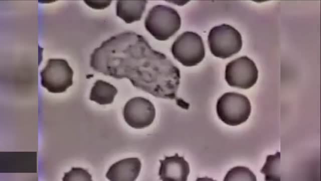
Blood Transfusion-Transmitted Diseases
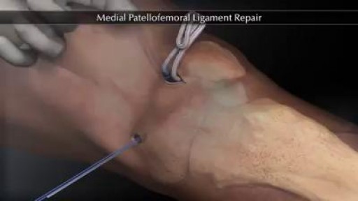
The medial patellofemoral ligament (MPFL) helps to keep the kneecap centered along the front of the knee, so that it tracks well during knee movements. MPFL injuries typically occur during a forceful traumatic kneecap dislocation. This injury is most common among young, active females. Depending on the severity of an MPFL injury, treatment may involve surgical reconstruction, followed by physical therapy. Physical therapists design treatment programs for individuals with MPFL injuries to help them gently restore their knee strength and function.
Scoliosis (pronounced sko-lee-o-sis) is a three-dimensional deformity that occurs when the spine becomes abnormally rotated and curved sideways. Most often this deformity has no known cause, in which case it is called idiopathic scoliosis. While the cause is unknown, idiopathic scoliosis does tend to run in families. The specific genes involved have not all been identified yet, and there could be factors beyond genetics as well

Letting children patients play the role of a dentist may be a good way to introduce them to the different types of instruments used in a dental clinic. This in turn may also reduce thier anxiety or fear of the dentist and make them more easy-going and compliant towards dental treatments. Ofcourse instruments should be clean and steril and care should be taken to not give them pointed or sharp objects.
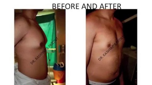
PREPARING FOR GYNECOMASTIA SURGERY - This maybe the first time you are planning to undergo surgery. It's natural to feel little anxious about the whole process. Get more information www.bestbreastsurgeryindia.com | www.themedspa.us/cosmetic-surgery/male-breast-reduction.html
The inflatable penile prosthesis consists of two attached cylinders -- a reservoir and a pump -- which are placed surgically in the body. The two cylinders are inserted in the penis and connected by tubing to a separate reservoir of saline. The reservoir is implanted under the rectus muscles in the lower abdomen. A pump is also connected to the system and sits under the loose skin of the scrotal sac, between the testicles. This penile prosthesis is referred to as a 3-piece inflatable penile prosthesis, due to the three different components. A 2-piece inflatable penile prosthesis consists of only two components: the attached cylinders and the combined reservoir and pump unit. Instead of the reservoir being placed behind the groin, it is combined with the pump into one housing unit that fits comfortably within the scrotum. The advantage of a 2-piece prosthesis in that the surgery is shorter and less complicated and there is no device parts in the abdomen. The disadvantage of the 2-piece prosthesis is that the smaller reservoir may not result in adequate erections in some men. To inflate the prosthesis, the man presses on the pump. The pump transfers saline from the reservoir to the cylinders in the penis, inflating them and causing an erection. Pressing on a deflation valve at the base of the pump returns the fluid to the reservoir, deflating the penis and returning it to the normal flaccid state.
Open appendectomy (simulated)

World's first osteotomy for spine deformity
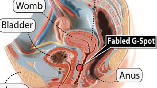
Watch that video to know What is G Spot?
Watch that Big Size Fibrodenoma Removal Under Local Anesthesia

Hidradenitis Suppurativa Surgery, Hidradenitis Suppurativa Photos, Hidradenitis Suppurativa Nz. http://hidradenitis-suppurativa-cure.good-info.co --- symptoms of hidradenitis suppurativa: First let us look at the symptoms of the disease Black heads: When they appear in a double barreled pattern Red bumps: When tender lesions, or bumps, which often contain pus and/or itching. Hard, painful lumps: When pea sized lumps cause pain and get inflamed These recur very often Don’t heal or improve for weeks Returns after treatment Appears in several different locations The lumps are accompanied by pain I am going to reveal to you step by step how to understand and deal with the problems of hidradenitis suppurativa. click here http://hidradenitis-suppurativa-cure.good-info.co

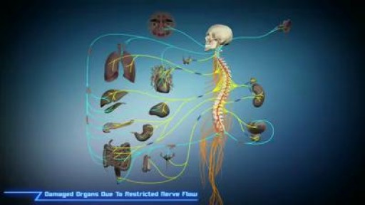
Types of Spinal Cord Injuries 1) Anterior cord syndrome 2) Central cord syndrome 3) Brown-Sequard syndrome 4) Tetraplegia 5) Paraplegia 6) Triplegia

Aumento De Gluteos, Metacrilato En Gluteos, Aumento De Gluteos Natural, Operacion De Nalgas.--- http://aumente-gluteos.plus101.com/ --- Con una combinación de dieta, ejercicio y mejoras artificiales, puedes cambiar la forma de los glúteos rápidamente, sin importar tu tipo de cuerpo. Aunque no verás un cambio significativo en una semana, si dedicas un tiempo y haces ejercicios enfocados en los tres músculos principales de los glúteos: el glúteo mayor, el glúteo medio y el glúteo menor, tendrás unos glúteos más grandes. Enfócate en consumir muchas proteínas. Las proteínas son esenciales para el crecimiento y el desarrollo de los músculos, por lo que es importante comer el tipo correcto de proteínas. La proteína combinada con el ejercicio correcto aumentará definitivamente el tamaño de los glúteos. Algunas fuentes saludables de proteínas incluyen los huevos, las pechugas de pollo sin piel, el salmón, el atún, el queso cottage, el pavo, los frijoles, las legumbres, la carne de res magra y las nueces de soya. En cuanto a la carne, busca una que sea magra y sin procesar. Cuando compres el pescado, trata de hornearlo en lugar de freírlo. Elige el tipo correcto de carbohidratos y grasas. Existen muchas dietas que dicen que eliminan por completo los carbohidratos y las grasas, pero lo mejor no es eliminar los alimentos de la dieta, sino sustituirlos por opciones más saludables. Evita el exceso de calorías y la mala alimentación, alejándote de los carbohidratos procesados, como las papas fritas y la pasta. Los carbohidratos saludables incluyen la quinua, el camote, el arroz integral, los granos de avena enteros y los panes integrales. Las fuentes de grasas saludables que pueden ayudarte a perder peso y a tonificar los glúteos son los aceites de pescado, el aceite de oliva extra virgen, la mantequilla de almendras y las nueces. Abastécete de vegetales. Los vegetales suelen ser una parte olvidada de la dieta para agrandar los músculos. Al agregar vegetales a cada comida te darás cuenta de que tus niveles de energía serán más constantes y por lo tanto, podrás hacer un entrenamiento más fuerte ya que no sentirás demasiado cansancio. Descubre por qué las cirugías y los implantes no son la solución más efectiva. Olvídate del quirófano y ahorra tu dinero, porque con mi método resolverás el problema de “Síndrome de los glúteos planos” rápidamente. ingresa ahora a: http://aumente-gluteos.plus101.com/
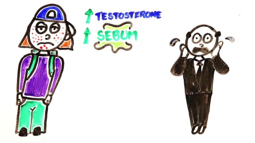
Watch that video to know How to Treat Pimples on Your Face?
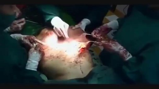
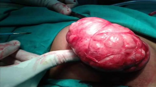
Watch that Huge Stomach Tumor Removal Medical Surgery
Watch that World's first osteotomy surgery for spine deformity
Draining a Blood Clot from the left thigh after knife stab wound sutured by some ER!
