- Physical Examination
- Surgical Examination
- Ophthalmology
- Clinical Skills
- Orthopedics
- Surgery Videos
- Laparoscopy
- Pediatrics
- Funny Videos
- Cardiothoracic Surgery
- Nursing Videos
- Plastic Surgery
- Otorhinolaryngology
- Histology and Histopathology
- Neurosurgery
- Dermatology
- Pediatric Surgery
- Urology
- Dentistry
- Oncology and Cancers
- Anatomy Videos
- Health and Fitness
- Radiology
- Anaesthesia
- Physical Therapy
- Pharmacology
- Interventional Radiology
- Cardiology
- Endocrinology
- Gynecology
- Emergency Medicine
- Psychiatry and Psychology
- Childbirth Videos
- General Medical Videos
- Nephrology
- Physiology
- Diet and Food Health
- Diabetes Mellitus
- Neurology
- Women Health
- Osteoporosis
- Gastroenterology
- Pulmonology
- Hematology
- Rheumatology
- Toxicology
- Nuclear Medicine
- Infectious Diseases
- Vascular Disease
- Reproductive Health
- Burns and Wound Healing
- Other
Top videos
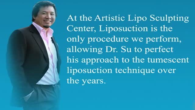
For patients looking to slim down their neck and achieve a more contoured and sculpted jaw line, then Tampa chin liposuction at the Artistic Lipo Sculpting Center is the answer! This one procedure can literally make patients look 10 years younger and 20 lbs lighter. Dr. Thomas Su is specialized in performing Liposuction procedures and consistently achieves stunning results for his Tampa patients. To find out more about Tampa neck lipo, visit http://www.artlipo.com/liposuction/liposuction-body-areas/lipo-chin---neck.html

Anatomy Tutorial During Trans Nasal Endoscopy
Laparoscopy in Hemodynamic Instable Patient
TMJ Surgery Temboro mandibular Joint HD
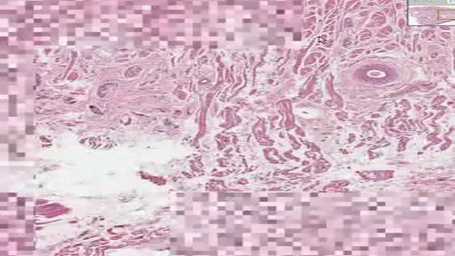
Histology of Tongue
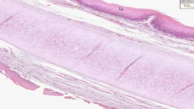
Histology of Epiglottis
Preventing Sexually Transmitted Diseases A sexually transmitted disease (STD) is an infection that is spread during sexual contact with another person. This includes touching, since some STDs can be spread from skin-to-skin contact. In general, STDs are highly preventable. Almost 20 million new STDs are diagnosed each year in the United States, according to the Centers for Disease Control and Prevention (CDC). However, a large number of those infections could be avoided if people made different decisions about their sexual health. The only guaranteed method to prevent STDs is to abstain from all sexual contact. This may not a practical solution for everyone. Fortunately, there are steps people can take to limit their risk of exposure.

The immune system is the body's defense against infectious organisms and other invaders. Through a series of steps called the immune response, the immune system attacks organisms and substances that invade body systems and cause disease.
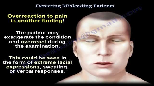
Factitious disorder is the term used to describe a pattern of behavior centered on the exaggeration or outright falsifications of one’s own health problems or health problems of others. Some people with this disorder fake or exaggerate physical problems; others fake or exaggerate psychological problems or a combination of physical and psychological problems. Factitious disorder differs from a pattern of falsified or exaggerated behavior called malingering. While malingerers make their claims out of a motivation for personal gain, people with factitious disorder have no such motivation.
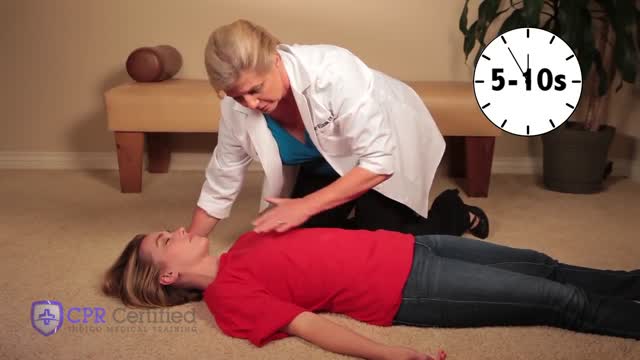
Anybody can and anybody should learn how to perform CPR (Cardiopulmonary resuscitation): According to the American Heart Association, a stunning 70% of Americans don’t know how what to do if somebody is experiencing a cardiac emergency because they don’t know how to administer CPR or they forgot the exact technique. This is especially alarming since almost 90% of cardiac arrests occur at home — where patients depend on the immediate respiratory care response of their family members. In brief, knowing how to perform CPR can save the life of a loved one someday.
When is endoscopy used? Endoscopes were first developed to look at parts of the body that couldn’t be seen any other way. This is still a common reason to use them, but endoscopy now has many other uses too. It’s often used in the prevention, early detection, diagnosis, staging, and treatment of cancer. To prevent and screen for cancer Some types of endoscopes are used to look for cancer in people who have no symptoms. For example, colonoscopy (KO-lun-AH-skuh-pee) and sigmoidoscopy (SIG-moid-AH-skuh-pee) are used to screen for colon and rectal cancer. These procedures can also help prevent cancer because they let doctors find and remove polyps (growths) that might become cancer if left alone. To find cancer early Endoscopy can sometimes be used to find cancer early, before it has had a chance to grow or spread. Looking for causes of symptoms When people go to the doctor with certain symptoms, endoscopy can sometimes be used to help find a cause. For instance: Laryngoscopy to look at the vocal cords in people with long-term hoarseness Upper endoscopy in people having trouble swallowing Colonoscopy in people with anemia (low red blood cell counts) with an unknown cause Colonoscopy in people with blood in their stool Looking at problems found on imaging tests Imaging tests such as x-rays and CT scans can sometimes show physical changes within the body. But these tests may only give information about the size, shape, and location of the problem. Doctors use endoscopes to see more details, like color and surface texture, when trying to find out what’s going on. Newer methods of endoscopy that include high magnification are being tested to find out whether they are more useful in detecting cancer and other abnormal cells on the inner surfaces of the body. To diagnose and find out the stage (extent) of cancer To get a tissue sample Going one step further, most types of endoscopes have tools on the end that the doctor can use to take out small tissue samples. This procedure is called a biopsy (BY-op-see). Samples can be taken from suspicious areas and then looked at under a microscope or tested in other ways to see if cancer is there. A biopsy is usually the best way to find out if a growth or change is cancer or something else. Getting a closer look In some cases endoscopes are used to help find out how far a cancer has spread. Thoracoscopy (THOR-uh-KAHS -kuh-pee) and laparoscopy (LAP-uh-RAHS-kuh-pee) can be very useful in finding out if cancer has spread into the thorax (chest) or abdomen (belly). The surgeon can look into these places making only a small incision (cut) in the skin.
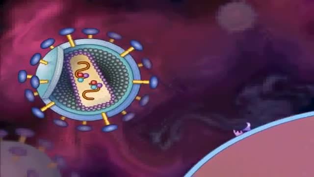
HIV stands for human immunodeficiency virus. If left untreated, HIV can lead to the disease AIDS (acquired immunodeficiency syndrome). Unlike some other viruses, the human body can’t get rid of HIV completely. So once you have HIV, you have it for life. HIV attacks the body’s immune system, specifically the CD4 cells (T cells), which help the immune system fight off infections. If left untreated, HIV reduces the number of CD4 cells (T cells) in the body, making the person more likely to get infections or infection-related cancers. Over time, HIV can destroy so many of these cells that the body can’t fight off infections and disease. These opportunistic infections or cancers take advantage of a very weak immune system and signal that the person has AIDS, the last state of HIV infection. No effective cure for HIV currently exists, but with proper treatment and medical care, HIV can be controlled. The medicine used to treat HIV is called antiretroviral therapy or ART. If taken the right way, every day, this medicine can dramatically prolong the lives of many people with HIV, keep them healthy, and greatly lower their chance of transmitting the virus to others. Today, a person who is diagnosed with HIV, treated before the disease is far advanced, and stays on treatment can live a nearly as long as someone who does not have HIV.
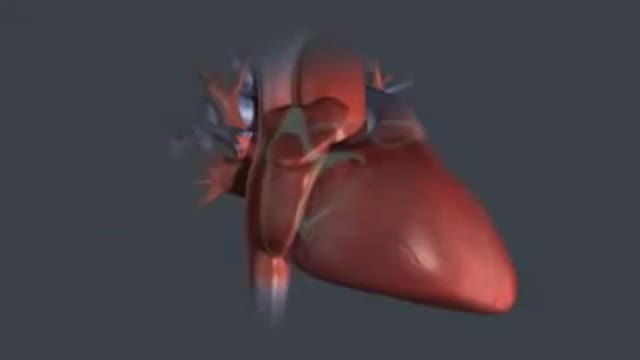
Every 10 minutes, someone is added to the national transplant waiting list, and every day, 22 people on average die waiting for a match, according to the United Network for Organ Sharing. But, thanks to innovations in bioengineering, all of that could change. Conceived nearly 60 years ago, the total artificial heart (TAH) has helped sustain the sickest biventricular failure patients waiting for a transplant. While the design of the primary TAH used today has mostly remained stagnant since the ’80s, when it was first implanted in a patient, new models and clinical trials may lead to a better device and, one day, a permanent solution. “We are still many years away from that,” Dr. Nader Moazami, director of the Cardiac Transplantation and Ventricular Assist Device Therapy Program at the Cleveland Clinic, told FoxNews.com of a permanent artificial heart. “Although tremendous strides have been made, biocompatibility will always remain a challenge.”
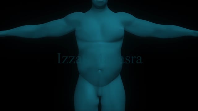
Bariatric surgical procedures cause weight loss by restricting the amount of food the stomach can hold, causing malabsorption of nutrients, or by a combination of both gastric restriction and malabsorption. Bariatric procedures also often cause hormonal changes. Most weight loss surgeries today are performed using minimally invasive techniques (laparoscopic surgery). The most common bariatric surgery procedures are gastric bypass, sleeve gastrectomy, adjustable gastric band, and biliopancreatic diversion with duodenal switch. Each surgery has its own advantages and disadvantages.
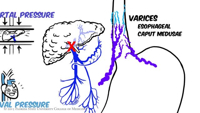
Portal hypertension is an increase in the blood pressure within a system of veins called the portal venous system. Veins coming from the stomach, intestine, spleen, and pancreas merge into the portal vein, which then branches into smaller vessels and travels through the liver.
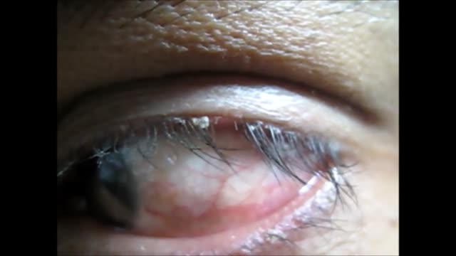
Blepharitis is an inflammation of the eyelids in which they become red, irritated and itchy and dandruff-like scales form on the eyelashes. It is a common eye disorder caused by either bacteria or a skin condition, such as dandruff of the scalp or acne rosacea. It affects people of all ages. Although uncomfortable, blepharitis is not contagious and generally does not cause any permanent damage to eyesight.

Here's Why Your Skin Doesn't Rip Easily
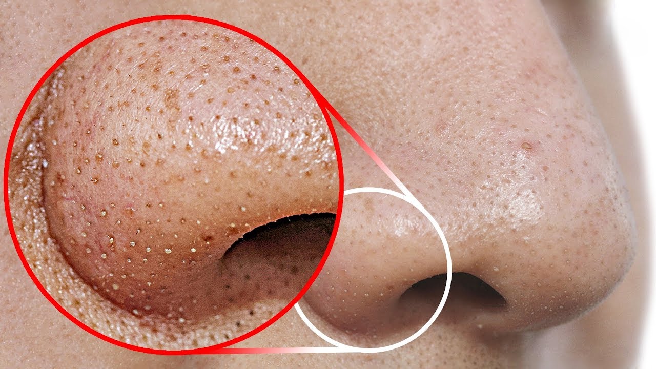
Watch that video to know How to Get Rid of Blackheads From Your Nose
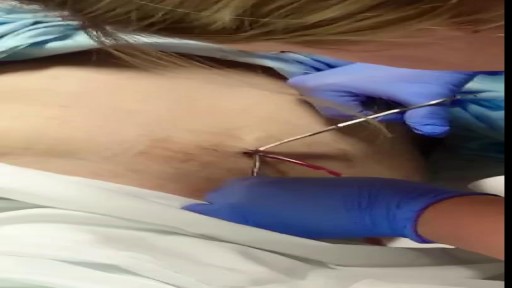
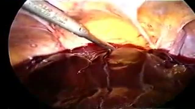
Removal of drain tube after spleen surgery
