Top videos

Horrible Skin Jiggers Removal
Horrible Skin Jiggers Removal hooda 77,468 Views • 3 years ago

Watch that video of Horrible Skin Jiggers Removal

Dieta Iposodica Per Acufeni, Agopuntura Efficacia Contro Acufeni, Ronzio Orecchie Nel Silenzio
Dieta Iposodica Per Acufeni, Agopuntura Efficacia Contro Acufeni, Ronzio Orecchie Nel Silenzio marin vinasco 2,107 Views • 3 years ago

Dieta Iposodica Per Acufeni, Agopuntura Efficacia Contro Acufeni, Ronzio Orecchie Nel Silenzio---- http://acufeni-cura.plus101.com/ --- I sintomi dell'acufene, I sintomi dell'acufene possono causare molti fastidi e problemi a chi ne soffre. Inoltre anche i sintomi possono causare confusione. Prendiamo un esempio per capire meglio. Stai conducendo una vita perfetta, vai al lavoro e torni a casa ogni giorno, ma improvvisamente incominci a sentire strani rumori all'orecchio. Ovviamente credi che ci sia una sorgente di tali rumori. Stranamente nessun altro sembra sentirli. Puoi spaventarti e pensare che quei rumori provengano dal tuo corpo, così vai dal dottore. Dopo qualche esame il dottore conferma che è tutto a posto. Il problema è che tu i rumori continui a sentirli. Incominciano a darti sui nervi e ti influenzano nel lavoro. E ancora peggio, i rumori sembrano farsi più forti la notte, privandoti del tuo prezioso sonno. Si tratta di acufene. I rumori che senti sono i sintomi principali e possono essere di vario tipo. Puoi sentire strani scricchilii, ronzii, brusii, fischi o sibili all'orecchio. Non è mai lo stesso per tutti. Alcuni li sentono di tanto in tanto, altri per tutto il tempo. alcuni raccontano di forti attacchi. Ed altri sono stremati da trapanamenti costanti. Ma una cosa è certa - molte persone negli USA e altrove hanno gli acufeni. Quindi non sei davvero l'unico in queste condizioni. La maggior parte dei sintomi dell'acufene non sono altro che rumori fantasma Molte persone sono confuse se non riescono ad indivuare la fonte del rumore alle orecchie. Alcuni si spaventano. Spesso queste persone sono messe in ridicolo da chi non sente quei rumori. Ma per chi soffre di acufeni i suoni sono assolutamente reali. Di fatto sono rumori fantasma, una percezione delle orecchie. Vi sono eccezioni. In almeno un caso il rumore può essere reale. In altre parole c'è una effettiva fonte del rumore che senti. Questo è l'acufene pulsatile. In questo caso sei in grado di sentire il battito del tuo cuore e ciò ti può far impazzire perchè lo senti in continuazione. Vi è un'altra differenza. C'è sicuramente più di una persona che sente questi rumori e questo è il tuo dottore. Avrà bisogno di uno strumento di ascolto per sentirli. Prendi nota - non è come sentire il cuore con uno stetoscopio. in questo caso il dottore utilizza uno strumento di ascolto per sentire il rumore all'orecchio, non nel petto. Che cosa causa l'acufene pulsatile? Potresti sentire il tuo cuore in caso di pressione alta, danni alle arterie e anche cambiamenti della circolazione del sangue. A volte, una grande dose di stress, ansia o depressione può causare questa situazione. Non Importa Quale Sia La Causa, Tipo O Gravità Del Tuo Acufene, Puoi Iniziare Ad Utilizzare Questo Efficare Sistema PROPRIO ORA Ed Ottenere Un Sollivo E Una Liberazione Permanente ISTANTANEA Dai Tuoi Acufeni! inserisci ora: http://acufeni-cura.plus101.com/

Flecken Auf Der Haut, Braune Flecken Auf Der Haut Pilz, Homöopathie Bei Pigmentflecken
Flecken Auf Der Haut, Braune Flecken Auf Der Haut Pilz, Homöopathie Bei Pigmentflecken marin vinasco 2,580 Views • 3 years ago

Flecken Auf Der Haut, Braune Flecken Auf Der Haut Pilz, Homöopathie Bei Pigmentflecken--- http://vitiligo-heilung.info-pro.co --- Vitiligo Heilung für weiß gefleckte Haut, Zuerst treten die weißen Flecken punktuell auf, schließlich verbreiten sie sich über den ganzen Körper: Die Weißfleckenkrankheit ist belastend – und jetzt heilbar. Das Hautleiden ist weder gefährlich noch ansteckend – aber psychisch sehr belastend. Viele Betroffene trauen sich nicht mehr, in öffentliche Bäder zu gehen oder kurze Kleidung zu tragen. Schätzungen zufolge leiden bis zu zwei Prozent der Weltbevölkerung unter Vitiligo („Scheckhaut“). Statistisch gesehen sind die Hautpartien an Unterarmen, Handgelenken, Händen, Fingern, Ellbogen, Füßen und Genitalien am häufigsten betroffen. Die genauen Ursachen sind nicht bekannt. Wissenschaftler vermuten, dass Stress die Pigmentstörung auslösen kann. Klar ist aber, dass die Hautzellen einen zu hohen Anteil an Wasserstoffperoxid aufweisen. Es verhindert die Bildung des Hautfarbstoffs Melanin. Zudem ist bei den Betroffenen das Enzym Katalase beschädigt, das normalerweise den Abbau von Wasserstoffperoxid steuert. Die bessere Behandlungswahl bei Vitiligo Ein weitaus effektiverer und sicherer Weg zur Behandlung von Vitiligo ist die Nutzung holistischer Methoden. Dies umfasst die Verwendung von Kräuterextrakten - welche die weitere Ausbreitung der weißen Flecken behindern - zusammen mit der Einnahme bestimmter Vitaminergänzungen. Die Methode stimuliert den Hautpigmentierungsprozess. Damit diese natürliche Therapie aber in vollstem Umfange wirken kann, muss der Patient gewillt sein, Änderungen in seinem Lebensstil und seinen Essgewohnheiten einzuführen und konsequent zu befolgen, um so den Heilungsprozess zu beschleunigen. Erfahren Sie mehr darüber, indem Sie diese Webseite besuchen: http://vitiligo-heilung.info-pro.co

Eye Color Change Surgery with Implant
Eye Color Change Surgery with Implant Scott 9,374 Views • 3 years ago

Eye Color Change Surgery with Implant

How to treat acne scars?
How to treat acne scars? samer kareem 3,240 Views • 3 years ago

The first step is to see if you have pigmentation issues -not really classed as acne scars, but this is controversial, or if you have contour changes. The best thing to do is to examine under tangential or angled lighting, as this will reveal all. Once this is done, scars can be subtyped and mapped, with high resolution photos. The second aspect, which is equally as important is to examine scars upon animanation, namely when you speak, smile and move your face. This will give me an idea of the amount of tethering and anchored acne scars. Time and time again I get request for ‘what is the treatment’ with static photos, an impossible task to answer correctly. Acne scar assessment has to be done live- with the patient in front of you, and lighting from all angles. Photos do not map scars as well a real time examination under magnification and lighting.

Management of traumatic rupture of the posterior capsule
Management of traumatic rupture of the posterior capsule Mohamed Ibrahim 13,998 Views • 3 years ago

This video shows management of rupture of the posterior capsule post blunt trauma in a child aged 8. Pre-operative suspicion of PCR was strong because of a flat anterior. So we were careful in our approach from the very beginning. CCC was performed and then dry aspiration of lens matter initiated. Sice vitreous showed, so anterior vitrectomy was done along with systematic removal of the lens matter. An acrysof multi-component lens was implanted into the sulcus and optic captured into the CCC.Outcome was very good.

Tourniquet
Tourniquet DrHouse 8,621 Views • 3 years ago

how to apply a tourniquet

Extra Ocular Muscles exam
Extra Ocular Muscles exam Surgeon 14,463 Views • 3 years ago

assessment of the extra-ocular muscles

Infatnt CPR
Infatnt CPR Mohamed 14,023 Views • 3 years ago

Infant Cardio-pulmonary Resuscitation

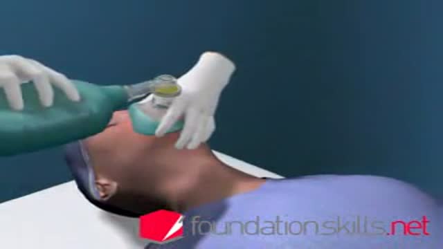
Endotracheal tube in children
Endotracheal tube in children DrHouse 17,750 Views • 3 years ago

How to insert Endotracheal tube in children

Loyola Respiratory System Exam Part 2
Loyola Respiratory System Exam Part 2 Loyola Medicine 21,858 Views • 3 years ago

Loyola Respiratory System Exam Part 2 A video from Loyola Medical School, Chicago showing the medical and clinical examination of the respiratory system.

How to read ECG Part 1
How to read ECG Part 1 M_Nabil 76,107 Views • 3 years ago

How to read ECG:
Part 1 Shows:
1-All
2-Introduction
3-Rate and Axis
4-Chamber Hypertrophy
5-Bundle Branch Block
6-Myocardial Infarction

Esophageal En Bloc Mucosectomy
Esophageal En Bloc Mucosectomy M_Nabil 12,056 Views • 3 years ago

En Bloc Esophageal Mucosectomy, an experimental technique for the endolumenal management of Barrett's related dysplasia and neoplasia. High grade dysplasia is in indication for esophagectomy; however esophagectomy has a mortality rate up to 12 percent, and up to 56 percent of patients may develop s...erious post-operative complications. Multiple ablated lesions can progress under the neo-squamous layer, leading to buried Barrett's mucosa. With conventional piecemeal EMR, cautery effect limits evaluation in areas of interest, Barrett's epithelium is left behind, tissue is not evaluated in situ and invasive lesions may be missed due to incomplete sampling. A new technique, en bloc esophageal mucosectomy, or EEM, was developed. The technique begins with conventional EMR in the proximal esophagus to access the submucosal space. Conventional EMR is being performed here. The mucosa is resected using an electrothermal snare. The mucosal defect from the first EMR is seen here. EMR is then repeated on the opposing wall. Sequential EMR creates a complete concentric mucosal defect. In the following sequence the completely detached column of mucosa can be seen, bounded by submucosa and muscularis propria layers. Here in the stomach, the endoscope is retroflexed and is covered by a sleeve of esophageal mucosa which has been freed to the GE junction and inverted. This sequence demonstrates a double snare technique. This snare is alongside the endoscope. The snare has been passed through the working channel. The working channel snare is pulled back, and the snare alongside the scope is used to grasp the mucosal column. With tension on the column the working channel snare can be threatened and advanced. This sequence shows the snare as it is being passed down to the GE junction. At the GE junction, the snare is tightened and cautery is applied. This frees the column of mucosal tissue from the remaining attachment. The endoscope is then withdrawn. Then detached mucosal column can be grasped with a snare and retrieved. In the following sequence, the long column of mucosa is being withdrawn via the overtube. Here, endoscopic forceps have been passed through the column to demonstrates the concentric nature of the specimen. The length of mucosa can be seen here alongside 2 conventional EMR specimens. Approximately 15cm of tissues was removed in this case. On endoscopy immediately following the resection, there is no bleeding or evidence of perforation in the area of resection. The endoscope is advanced and the exposed submucosa can be appreciated down to the GE junction. This is the low power view of the histologic specimen generated by EEM. Metaplastic tissue adjacent to a dysplastic focus would be completely removed. With a high power view, the layers of the esophagus can be appreciated. The epithelium, lamina propria, muscularis mucosa and submucosa are visible, with no cautery artifact in the area of interest. The technique would remove metplasia, low grade dysplasia, high grade dysplasia, and intramucosal carcinoma, as well a T 1 a lesions. All the animals in this series tolerated the procedure well. A total of five non-survival procedures and 4 survival procedures were performed. In the survival procedures, all four swine thrived in the post-operative period. Two swine were then survived for 9 days following the procedure. On post —op day nine, after passing into the upper esophagus, the proximal margin of the mucosectomy is seen here. Healing appears to be occurring. There is no evidence of leak, and no stricting is seen at 9 days down to the GE junction Passing into the stomach, some residual feed can be seen. Two swine were then survived for 13 days. On this follow-up endoscopy, the area of the mucosectomy is again healing. There was a loose stricture in both animals and both were easily traversed with a 9.8 mm gastroscope. There was a gross appearance of re-epitheliazation in some areas. It is notable that the stricture was present in the proximal esophagus with no narrowing distally. At necropsy there was not eviden

subfrontal approach to the anterior skull base with combined Le fort osteotomy
subfrontal approach to the anterior skull base with combined Le fort osteotomy M_Nabil 13,530 Views • 3 years ago

Access to processes within the skull base with lateral extension to the pterygopalatine fossa are reached by combined subfrontal osteotomy and Le Fort I osteotomy

Needle Insertion Transversus Abdominus Block
Needle Insertion Transversus Abdominus Block Doctor 16,859 Views • 3 years ago

Needle Insertion Transversus Abdominus Block

Vijay_Bose_Patient - Hip Resurfacing
Vijay_Bose_Patient - Hip Resurfacing Dr. Vijay Bose 8,316 Views • 3 years ago

The BMHR uses the same socket (hydroxyapatite-coated metal uncemented cup) and bearing(metal on metal) as the BHR. The modular head component fits onto a hydroxyapatite proximal porous coated cobalt chrome stem. It is an uncemented short stemmed prosthesis. It was invented by Prof. Dereck Mc.Minn a year ago and is performed by very few surgeons the world over. In India it is being done only at the Asian Regional Center for Hip Resurfacing in Chennai.

Liposuction with Abdominoplasty and Body Lifting
Liposuction with Abdominoplasty and Body Lifting Doctor 12,033 Views • 3 years ago

Liposuction for weight loss with Abdominoplasty and Body Lifting

Dental Implant Tooth failure
Dental Implant Tooth failure Mohamed Ibrahim 12,605 Views • 3 years ago

Although the success rate of dental implant is very high, there are about 10% of failure in all dental implantation. The cause is usually related to the bone quality of the implant site. Once the implant failed, it can be easily removed, wait for 6 weeks and re-implant again. It is easier to remove a failed implant than a natural tooth.

Endovascular Repair for Abdominal Aortic Aneurysm
Endovascular Repair for Abdominal Aortic Aneurysm Emery King 17,711 Views • 3 years ago

A DMC patient suffering from an abdominal aortic aneurysm receives an endovascular graft to alleviate the potentially deadly problem, performed by DMC cardiac specialist Dr. Ali Kafi. ~ Detroit Medical Center

Hawkin's Test
Hawkin's Test Anatomist 17,625 Views • 3 years ago

Hawkin's Test

Showing 316 out of 373