
- Physical Examination
- Surgical Examination
- Ophthalmology
- Clinical Skills
- Orthopedics
- Surgery Videos
- Laparoscopy
- Pediatrics
- Funny Videos
- Cardiothoracic Surgery
- Nursing Videos
- Plastic Surgery
- Otorhinolaryngology
- Histology and Histopathology
- Neurosurgery
- Dermatology
- Pediatric Surgery
- Urology
- Dentistry
- Oncology and Cancers
- Anatomy Videos
- Health and Fitness
- Radiology
- Anaesthesia
- Physical Therapy
- Pharmacology
- Interventional Radiology
- Cardiology
- Endocrinology
- Gynecology
- Emergency Medicine
- Psychiatry and Psychology
- Childbirth Videos
- General Medical Videos
- Nephrology
- Physiology
- Diet and Food Health
- Diabetes Mellitus
- Neurology
- Women Health
- Osteoporosis
- Gastroenterology
- Pulmonology
- Hematology
- Rheumatology
- Toxicology
- Nuclear Medicine
- Infectious Diseases
- Vascular Disease
- Reproductive Health
- Burns and Wound Healing
- Kidney Zone Podcast
- Other
Top videos


http://without-glasses.good-info.co How To Improve Eyesight Naturally With Food , How To Improve Eyesight Naturally With Exercises Food. Naturally PERFECT your Vision to 20/20 If you are one of the millions of Americans who suffer from visual problems such as Myopia and Hyperopia then this video will SHOCK you! In the following free video you'll discover: How you can 100% naturally and safely cure almost any visual problem. Why your glasses and contacts are in fact WORSENING your eye condition. The real TRUTH about the Eyecare industry This revolutionary program that you'll soon discover has dared to challenge the billion-dollar Eyecare industry. It reveals this amazing secret to getting 20/20 vision and it doesn't matter what your eye problems are, whether short or long sightedness, Presbyopia, Glaucoma.. whatever! It should help with everything. more information in. http://without-glasses.good-info.co
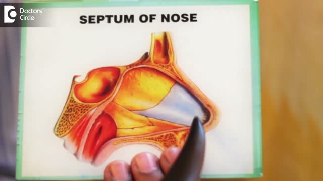
A deviated septum occurs when the thin wall (nasal septum) between your nasal passages is displaced to one side. In many people, the nasal septum is displaced — or deviated — making one nasal passage smaller. When a deviated septum is severe, it can block one side of your nose and reduce airflow, causing difficulty breathing. The additional exposure of a deviated septum to the drying effect of airflow through the nose may sometimes contribute to crusting or bleeding in certain individuals. Nasal obstruction can occur from a deviated nasal septum, from swelling of the tissues lining the nose, or from both. Treatment of nasal obstruction may include medications to reduce the swelling or nasal dilators that help open the nasal passages. To correct a deviated septum, surgery is necessary.
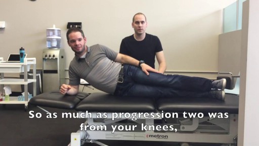
We get excited when people graduate! May it be graduating from physiotherapy or even graduating onto a new progression of an exercise! Today you move onto new challenges as Mike & Tyler demonstrate the final side plank progression. Kitchener Massage Therapy - http://www.strivept.ca/massage-therapy-kitchener.html
Beta-blockers, also known as beta antagonists, beta-adrenergic blocking agents, or beta-adrenergic antagonists, are drugs that are prescribed to treat several different types of conditions, including hypertension (high blood pressure), angina, some abnormal heart rhythms, heart attack (myocardial infarction), anxiety, ...Jul 27, 2015
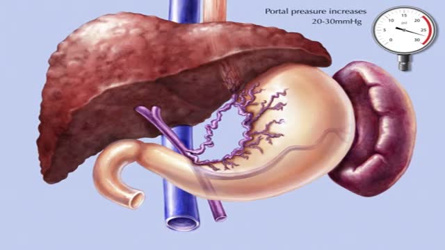
Transjugular intrahepatic portosystemic shunt or transjugular intrahepatic portosystemic stent shunting (commonly abbreviated as TIPS or TIPSS) is an artificial channel within the liver that establishes communication between the inflow portal vein and the outflow hepatic vein.
To treat pregnancy acne, start with self-care: Wash problem areas with a gentle cleanser. Twice a day, use your hands to wash your face with a mild soap and warm water. ... Shampoo regularly. ... Don't pick or squeeze blemishes. ... Avoid irritants. ... Watch what touches your skin.

Symptoms Of Anxiety, Anxiety Disorder Symptoms, What Is Social Anxiety, Zoloft For Anxiety --- http://panic-attacks-anxiety.good-info.co --- Panic attacks and anxiety While there are times for doctors, I want you to consider this: MOST of your anxiety is under the radar… Masquerading as “just feeling a bit nervous”... or “just a tad irritable thanks to this diet plan”... or, “let’s skip the party and just stay home tonight.” And, I’m telling you, that anxiety not only destroys your fat burning power: It often leads to all-out panic disorder, if you just ignore it. Fortunately for you, there’s a 60-Second Solution that restores your calm, removes those anxious feelings, and allows you to keep burning body fat for energy. Now, this exact same technique works for all-out panic and even more serious anxiety issues, too… The man who delivers this presentation had panic attacks in “everyday” situations… and he too had feelings of anxiety whenever he tried to diet-off body fat… This Simple Trick Stops Panic Attacks And Anxiety Click Here: http://panic-attacks-anxiety.good-info.co
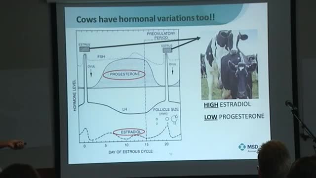
Postpartum endometritis refers to infection of the decidua (ie, pregnancy endometrium). The infection may also extend into the myometrium (called endomyometritis) or involve the parametrium (called parametritis).

Reactive arthritis is a painful form of inflammatory arthritis (joint disease due to inflammation). It occurs in reaction to an infection by certain bacteria. Most often, these bacteria are in the genitals (Chlamydia trachomatis) or the bowel (Campylobacter, Salmonella, Shigella and Yersinia). Chlamydia most often transmits by sex. It often has no symptoms, but can cause a pus-like or watery discharge from the genitals. The bowel bacteria can cause diarrhea. If you develop arthritis within one month of diarrhea or a genital infection – especially with a discharge – see a health care provider. You may have reactive arthritis. - See more at: https://www.rheumatology.org/i-am-a/patient-caregiver/diseases-conditions/reactive-arthritis#sthash.PukaaQhj.dpuf
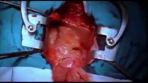
Beating Heart during surgery

Gastroschisis is a birth defect that develops in a baby while a woman is pregnant. This condition occurs when an opening forms in the baby's abdominal wall. The baby's bowel pushes through this hole. It then develops outside of the baby's body in the amniotic fluid.
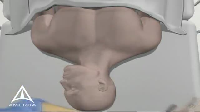
Central Line Placement 3D Animation
A blood transfusion is a routine medical procedure that can be lifesaving. During a blood transfusion, donated blood is added to your own blood. A blood transfusion may also be done to supplement various components of your blood with donated blood products. In some cases, a blood transfusion is done with blood that you've donated ahead of time before you undergo elective surgery. During a typical blood transfusion, certain parts of blood are delivered through an intravenous (IV) line that's placed in one of the veins in your arm. A blood transfusion usually takes one to four hours, though in an emergency it can be done much faster.
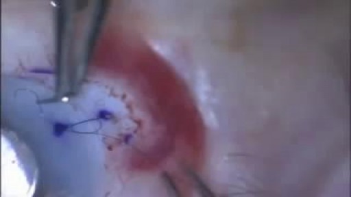
Video demonstrates the fundamental components of placing your first suture.
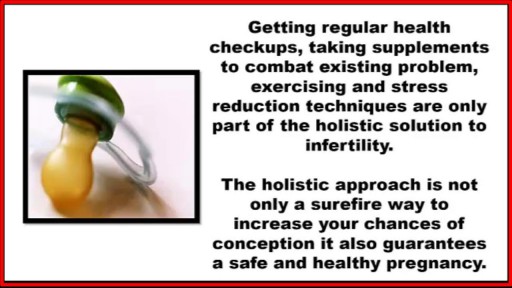
http://how-to-get-pregnant.info-pro.co ----- How To Get Pregnant, Ways To Get Pregnant, Best Days To Get Pregnant, Easiest Way To Get Pregnant. Signs of Infertility. What exactly is infertility? The problems with either conceiving a child, or with carrying out the pregnancy to its eventual fruitful end, fall under the definition of infertility. Infertility is the incapability of an individual to become pregnant, in case of females, or the incapability to induce pregnancy, in case of the males. The inability of an individual to carry out a pregnancy to its full term is also dubbed infertility. How does one recognize infertility? What are the signs of infertility? Signs of infertility are not always evident. Most people go through life without knowing there is a problem with their reproductive systems, attributing failed pregnancies to providence. In fact, miscarriages are the most common indicator of infertility. Signs of infertility in women: In women, the signs of infertility are more readily recognized as compared to men. Endometriosis causes the lining of the uterus to grow outside the uterus. Bacterial infections may begin around the uterus and spread to other reproductive organs, resulting in infertility. Fibroids in the uterus are indicative of infertility. Tumors in the cervix often cause stenosis, or narrowing of the cervix, which is a common indicator of infertility. Ovulating before the tenth day and after the twentieth day of one's monthly cycle, pre-menstrual spotting, menopausal symptoms, etc. are indicative of luteal phase defect, and thus in turn are signs too.
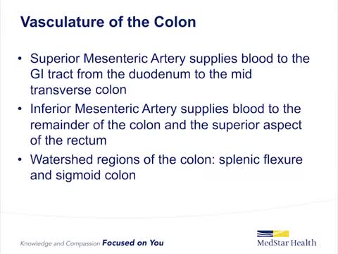
Ischemic colitis occurs when blood flow to part of the large intestine (colon) is reduced, usually due to narrowed or blocked blood vessels (arteries). The diminished blood flow doesn't provide enough oxygen for the cells in your digestive system. Ischemic colitis can cause pain and may damage your colon. Any part of the colon can be affected, but ischemic colitis usually causes pain on the left side of the belly area (abdomen). The condition can be misdiagnosed because it can easily be confused with other digestive problems. Ischemic colitis may heal on its own. But you may need medication to treat ischemic colitis or prevent infection, or you may need surgery if your colon has been damaged. Symptoms ShareTweet Oct. 13, 2015 References Products and Services Newsletter: Mayo Clinic Health Letter See also Abdominal pain Colonoscopy Color Blue Detects Colon Cancer CT scan CT scans: Are they safe? Diarrhea Ultrasound Advertisement Mayo Clinic does not endorse companies or products. Advertising revenue supports our not-for-profit mission. Advertising & Sponsorship PolicyOpportunitiesAd Choices Mayo Clinic Store Check out these best-sellers and special offers on books and newsletters from Mayo Clinic. NEW! – The Mayo Clinic Diet, Second Edition Treatment Strategies for Arthritis Mayo Clinic on Better Hearing and Balance Keeping your bones healthy and strong The Mayo Clinic Diet Online Ads by Swoop Psoriasis Treatment www.informationaboutpsoriasis.com Explore a Treatment Option for Moderate to Severe Plaque Psoriasis Immune Biomarker PD-L1 - Discover the Science iobiomarkers.bmsinformation.com Understanding Assay Results for PD-L1 is Crucial for Treatment Decisions. Biomarker PD-L1 Information - Easy to Download Resources iobiomarkers.bmsinformation.com Explore the Role of PD-L1 in Immuno-Oncology & the Evolving Biomarker Landscape.

Whether you need to boost your energy or curb an afternoon craving, staying hydrated is made easy with these tips.

Master perfect plank form and you .ll strengthen your core in no time.

Medical providers divide the spinal cord into four distinct regions. Knowing the region in which the injury is located is often the key to understanding diagnosis and treatment. The four spinal cord regions are: The cervical spinal cord: This is the topmost portion of the spinal cord, where the brain connects to the spinal cord, and the neck connects to the back. This region consists of eight vertebrae, commonly referred to as C1-C8. All spinal cord numbers are descending, so C1 is the highest vertebra, while C8 is the lowest in this region. The thoracic spinal cord: This section forms the middle of the spinal cord, containing twelve vertebrae numbered T1-T12. The lumbar spinal cord: This is a lower region of the spinal cord, where your spinal cord begins to bend. If you put your hand in your lower back, where your back bends inward, you're feeling your lumbar region. There are five lumbar vertebrae, numbered L1-L5.