- Physical Examination
- Surgical Examination
- Ophthalmology
- Clinical Skills
- Orthopedics
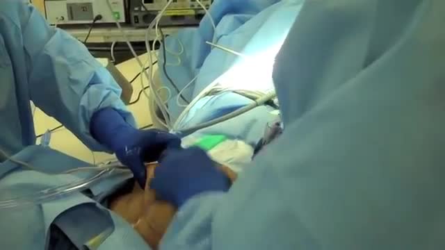
- Surgery Videos
- Laparoscopy
- Pediatrics
- Funny Videos
- Cardiothoracic Surgery
- Nursing Videos
- Plastic Surgery
- Otorhinolaryngology
- Histology and Histopathology
- Neurosurgery
- Dermatology
- Pediatric Surgery
- Urology
- Dentistry
- Oncology and Cancers
- Anatomy Videos
- Health and Fitness
- Radiology
- Anaesthesia
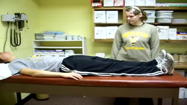
- Physical Therapy
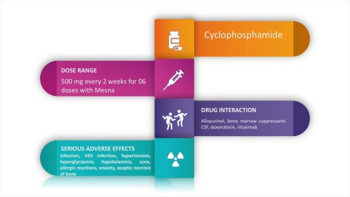
- Pharmacology
- Interventional Radiology
- Cardiology
- Endocrinology
- Gynecology
- Emergency Medicine
- Psychiatry and Psychology
- Childbirth Videos
- General Medical Videos
- Nephrology
- Physiology
- Diet and Food Health
- Diabetes Mellitus
- Neurology
- Women Health
- Osteoporosis
- Gastroenterology
- Pulmonology
- Hematology
- Rheumatology
- Toxicology
- Nuclear Medicine
- Infectious Diseases
- Vascular Disease
- Reproductive Health
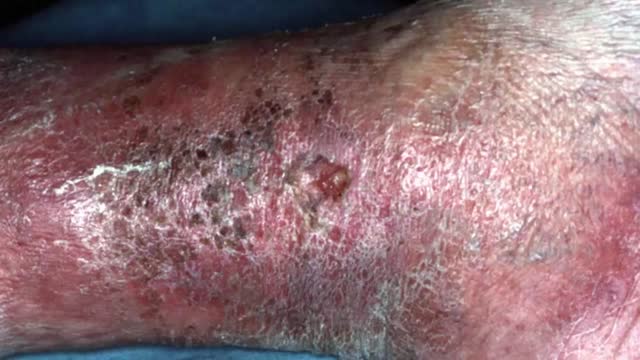
- Burns and Wound Healing
- Other
Top videos

An animation illustrating valvular heart surgery
@http://www.doctorsgate.blogspot.com/
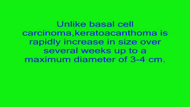
A video shows description of keratoacanthoma with multiple pictures.For more images,Diagrams, MNEMONICS , ALGORITHMS ..join us on http://www.doctorsgate.blogspot.com/
USMLE Step 2 CS - BPH - This is just preview video. To get full access please visit our website : www.usmletutoring.com
USMLE Step 2 CS - Hemetemesis This is just preview video. To get full access please visit our website : www.usmletutoring.com

http://www.mediplus.co.uk A new and safer method of inserting a Foley catheter suprapubically. The technique allows the insertion to be carried out in an Outpatient setting, thus saving time, cost and effort. By using the Seldinger technique, the product reduces the chances of bowel or bladder perforation and resultant morbidity.
The product has been chosen by The NHS National Technology Adoption Centre to help facilitate adoption of the product
Anatomy of the Heart
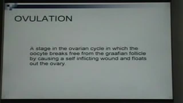
Ovulation
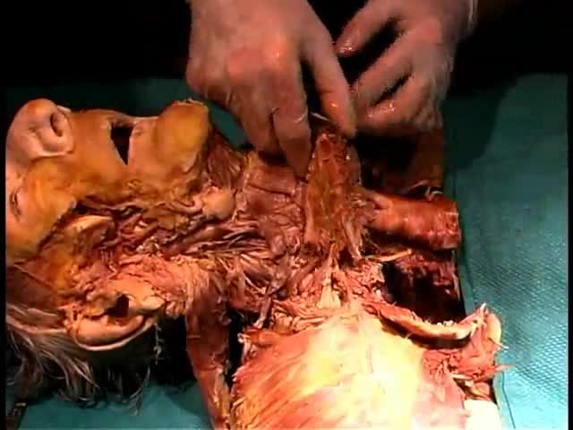
Anatomy of The Deep Neck
Rickets is the softening and weakening of bones in children, usually because of an extreme and prolonged vitamin D deficiency. Vitamin D promotes the absorption of calcium and phosphorus from the gastrointestinal tract. A deficiency of vitamin D makes it difficult to maintain proper calcium and phosphorus levels in bones, which can cause rickets. Adding vitamin D or calcium to the diet generally corrects the bone problems associated with rickets. When rickets are due to another underlying medical problem, your child may need additional medications or other treatment. Some skeletal deformities caused by rickets may require corrective surgery.
Hoover's sign of leg paresis is one of two signs named for Charles Franklin Hoover. It is a maneuver aimed to separate organic from non-organic paresis of the leg. The sign relies on the principle of synergistic contraction. ... Feeling this would indicate an organic cause of the paresis.
Yeast Infection Symptoms in Women and Men - Causes, Signs, photos, Pictures of Candidiasis Fungus
Good and Bad Foods to Eat
Encephalitis (en-sef-uh-LIE-tis) is inflammation of the brain. Viral infections are the most common cause of the condition. Encephalitis can cause flu-like symptoms, such as a fever or severe headache. It can also cause confused thinking, seizures, or problems with senses or movement. However, many cases of encephalitis result in only mild flu-like symptoms or even no symptoms. Severe cases of encephalitis, while relatively rare, can be life-threatening. Because the course of any single case of encephalitis can be unpredictable, it's important to get a timely diagnosis and treatment.

URBN Dental is at your service to provide professional dental tips and quality service. Do you ever wonder how often you should be changing your toothbrush (or toothbrush head if you are using an electric one)? Switching your electric head or tossing your toothbrush is recommended every three months. If you are sick or have a cold sore or canker sore, it’s highly suggested to also switch your brushes. The toothbrush bristles can contain a lot of harmful bacteria and need to be replaced to decrease your risk of potential systemic illnesses. Also the bristles themselves can get worn and frayed and will decrease the efficiency of the toothbrush. Without replacing the toothbrush bristles, you suffer from a greater risk of encountering gum disease and cavities, so be sure you swap those bristles! Schedule a dental appointment now to learn more! Click on our website to book today: https://www.urbndental.com/
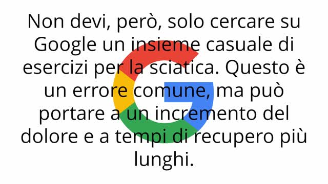
http://sciatica-rimedi.good-info.co Nervo Sciatico, Accavallamento Nervi, Lombosciatalgia Sintomi E Cure, Dolore Coscia, Sciatica. Come curare la sciatica a casa Se hai avuto abbastanza sciatalgia a dirigere la tua vita, non disperare! ti mostrerò tre dei più comuni trattamenti casalinghi per la sciatica, e come usarli per ridurre il dolore in modo rapido. La parte migliore di questi trattamenti è che possono curare la sciatica, e non solo coprire il dolore. Quindi, cominciamo... 1. Programma di esercizi a casa I programmi di esercizio sono una componente importante di qualsiasi piano di trattamento della sciatica. Con l'allungamento e il rafforzamento di parti del corpo che possono causare l'irritazione del nervo sciatico, è possibile ridurre il dolore e accelerare il recupero. Gli esercizi più efficaci dipendono dalla ragione di fondo per cui soffri di sciatica. La sciatica causata da un'ernia del disco, per esempio, non viene trattata con gli stessi esercizi della sciatica causata da stenosi spinale. È anche importante mantenere il corpo rilassato, per consentirgli di guarire. Un modo grandioso per farlo, senza aggravare la tua condizione, è camminare a ritmo sostenuto. Altre attività leggere possono avere un effetto simile, ma se qualcosa fa male è necessario fermarsi immediatamente. Suggerimento gratuito: è essenziale che non ci si riduca a letto a causa del dolore. Stare sdraiati a letto per più di due giorni ha dimostrato peggiorare la sciatica, perché i muscoli si irrigidiscono e si indeboliscono. 2. Bilancia la tua dieta Curare la sciatica in modo permanente, spesso significa trattare più che la semplice causa fisica. Per impedire che il dolore si ripresenti, dovrai anche migliorare la tua dieta. Uno dei modi più semplici per ridurre il dolore associato con sciatica è quello di bere più acqua. Quando si è disidratati, parti della colonna vertebrale si sgonfiano. Questo può causare ulteriore pressione sul nervo sciatico. Se possibile, si dovrebbe anche cercare di evitare alimenti infiammatori. Gli alimenti infiammatori sono troppi, per elencarli in questo articolo, ma qualsiasi alimento dotato di elevato contenuto di zucchero può, potenzialmente, portare a infiammazione e ad aumento del dolore. 3. Rimedi casalinghi I rimedi casalinghi possono fare una grande differenza per tua sciatalgia, spesso in tempi relativamente brevi. La cosa grandiosa dei rimedi casalinghi è che non richiedono prescrizione o ingredienti costosi. Uno dei più semplici rimedi casalinghi sono le noccioline. Questo perché le arachidi contengono un sacco di magnesio, che è cruciale per consentire muscoli di rilassarsi.
Systemic lupus erythematous is an autoimmune condition characterised by damage to organ systems due to autoantibodies and immune complex deposition. Genes, epigenetic changes and environment play a role in its pathogenesis. SLE is a truly multi system disease causing widespread clinical manifestations in almost all organ systems. Autoantibodies in SLE are numerous and mainly include ANA, dsDNA, Sm and others.
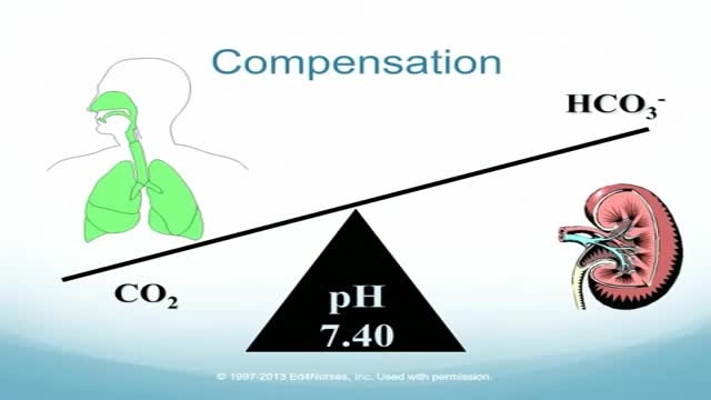
Four-Step Guide to ABG Analysis Is the pH normal, acidotic or alkalotic? Are the pCO2 or HCO3 abnormal? Which one appears to influence the pH? If both the pCO2 and HCO3 are abnormal, the one which deviates most from the norm is most likely causing an abnormal pH. Check the pO2. Is the patient hypoxic?
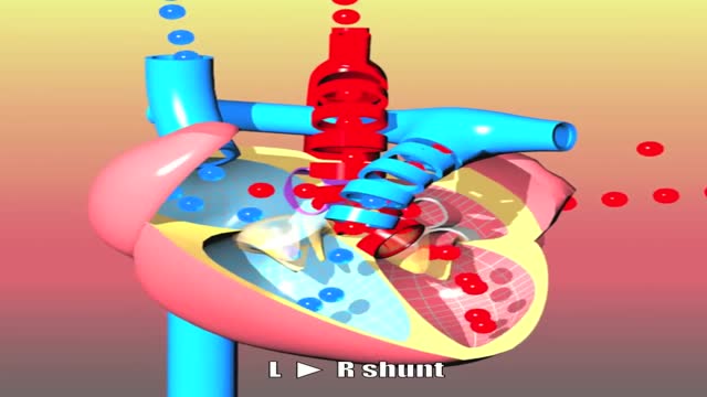
Congenital heart defects I: ASD, VSD, AS, PS, PDA and PFO
The Babies Hooked On Heroin |
Laparoscopic use of Palmer's Point
