- Physical Examination
- Surgical Examination
- Ophthalmology
- Clinical Skills
- Orthopedics
- Surgery Videos
- Laparoscopy
- Pediatrics
- Funny Videos
- Cardiothoracic Surgery
- Nursing Videos
- Plastic Surgery
- Otorhinolaryngology
- Histology and Histopathology
- Neurosurgery
- Dermatology
- Pediatric Surgery
- Urology
- Dentistry
- Oncology and Cancers
- Anatomy Videos
- Health and Fitness
- Radiology
- Anaesthesia
- Physical Therapy
- Pharmacology
- Interventional Radiology
- Cardiology
- Endocrinology
- Gynecology
- Emergency Medicine
- Psychiatry and Psychology
- Childbirth Videos
- General Medical Videos
- Nephrology
- Physiology
- Diet and Food Health
- Diabetes Mellitus
- Neurology
- Women Health
- Osteoporosis
- Gastroenterology
- Pulmonology
- Hematology
- Rheumatology
- Toxicology
- Nuclear Medicine
- Infectious Diseases
- Vascular Disease
- Reproductive Health
- Burns and Wound Healing
- Other
Top videos
Most people have general anesthesia right before surgery. This means you will be asleep and pain-free. Other kinds of anesthesia, like regional anesthesia or a block, may also be used for this surgery. The tissue to replace your damaged ACL will come from your own body or from a donor. A donor is a person who has died and chose to give all or part of his or her body to help others. Tissue taken from your own body is called an autograft. The two most common places to take tissue from are the knee cap tendon or the hamstring tendon. Your hamstring is the muscle behind your knee. Tissue taken from a donor is called an allograft. The procedure is usually performed with the help of knee arthroscopy. With arthroscopy, a tiny camera is inserted into the knee through a small surgical cut. The camera is connected to a video monitor in the operating room. Your surgeon will use the camera to check the ligaments and other tissues of your knee. Your surgeon will make other small cuts around your knee and insert other medical instruments. Your surgeon will fix any other damage found, and then will replace your ACL by following these steps: The torn ligament will be removed with a shaver or other instruments. If your own tissue is being used to make your new ACL, your surgeon will make a larger cut. Then, the autograft will be removed through this cut. Your surgeon will make tunnels in your bone to bring the new tissue through. This new tissue will be in the same place as your old ACL. Your surgeon will attach the new ligament to the bone with screws or other devices to hold it in place. As it heals, the bone tunnels fill in. This holds the new ligament in place. At the end of the surgery, your surgeon will close your cuts with sutures (stitches) and cover the area with a dressing. You may be able to view pictures after the procedure of what the doctor saw and what was done during the surgery.
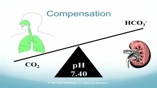
Four-Step Guide to ABG Analysis Is the pH normal, acidotic or alkalotic? Are the pCO2 or HCO3 abnormal? Which one appears to influence the pH? If both the pCO2 and HCO3 are abnormal, the one which deviates most from the norm is most likely causing an abnormal pH. Check the pO2. Is the patient hypoxic?
The Babies Hooked On Heroin |
Laparoscopic use of Palmer's Point
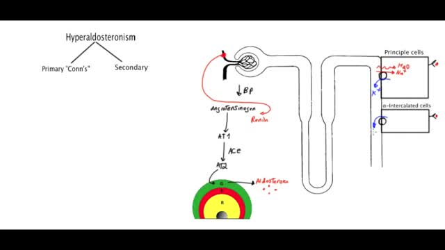
Primary aldosteronism, also known as primary hyperaldosteronism or Conn's syndrome, is excess production of the hormone aldosterone by the adrenal glands resulting in low renin levels. Often it produces few symptoms. Most people have high blood pressure which may cause poor vision or headaches.
Cardiovascular disease (CVD) is a general term that describes a disease of the heart or blood vessels. Blood flow to the heart, brain or body can be reduced as the result of a blood clot (thrombosis), or by a build-up of fatty deposits inside an artery that cause the artery to harden and narrow (atherosclerosis).
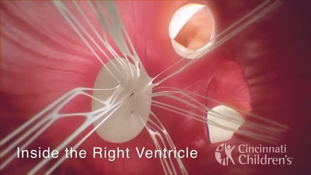
Interrupted aortic arch (IAA) is the absence or discontinuation of a portion of the aortic arch, the section of the aorta that turns downward toward the lower half of the body. Once the diagnosis of this rare defect is suspected and confirmed, treatment and surgical intervention are vitally important. Heart models and animation were developed by the Cincinn
Our nervous system is involved in everything our body does, from maintaining our breath to controlling our muscles. Our nerves are vital to all we do; therefore, nerve pain and damage can heavily influence our quality of life. In Discovery News' latest video, "Why Can't We Reverse Nerve Damage?" host Lissette Padilla explains the central nervous system (CNS) has certain proteins that inhibit cell regeneration, because each cell in the nervous system has a unique function on the pathway, like a circuit, and can't be replaced.
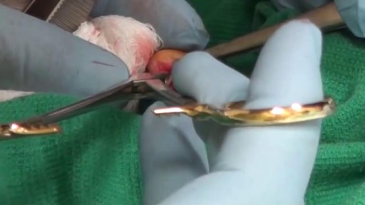
Simple interrupted suturing is the most basic and most important of the suturing techniques.
your DNA Journey
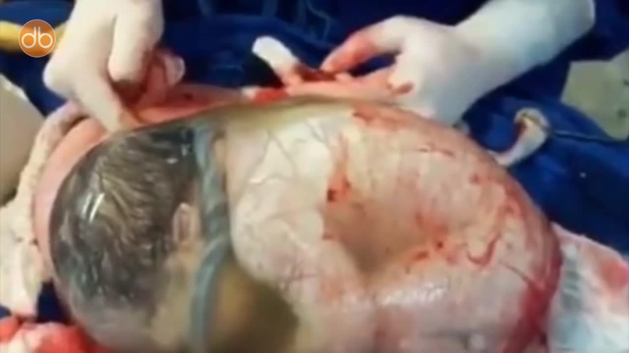
Baby Born Still Inside The Amniotic Sac
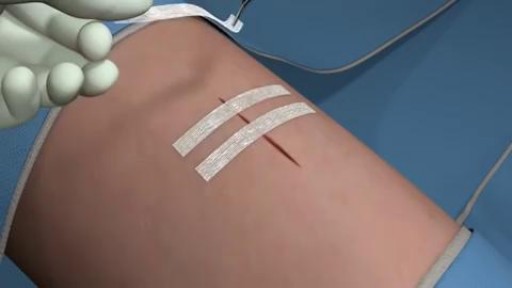
The Steri-Strip™ brand offers an extended line of adhesive skin closures to meet your needs. Our versatile, cost-saving, non-invasive Steri-Strips™ have many applications and come in a variety of sizes. Options include reinforced, elastics, "blend-tone," an antimicrobial and a waterproof wound closure system.
Stuck with an Embroidery Needle
why you should keep your ears clean.
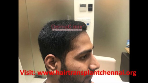
4000 Grafts Hair Transplant Surgery Result after 7months by Dr. Ariganesh Chandrasegaran (MD, PGI Chandigarh) at DermaClinix. DermaClinix is known for the best hair transplant in Chennai as well as across India. Dramatic transformation by our flagship patented procedure B.E.S.T. (Bio-Enhanced Simultaneous Transplant Technique) FUE which helps in achieving almost complete survival with early and natural looking results. Book Your Free Consultation: +91 8939636222, 04428282606. More at https://www.hairtransplantchennai.org

http://combatir-la-ansiedad.good-info.co/ --- Como Calmar La Ansiedad, Ataque De Ansiedad Que Hacer, Remedios Naturales Para La Ansiedad. Como ir curando tu ansiedad... ¿De seguro te sientes desesperado por no poder saber como poder curar tu ansiedad cierto? No te preocupes, esto se puede solucionar. Muchas veces probamos todo tipo de tratamientos que al final muchas veces hace que todo se haga mucho mas dificil. Tanto que inclusive al final sentimos que hemos empeorado todo ¿cierto? Sin la guia adecuada, parece como si nunca fueras encontrar el metodo correcto para poder vivir una vida libre de ansiedad. Afortunadamente actualmente existe una guia la cual te ahorrara el camino dificil y asi podras ir eliminando todos esos sintomas que te hace sentir miedo y ansiedad. para que asi puedas vivir una vida en paz y llena de alegria. Si quieres conocer más tan solo visita este enlace. http://combatir-la-ansiedad.good-info.co
Fibromialgia Remedios Naturales, Como Curar La Fibromialgia, Medicamento Para Fibromialgia. http://fibromialgia-cura.info-pro.co/ -- Medicina Natural Para La Fibromialgia. Se estima que 5 millones de estadounidenses sufren de fibromialgia. Los dolores profundos y crónicos pueden tener un enorme impacto en la salud física y emocional. Desafortunadamente, los tratamientos son pocos y distantes entre sí, y los que existen a menudo vienen con la posibilidad de efectos secundarios desagradables. La medicina natural para la fibromialgia puede ser una bendición para los enfermos que la padecen. Estas terapias complementarias, suelen ser efectivas y pueden mejorar la calidad de vida y rejuvenecer un cuerpo atormentado por el dolor crónico. El masaje es a menudo uno de los métodos más eficaces para reducir los síntomas de fibromialgia. Alivia la rigidez, mejora el rango de movimiento, reduce el dolor y ayuda a controlar el estrés. Una técnica llamada liberación miofascial es especialmente adecuado para la fibromialgia el dolor calmante. La fascia es un tejido conectivo delgada que cubre y se extiende a lo largo del músculo. Los pacientes con fibromialgia sufren comúnmente de apriete de la fascia que contribuye al dolor y la fatiga muscular. La liberación miofascial es una técnica suave que relaja la fascia y reduce el dolor asociado. Las terapias naturales pueden ayudar desde dentro también. La investigación ha encontrado que muchos enfermos de fibromialgia tienen niveles bajos de vitamina D y magnesio. 100% natural aliviar el dolor y mejorar tu calidad de vida solo haciendo click aqui. http://fibromialgia-cura.info-pro.co

Chicken Skin, Chicken Skin On Legs, Keratosis Pilaris Treatment Over The Counter, Kp Vitamin A--- http://banishmybumps.plus101.com/ --- What Causes KP? The particular cause of Keratosis Pilaris is unidentified. Usually, it happens when there is a difficulty on the production of the keratin that is called hyper keratinizatinization. Keratosis Pilaris is believed to be inherited partially in origin. About fifty percent up to seventy percent of patients with this type of skin condition have identified genetic predisposition and has a higher rate of affected members of the family. Most people have a solid family history of follicular pilaris. The primary cause may be related partly to hypersensitivity reactions as well as the overall skin dryness. This skin condition is closely related also to dry skin, allergies, asthma, eczema, atopic dermatitis and rhinitis. The red bumps in Keratosis Pilaris seem to rise from the unnecessary build-up of keratin dry skin particles and very small at the hair follicles opening. The skin as observed under the hand held microscope demonstrates mild condensing, plugging and hyperkeratosis of your hair follicle. Your upper skin layers may have a number of dilation of the minor superficial blood vessels. In this manner, it gives the skin a flushed or reddish appearance. Keratosis Pilaris is not infectious, therefore people surrounding you are safe from this irritating skin condition. The condition is worse during winter. Summer season is normally a reprieve as it brings moisture and usually allows the skin condition to smooth up more than normal. The aggravating is associated directly with lower moisture, tight clothes and skin dryness that cause to rub the red bumps all the times. Many people stated that this skin condition worsened during the period of pregnancy before and after child birth. Medical doctors and patients as well, believed that keratosis pilaris is also related to food intake. So, large intakes of spicy foods increase the form of red bumps that make them more visible. In some way, patients that eliminated or reduced milk as well as the derivatives from their balance diet stated the improvement of their condition. Keratosis Pilaris can be difficult to treat, but it is possible to seek permanent relief. New research shows that all-natural treatment systems, such as BanishMyBumps, are successful. You can learn more at http://banishmybumps.plus101.com/
Watch that Thyroid Removal Surgery
