- Physical Examination
- Surgical Examination
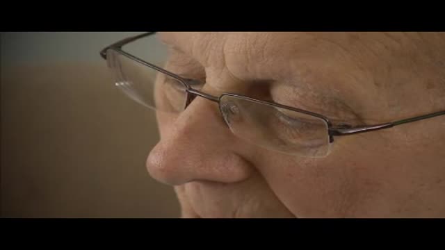
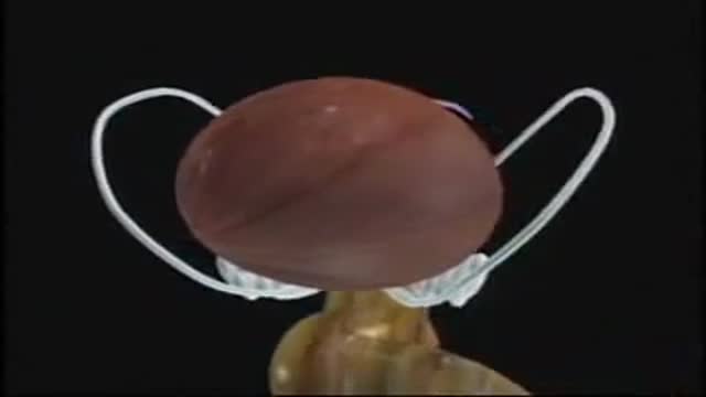
- Ophthalmology
- Clinical Skills
- Orthopedics
- Surgery Videos
- Laparoscopy
- Pediatrics
- Funny Videos
- Cardiothoracic Surgery
- Nursing Videos
- Plastic Surgery
- Otorhinolaryngology
- Histology and Histopathology
- Neurosurgery
- Dermatology
- Pediatric Surgery
- Urology
- Dentistry
- Oncology and Cancers
- Anatomy Videos
- Health and Fitness
- Radiology
- Anaesthesia
- Physical Therapy
- Pharmacology
- Interventional Radiology
- Cardiology
- Endocrinology
- Gynecology
- Emergency Medicine
- Psychiatry and Psychology
- Childbirth Videos
- General Medical Videos
- Nephrology
- Physiology
- Diet and Food Health
- Diabetes Mellitus
- Neurology
- Women Health
- Osteoporosis
- Gastroenterology
- Pulmonology
- Hematology
- Rheumatology
- Toxicology
- Nuclear Medicine
- Infectious Diseases
- Vascular Disease
- Reproductive Health
- Burns and Wound Healing
- Other
Top videos
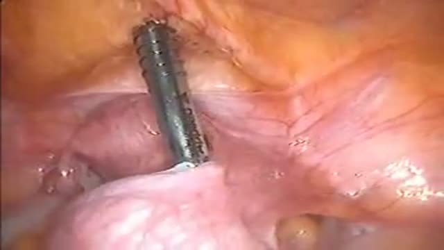
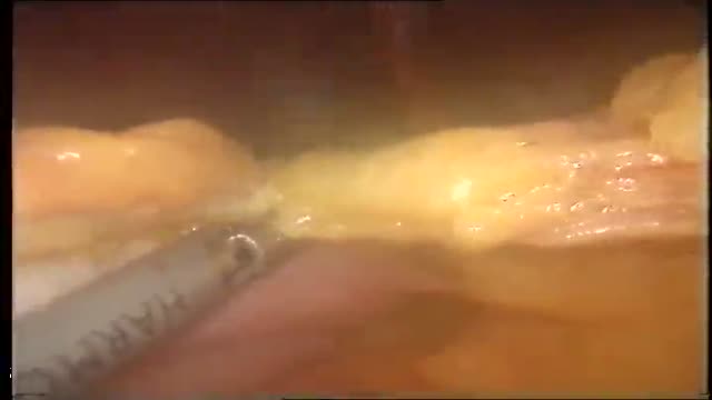
Laparoscopic Removal of Ovarian Cyst
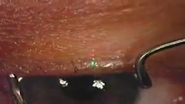
In PRK the epithelium (top layer of the cornea) is scraped off and then the laser treatment is applied. A contact lens is used as a "bandage" to decrease discomfort. The epithelium then grows back over the bare area during the next few days.
In LASEK the epithelium is exposed to 20% alcohol which helps separate epithelium from the cornea. The epithelium is pushed to one side and laser treatment applied. The epithelial layer is replaced back onto the eye and held in place with a contact lens. The contact lens is then removed a few days later. LASEK is hence a "no knife"/flap operation.
Epi-Lasik is a similar procedure that uses a keratome like that used for Lasik, but engineered to only separate the epithelium. The epithelium is left on a hinge, laser treatment applied and flap replaced.
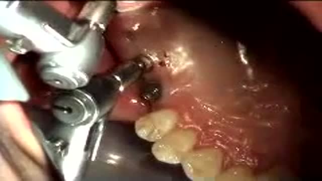
The sinus is a hollow area in the back part of the mouth, when people lost thier teeth in this area, the bone will quickly resorbed, One way we can place implant into this area is by put graft materials in the sinus and hoped that the bone will take and allow us to place implant into the grafted bone. The grafting increases the time and the risk of successful implantation.
Visit http://www.nasalcleanse.com/index.php after watching our video on NasalCare nasal irrigation versus sinus sprays for sinusitis & sinus congestion relief. Learn how & why this natural sinus remedy really works! Unlike the temporary relief offered by chemical-laden nasal sprays, our patented NasalCare® Nasal Rinse System ensures comfortable and effective delivery throughout the nasal passages, preventing sinus infection, allergy and post nasal drip. A soothing mix of sea salt and Aloe Vera extract washes away nasal irritants and the common causes of colds and flu without the potential addiction that comes with nasal spray use. NasalCare also acts as a sinus wash for allergy treatment. Used for centuries in the Orient as a preventative measure for all sinus conditions, nasal irrigation is just catching on here. Catch us now and stop catching colds and the flu – the natural way! Order online at: http://www.nasalcleanse.com/index.php.
AL EMADI HOSPITAL-QATAR-DOHA
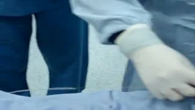
LIPOSUCTION IN QATAR surgery
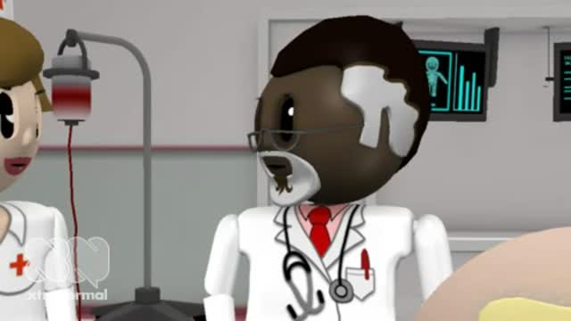
A funny animation showing A Stupid Surgeon and MedicalVideos.US
Jane Seymour sheds light on atrial fibrillation and AF-related stokes.
Patients open up about living with cancer.

Brain Worm Infection

Hip Replacement Surgery
Robotic Prostatectomy Cornell Athermal Robotic Technique
Histology of Fibrocartilage
Inside the living body
What is myositis? Myositis means muscle inflammation, and can be caused by infection, injury, certain medicines, exercise, and chronic disease. Some of the chronic, or persistent, forms are idiopathic inflammatory myopathies, and those are the diseases we discuss here. "Idiopathic" means that the cause is unknown.

Sed rate, or erythrocyte sedimentation rate ( ESR ), is a blood test that can reveal inflammatory activity in your body. A sed rate test isn't a stand-alone diagnostic tool, but it can help your doctor diagnose or monitor the progress of an inflammatory disease. ... Inflammation can cause the cells to clump.

Amnesia refers to the loss of memories, such as facts, information and experiences. Though having no sense of who you are is a common plot device in movies and television, real-life amnesia generally doesn't cause a loss of self-identity. Instead, people with amnesia — also called amnestic syndrome — are usually lucid and know who they are, but may have trouble learning new information and forming new memories. Amnesia can be caused by damage to areas of the brain that are vital for memory processing. Unlike a temporary episode of memory loss (transient global amnesia), amnesia can be permanent. There's no specific treatment for amnesia, but techniques for enhancing memory and psychological support can help people with amnesia and their families cope.
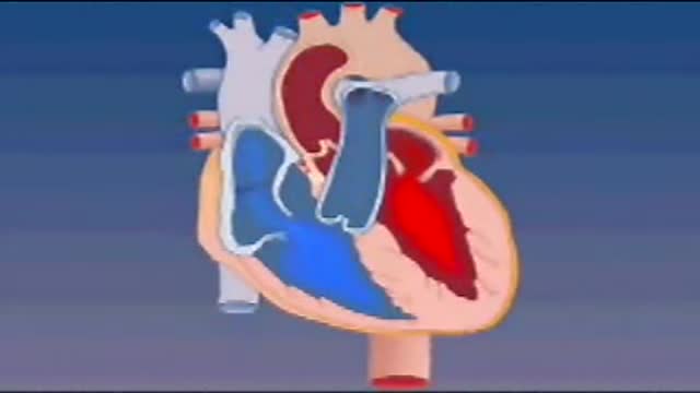
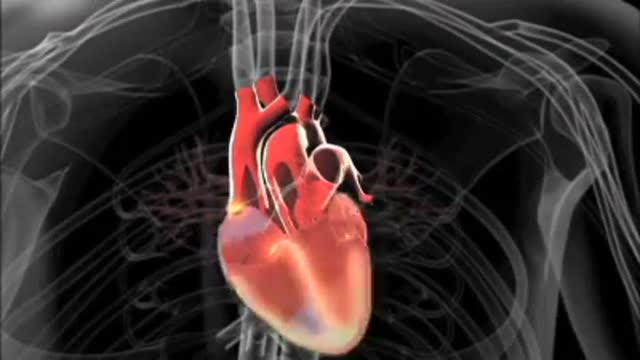
Blood enters the heart through two large veins, the inferior and superior vena cava, emptying oxygen-poor blood from the body into the right atrium. As the atrium contracts, blood flows from your right atrium into your right ventricle through the open tricuspid valve.

What is your mental age?

Gastroparesis -- literally “paralyzed stomach” -- is a serious condition manifested by delayed emptying of stomach contents into the small intestine after a meal. There is no cure for gastroparesis, but treatment can speed gastric emptying and relieve gastrointestinal symptoms such as nausea and vomiting.
