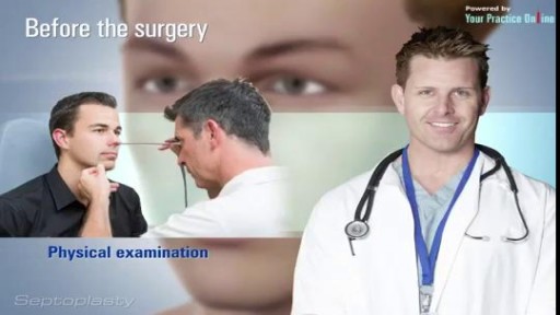
- Physical Examination
- Surgical Examination
- Ophthalmology
- Clinical Skills
- Orthopedics
- Surgery Videos
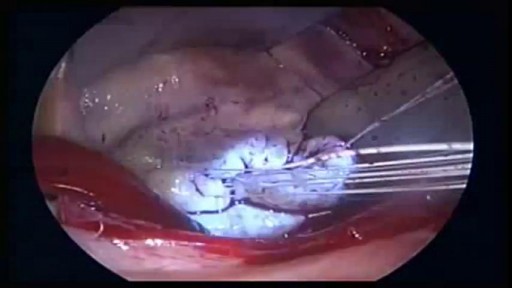
- Laparoscopy
- Pediatrics
- Funny Videos
- Cardiothoracic Surgery
- Nursing Videos
- Plastic Surgery
- Otorhinolaryngology
- Histology and Histopathology
- Neurosurgery
- Dermatology
- Pediatric Surgery
- Urology
- Dentistry
- Oncology and Cancers
- Anatomy Videos
- Health and Fitness
- Radiology
- Anaesthesia
- Physical Therapy
- Pharmacology
- Interventional Radiology
- Cardiology
- Endocrinology
- Gynecology
- Emergency Medicine
- Psychiatry and Psychology
- Childbirth Videos
- General Medical Videos
- Nephrology
- Physiology
- Diet and Food Health
- Diabetes Mellitus
- Neurology
- Women Health
- Osteoporosis
- Gastroenterology
- Pulmonology
- Hematology
- Rheumatology
- Toxicology
- Nuclear Medicine
- Infectious Diseases
- Vascular Disease
- Reproductive Health
- Burns and Wound Healing
- Other
Top videos
Robot helps disabled patients regain control of their hands 1
How to Treat Cuts & Scrapes | First Aid Training
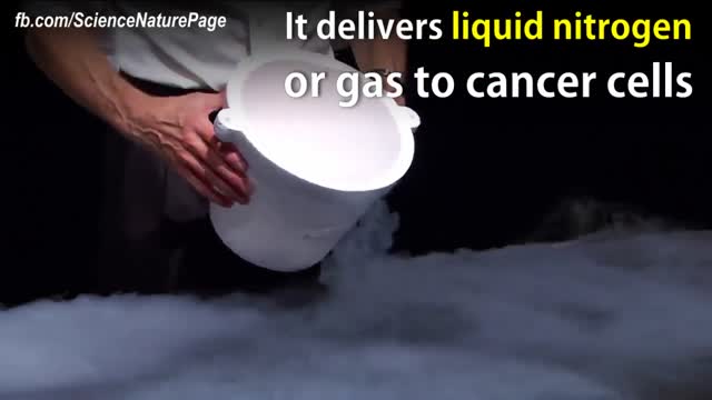
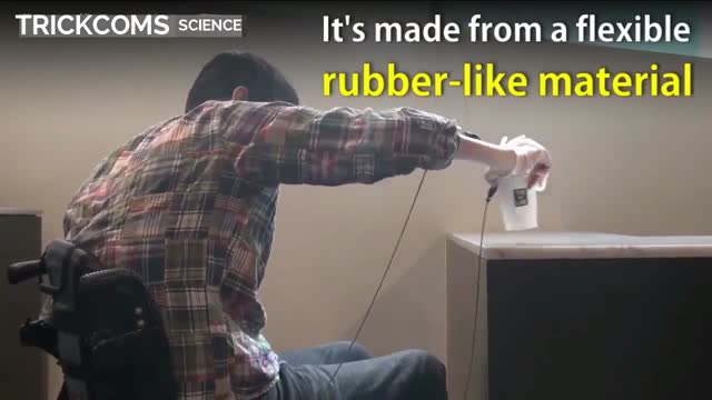
In a small but promising Phase II clinical trial of breast cancer treatment, cryoablation killed 25 early-stage tumors in 13 women. The tumors ranged in size from .5 cm (very small) to 5.8 cm (very large), with an average size of 1.7cm. Patients were first given a local anesthesia with mild sedation before physicians used ultrasound with or without computed tomography (CT) imaging to guide needle-like probes to deliver very low temperature gas to the tumor site. The ultra-cold gas forms a ball of ice around the probe tip, then expands and destroys surrounding tumor cells. A harmless saline solution was first injected into the chest wall and skin of the breast to protect the tissue surrounding the tumor from the freezing effects. Patients experienced very little pain and most healed completely within six months with no complications and with little or no scarring. The cryotherapy margins of each participant were biopsied immediately after the procedures, and all were negative, with no evidence of cancerous tissue. All 13 patients were without recurrence at an average of 18 months and up to five years following the procedure. These results are promising, but larger studies with lengthier follow-up are needed to determine whether cryotherapy as effective as lumpectomy. A study involving cryoablation of mouse tumors at the University of Michigan Comprehensive Cancer Center found that the freezing procedure also works like a vaccine, boosting the immune system to reduce the likelihood of recurrence. Just how quickly the tumor was frozen made a difference: a 30-second freeze killed tumors and also boosted the immune system, inhibiting metastases to the lungs. A slower freezing lasting several minutes destroyed tumors just as effectively, but actually suppressed the immune system, resulting in greater metastases to the lungs.
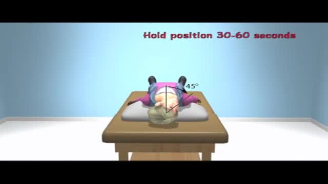
Benign paroxysmal positional vertigo (BPPV) is one of the most common causes of vertigo — the sudden sensation that you're spinning or that the inside of your head is spinning. Benign paroxysmal positional vertigo causes brief episodes of mild to intense dizziness. Benign paroxysmal positional vertigo is usually triggered by specific changes in the position of your head. This might occur when you tip your head up or down, when you lie down, or when you turn over or sit up in bed. Although benign paroxysmal positional vertigo can be a bothersome problem, it's rarely serious except when it increases the chance of falls. You can receive effective treatment for benign paroxysmal positional vertigo during a doctor's office visit
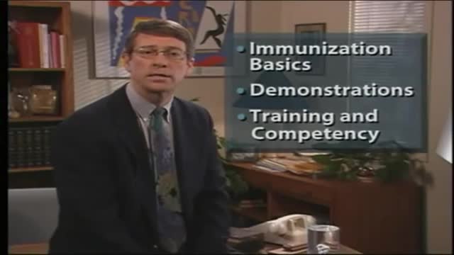
Immunization Techniques
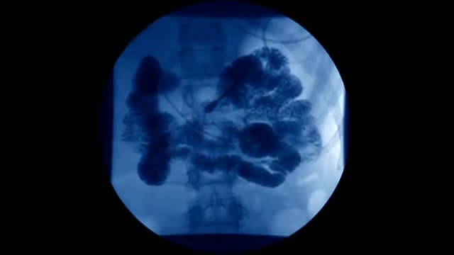
Inside the living body
What is myositis? Myositis means muscle inflammation, and can be caused by infection, injury, certain medicines, exercise, and chronic disease. Some of the chronic, or persistent, forms are idiopathic inflammatory myopathies, and those are the diseases we discuss here. "Idiopathic" means that the cause is unknown.

Sed rate, or erythrocyte sedimentation rate ( ESR ), is a blood test that can reveal inflammatory activity in your body. A sed rate test isn't a stand-alone diagnostic tool, but it can help your doctor diagnose or monitor the progress of an inflammatory disease. ... Inflammation can cause the cells to clump.

Amnesia refers to the loss of memories, such as facts, information and experiences. Though having no sense of who you are is a common plot device in movies and television, real-life amnesia generally doesn't cause a loss of self-identity. Instead, people with amnesia — also called amnestic syndrome — are usually lucid and know who they are, but may have trouble learning new information and forming new memories. Amnesia can be caused by damage to areas of the brain that are vital for memory processing. Unlike a temporary episode of memory loss (transient global amnesia), amnesia can be permanent. There's no specific treatment for amnesia, but techniques for enhancing memory and psychological support can help people with amnesia and their families cope.
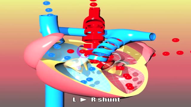
Congenital heart defects I: ASD, VSD, AS, PS, PDA and PFO
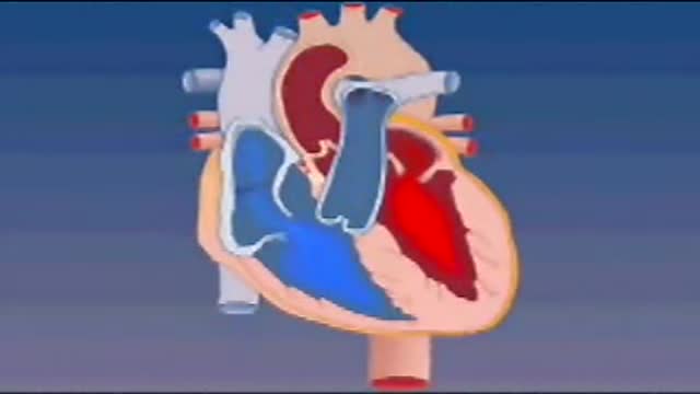
Blood enters the heart through two large veins, the inferior and superior vena cava, emptying oxygen-poor blood from the body into the right atrium. As the atrium contracts, blood flows from your right atrium into your right ventricle through the open tricuspid valve.

Gastroparesis -- literally “paralyzed stomach” -- is a serious condition manifested by delayed emptying of stomach contents into the small intestine after a meal. There is no cure for gastroparesis, but treatment can speed gastric emptying and relieve gastrointestinal symptoms such as nausea and vomiting.
See the effects of cannabis first hand, unedited, on Parkinson's tremor dyskinesia, and voice.
Face transplant allows this man to live a normal life. Hats off to all the surgeons involved!
There are several ways to do minimally invasive aortic valve surgery. Techniques include min-thoracotomy, min-sternotomy, robot-assisted surgery, and percutaneous surgery. To perform the different procedures: Your surgeon may make a 2-inch to 3-inch (5 to 7.5 centimeters) cut in the right part of your chest near the sternum (breastbone). The muscles in the area will be divided. This lets the surgeon reach the heart and aortic valve. Your surgeon may split only the upper portion of your breast bone allowing exposure to the aortic valve. For robotically-assisted valve surgery, the surgeon makes 2 to 4 tiny cuts in your chest. The surgeon uses a special computer to control robotic arms during the surgery. A 3D view of the heart and aortic valve are displayed on a computer in the operating room.
3D Medical
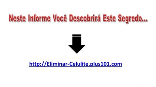
http://eliminar-celulite.plus101.com --- Eliminar Celulite, O Que Fazer Para Acabar Com A Celulite, Como Tirar Celulite Das Pernas. Mas as razões que vou compartilhar são diferentes das que a maioria das outras fontes está tentando fazê-la acreditar. Há um mito fazendo com que algumas mulheres acreditem que certos alimentos e nutrientes irão “eliminar as toxinas que estão causando a celulite”. ISSO É TOTALMENTE FALSO, porque não há toxinas em ou sob sua pele. Se houvesse toxinas se acumulando e ficando presas sob sua pele, você estaria morta. Simples assim. Nosso corpo foi feito para remover toxinas com muita eficácia. Este processo fisiológico acontece 24 horas por dia, 7 dias por semana, sem parar, o tempo todo. Então, a ideia não comprovada de que “toxinas” são a causa de sua celulite significa que a celulite não pode ser revertida ao “eliminá-las” com alguns alimentos, porque elas não estão lá, para começar. Mas não se preocupe, porque eis o que o planejamento alimentar apropriado pode fazer para reverter, ou prevenir, a raiz da causa da celulite em suas pernas, bumbum, quadris e coxas. Uma verdadeira dieta contra a celulite fornece nutrientes em quantidades que impactam positivamente a regulagem e equilíbrio dos hormônios femininos. Esta é a razão principal de o Planejamento Alimentar/Dieta Contra Celulite do "Adeus Celulite" só estar disponível para mulheres que começam com o Método de Exercícios SYMULAST do programa Adeus Celulite. Então se você estiver interessada, vá para: http://eliminar-celulite.plus101.com
Watch that video of a Black Salve Left an Inch Hole In Man's Hole
Eye Brow Transplant Procedure

For that matter, every healthcare professional undergoes this emotional hardship..
