- Physical Examination
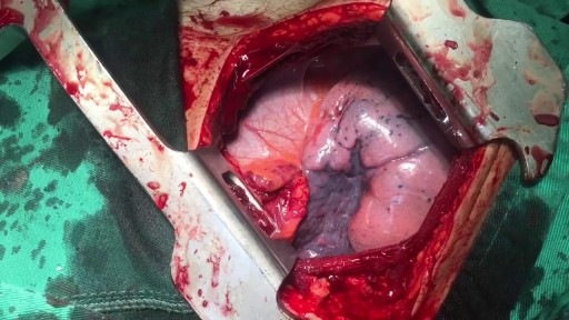
- Surgical Examination
- Ophthalmology
- Clinical Skills
- Orthopedics
- Surgery Videos
- Laparoscopy
- Pediatrics
- Funny Videos
- Cardiothoracic Surgery
- Nursing Videos
- Plastic Surgery
- Otorhinolaryngology
- Histology and Histopathology
- Neurosurgery
- Dermatology
- Pediatric Surgery
- Urology
- Dentistry
- Oncology and Cancers
- Anatomy Videos
- Health and Fitness
- Radiology
- Anaesthesia
- Physical Therapy
- Pharmacology
- Interventional Radiology
- Cardiology
- Endocrinology
- Gynecology
- Emergency Medicine
- Psychiatry and Psychology
- Childbirth Videos
- General Medical Videos
- Nephrology
- Physiology
- Diet and Food Health
- Diabetes Mellitus
- Neurology
- Women Health
- Osteoporosis
- Gastroenterology
- Pulmonology
- Hematology
- Rheumatology
- Toxicology
- Nuclear Medicine
- Infectious Diseases
- Vascular Disease
- Reproductive Health
- Burns and Wound Healing
- Other
Top videos
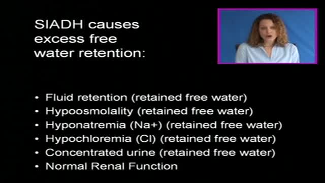
The syndrome of inappropriate antidiuretic hormone (ADH) secretion (SIADH) is defined by the hyponatremia and hypo-osmolality resulting from inappropriate, continued secretion or action of the hormone despite normal or increased plasma volume, which results in impaired water excretion.
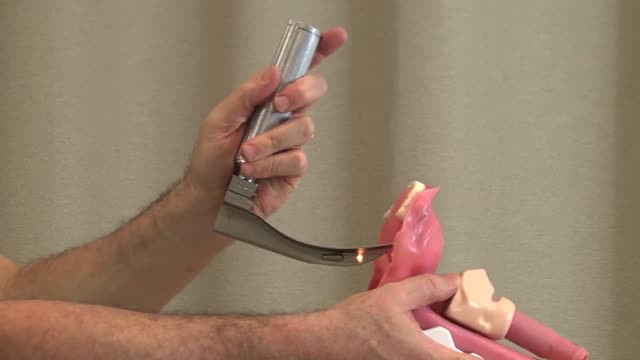
Direct Laryngoscopy: MICU Fellows Airway Course
How To Cleanse Colon


If you suspect that you have sleep apnea, the usual first step is to discuss your suspicions with your primary care physician. If you don’t have a primary care physician, you can go directly to a clinician who is a sleep specialist. But check your health care insurance coverage first. Some policies require you to see a primary care physician first, and some policies limit the sleep centers and testing facilities whose services they will pay for. Unfortunately, you may discover that your policy offers limited or no coverage for the diagnosis and treatment of sleep apnea, in which case you may wish to switch insurers if and when you can.
For strong lungs, chew 3 to 5 peppermint leaves each day. To treat congestion, add a few drops of peppermint oil to a pot of hot water and inhale the steam. You can also drink 2 cups of peppermint tea daily. To make the tea, add 1 teaspoon of dried peppermint leaves to a cup of hot water.
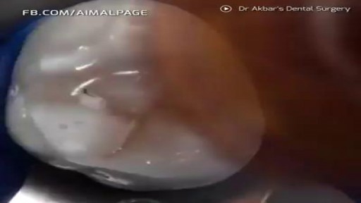
Watching A Dentist Fix Tooth Decay Is Beyond Satisfying.
The first transplant of a bionic eye on a patient with a rare disease.

RLS can make it hard or impossible for you to get enough sleep. Try these home remedies:
watch that video of Pulling out 1 foot long foot of gauze out from face
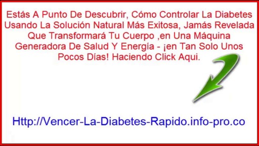
http://vencer-la-diabetes-rapido.info-pro.co/ Como Controlar La Diabetes Tipo 2 Naturalmente Sin Medicamentos, Pre Diabetes Y Diabetes Tipo 1. https://youtu.be/BOSkQ5MnjT0 Que es la Insulina? Una definición practica sin adentrarnos en terminos estrictamente medicos es que la insulina es una hormona formada por 51 aminoácidos. Dentro del páncreas, las células beta producen la hormona llamada insulina. Con cada comida, las células beta liberan insulina para ayudar al cuerpo a utilizar o almacenar en la sangre la glucosa que se obtiene de los alimentos. Su déficit provoca la diabetes mellitus y su exceso provoca hiperinsulinismo con hipoglucemia. En las personas con diabetes tipo 1, el páncreas no produce insulina. Las células beta han sido destruidas y se necesitan inyecciones de insulina para utilizar la glucosa de las comidas. Las personas con diabetes tipo 2 producen insulina, pero sus cuerpos no responden bien a la misma. Algunas personas con diabetes tipo 2 necesitan medicamentos para la diabetes o inyecciones de insulina para ayudar a su cuerpo a utilizar la glucosa para obtener energía. * La insulina no se puede tomar como una píldora, ya que se descompone durante la digestión al igual que la proteína en los alimentos. Se debe inyectar en la grasa debajo de la piel para que llegue a la sangre. Existen diferentes tipos de insulina en función de la rapidez con que trabajan, y en funcion de su duración. La insulina viene en diferentes concentraciones, la más común es U-100. Tipos de insulina: * De Acción Rápida: Comienza a trabajar unos 15 minutos después de la inyección, con picos en aproximadamente 1 hora, y continúa trabajando por un tiempo de 2 a 4 horas. Tipos: Insulina glulisina (Apidra), la insulina lispro (Humalog) y la insulina aspart (NovoLog). * Regular o de Acción Corta: Generalmente llega al torrente sanguíneo a los 30 minutos después de la inyección, picos de entre 2 a 3 horas después de la inyección, y es efectiva durante aproximadamente 3 a 6 horas. Tipos: Humulin R, Novolin R * De Acción Intermedia: Generalmente llega al torrente sanguíneo de aproximadamente 2 a 4 horas después de la inyección, picos de 4 a 12 horas y eseficaz durante aproximadamente 12 a 18 horas. Tipos: NPH (Humulin N, Novolin N) * De Acción Prolongada: Alcanza el torrente sanguíneo varias horas después de la inyección y tiende a disminuir los niveles de glucosa de manera bastante uniforme durante un período de 24 horas. Tipos: La insulina detemir (Levemir) y la insulina glargina (Lantus) Nota: Esta información debes consultarla siempre con tu medico especialista. La insulina Tiene 3 Características: El inicio: Es el tiempo antes de que la insulina alcance el torrente sanguíneo y se inicie la reducción de la glucosa en sangre. Pico: Es el tiempo durante el cual la insulina está surtiendo el máximo efecto en términos de reducción de la glucosa en sangre. La duración: Es cuánto tiempo la insulina continúa reduciendo la glucosa sanguínea.

Live in Caregiver Toronto - https://medwayhealthcare.com/ Foot Care Nurse - https://medwayhealthcare.com/foot-care/ Respite Care - https://medwayhealthcare.com/respite-care/
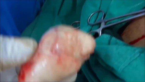
Watch that video of A Big Size Fibrodenoma Removal Surgery
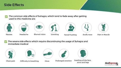
For More Information Kindly Visit : https://www.medexpressrx.com/suhagra.aspx
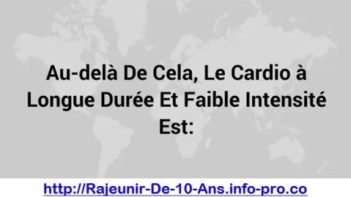
Comment Avoir Un Ventre Plat, Rajeuni, Rajeunir De 10 Ans En 3 Mois, Bruleur De Graisse Musculation --- http://rajeunir-de-10-ans.info-pro.co --- 5 Raisons pourquoi le Cardio Long-Lent n’est PAS bon. En passant à travers les e-mails de clients au cours des derniers jours, j'ai remarqué que beaucoup de gens font encore du cardio longue durée, à faible intensité, beurk! Voici l'affaire: si vous cherchez à obtenir un bénéfice maximal du temps que vous mettez dans vos séances d'entraînement, le cardio de longue durée à faible intensité n'est pas la voie à suivre, et pour de nombreuses raisons. Voici mon top 5: 1. Les calories brûlées minimales: 45 minutes sur le tapis roulant peuvent brûler un énorme 300 calories si vous êtes chanceux, l'équivalent de 50 grammes de graisse. En vous exerçant dix heures par semaine et vous pourriez perdre un demi-kilo! Ce qui m'amène à mon prochain point: 2. Beaucoup trop de temps consacré: Je ne sais pas pour vous, mais je n'ai pas des heures et des heures de mon temps à mettre dans l’entraînement chaque semaine. En fait, je n’ai le temps pour faire que quelques heures d'exercice par semaine, et vous savez quoi? C'est tout ce dont vous avez besoin. En fait, la recherche a montré que plus de 90 minutes par semaine peuvent être nuisibles! (Plus d'infos ici) http://rajeunir-de-10-ans.info-pro.co Au-delà de cela, le cardio à longue durée et faible intensité est: 3. Ennuyeux que diable: Assis sur un vélo d'exercice à regarder le mur en face de moi pendant 45-60 minutes? Non merci. Mais peut-être pire encore est le fait que le cardio de longue durée et à faible intensité ne fournit: Ces 5 étapes Révèlent Les Choses Que Vous Devez ABSOLUMENT ÉVITER Si Vous Voulez Ralentir Le Processus De Vieillissement, Récupérer Votre Santé Et Atteindre Un Corps Idéal. Cliquez Ici: http://rajeunir-de-10-ans.info-pro.co

cleral tattooing is the practice of tattooing the sclera, or white part of the human eye. The dye is not injected into the tissue, but between two layers of the eye, where it spreads out over a large area. The process is not common
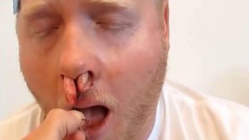
Nose Packing Application & Removal
The experts at stalbertphysiotherapy.com have now served over 12,420 patients in the St. Albert and Edmonton communities. Since 1992, they have helped patients find relief from pain from physical ailments caused from such things as disease, injuries, and deformities. The team of rehabilitation therapists are now offering no obligation appointments so potential patients can find out more about how the service might help them. Visit - https://stalbertphysiotherapy.com/contact/
