- Physical Examination
- Surgical Examination
- Ophthalmology
- Clinical Skills
- Orthopedics
- Surgery Videos
- Laparoscopy
- Pediatrics
- Funny Videos
- Cardiothoracic Surgery
- Nursing Videos
- Plastic Surgery
- Otorhinolaryngology
- Histology and Histopathology
- Neurosurgery
- Dermatology
- Pediatric Surgery
- Urology
- Dentistry
- Oncology and Cancers
- Anatomy Videos
- Health and Fitness
- Radiology
- Anaesthesia
- Physical Therapy
- Pharmacology
- Interventional Radiology
- Cardiology
- Endocrinology
- Gynecology
- Emergency Medicine
- Psychiatry and Psychology
- Childbirth Videos
- General Medical Videos
- Nephrology
- Physiology
- Diet and Food Health
- Diabetes Mellitus
- Neurology
- Women Health
- Osteoporosis
- Gastroenterology
- Pulmonology
- Hematology
- Rheumatology
- Toxicology
- Nuclear Medicine
- Infectious Diseases
- Vascular Disease
- Reproductive Health
- Burns and Wound Healing
- Other
Top videos

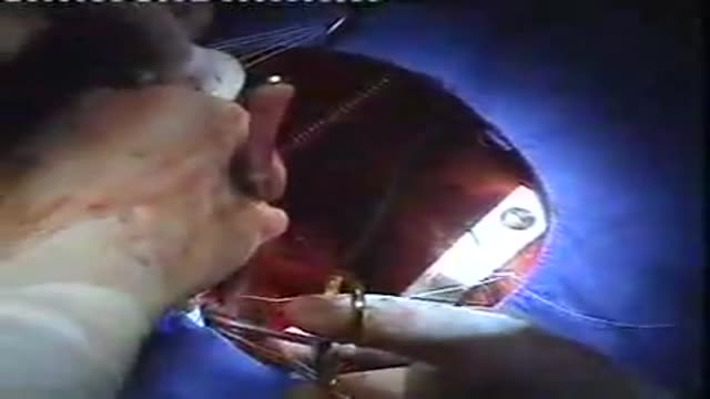
mitral valve replacement surgery

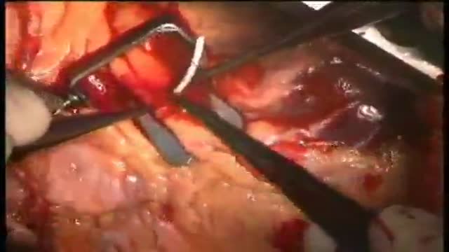
Carotid Endartrectomy, large atheroma removed completely, patient well after surgery.
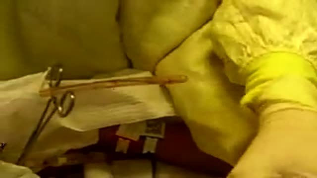
nurses removing chest tube from surgery after spontaneous pneumothorax
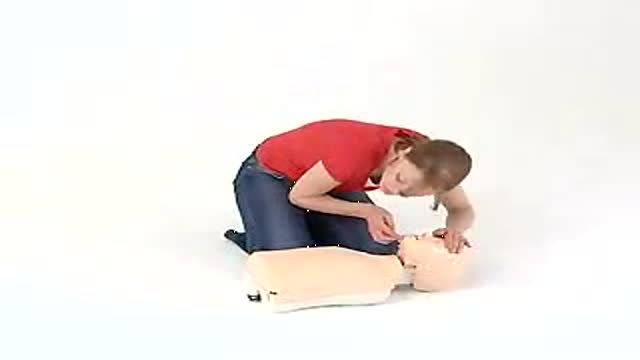
Child CPR
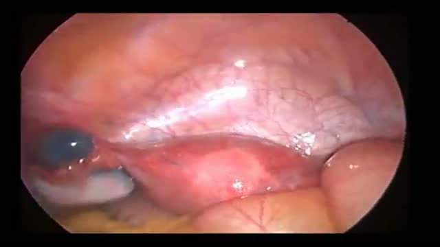
Fallopian Tube Diverticulus seen on Infertility workup Methylene Blue injected for tubal patency shows This. Edited by Dr Hemant Damle Prof & HOD Of Obs at SKN Medical College Pune India

Hip Replacement Surgery
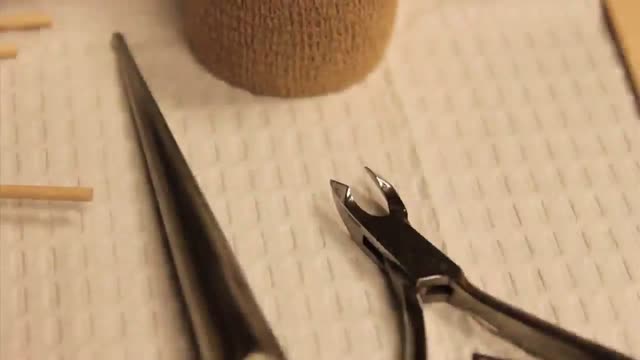
Ingrown Toenail Surgery HD
Beating Coronary Heart Surgery

The Micturition Reflex
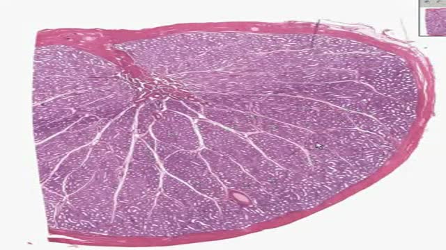
Histology of Kidney

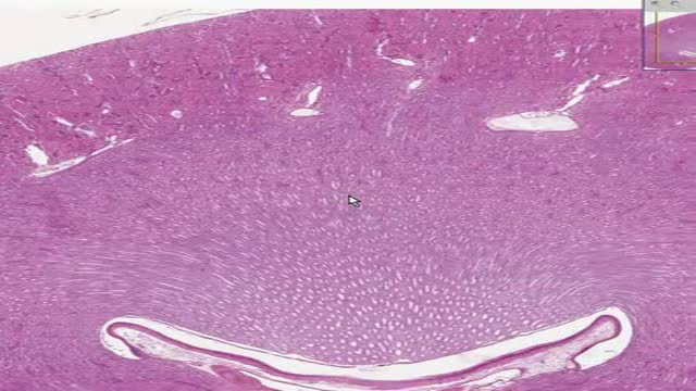
Histology of Spongy Bone
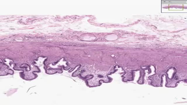
Histology of Gall Bladder
Histology of Adrenal
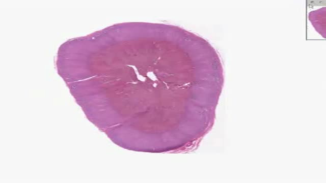
Histology of Testis

Intelligent People Have Fewer Friends, Here's Why...

What Your Handwriting Says About You

Wilms tumor, also known as nephroblastoma, is a cancer of the kidneys that typically occurs in children, rarely in adults. It is named after Dr. Max Wilms, the German surgeon (1867–1918) who first described it. Approximately 500 cases are diagnosed in the U.S. annually.

Asthma and COPD

If you suspect that you have sleep apnea, the usual first step is to discuss your suspicions with your primary care physician. If you don’t have a primary care physician, you can go directly to a clinician who is a sleep specialist. But check your health care insurance coverage first. Some policies require you to see a primary care physician first, and some policies limit the sleep centers and testing facilities whose services they will pay for. Unfortunately, you may discover that your policy offers limited or no coverage for the diagnosis and treatment of sleep apnea, in which case you may wish to switch insurers if and when you can.
