- Physical Examination
- Surgical Examination
- Ophthalmology
- Clinical Skills
- Orthopedics
- Surgery Videos
- Laparoscopy
- Pediatrics
- Funny Videos
- Cardiothoracic Surgery
- Nursing Videos
- Plastic Surgery
- Otorhinolaryngology
- Histology and Histopathology
- Neurosurgery
- Dermatology
- Pediatric Surgery
- Urology
- Dentistry
- Oncology and Cancers
- Anatomy Videos
- Health and Fitness
- Radiology
- Anaesthesia
- Physical Therapy
- Pharmacology
- Interventional Radiology
- Cardiology
- Endocrinology
- Gynecology
- Emergency Medicine
- Psychiatry and Psychology
- Childbirth Videos
- General Medical Videos
- Nephrology
- Physiology
- Diet and Food Health
- Diabetes Mellitus
- Neurology
- Women Health
- Osteoporosis
- Gastroenterology
- Pulmonology
- Hematology
- Rheumatology
- Toxicology
- Nuclear Medicine
- Infectious Diseases
- Vascular Disease
- Reproductive Health
- Burns and Wound Healing
- Other
Top videos

blood transfusion performance

Narcolepsy is a chronic sleep disorder characterized by overwhelming daytime drowsiness and sudden attacks of sleep. People with narcolepsy often find it difficult to stay awake for long periods of time, regardless of the circumstances. Narcolepsy can cause serious disruptions in your daily routine. Sometimes, narcolepsy can be accompanied by a sudden loss of muscle tone (cataplexy) that leads to weakness and loss of muscle control. Cataplexy is often triggered by a strong emotion, most commonly laughter. Narcolepsy is a chronic condition for which there's no cure. However, medications and lifestyle changes can help you manage the symptoms. Support from others — family, friends, employer, teachers — can help you cope with narcolepsy.

Human Glue used to repair a cut in the chin of a toddler

The ideal way to clean your teeth is no mystery; even small changes in your home dental care can lead longer-lasting teeth. Typically, by age of fifty, you will have crowns, bridges, partial prostheses, and sometimes, even a full prosthesis

Robotic Mitral Valve Repair

Dental Abscess extending into Submandibular space
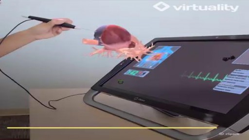
The future of education can be found within this AR tablet.

What are the best positions for labour? When your labour starts, you'll probably feel restless and want to move around and keep busy. Just take care that you don't get over-tired before your labour is properly under way. s your contractions get stronger, concentrate on them. Focus on what's happening to your body and your baby, and practise your breathing and relaxation exercises. Now is the time to find the positions and movements that help you to cope with your contractions. Your midwife should encourage and help you to keep moving around and find comfortable positions, preferably ones that are upright. You may think that you'll be most comfortable lying on the bed. But keeping as upright as possible will help: you to cope with your contractions you and your baby to cope better during labour You'll be able to keep moving by shifting your weight from one foot to another, or by rocking your pelvis. Some positions make it easier for your birth partner to massage your back, or breathe with you through the contractions. You could: Lean on a work surface, or on the back of a chair. Put your arms around your partner's neck or waist, and lean on him. Lean on the bed, with the height adjusted for your comfort, or on a window-sill. Kneel on a large cushion or pillow on the floor, and lean forwards on to the seat of a chair. Sit astride a chair, resting on a pillow placed across the top. Sit on the toilet, leaning forwards, or sit astride, leaning on to the cistern. Go on to all fours. Kneel on one leg, with the other leg bent..
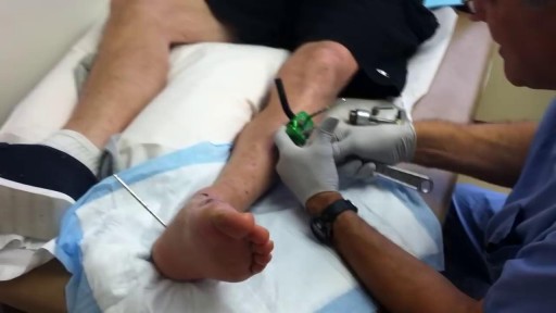
Live Surgery: Flexor Digitorum Profundus (FDP) Finger Tendon Repair

The Watchman can be inserted in less than an hour and could save your life.
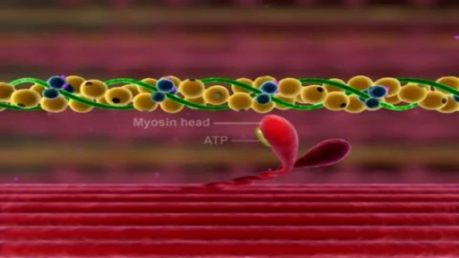
Muscle Contraction Part 3 The Cross Bridge Cycle

Pulsatile Tinnitus Cure, Constant Ear Ringing, Ear Wax Tinnitus, Whistling In Ear, Ringing In Ears. http://tinnitus-solution.info-pro.co First the good news - we know what causes tinnitus. And now the bad news - conventional medical science cannot cure it. Not permanently at least. Sure enough, your doctor would suggest a few remedies, and it may seem to you that the noises you hear are going down. As a result, you begin to relax believing that a pesky problem has been resolved. But suddenly the sounds return again. This is a very common problem actually. So let us turn to the causes instead, and see whether we can try to solve the issue from this end. Here Are Some of the Most Common Causes of Tinnitus Exposure to noise - Did your mom always tell you in your younger days to turn down the volume? She was right. Exposure to loud noise can give you tinnitus. In fact, rock musicians, and those who work with them, or in night clubs often have it. Those who work in construction sites also have tinnitus. So turn down that volume while you still can. You could begin to hear all kinds of noises if you have been exposed to just a single high-pitched noise. Or it could be due to a continuous attack of loud noises close to your ear. This is what happens. Prolonged exposure to noise can damage the Cochlea and cause tinnitus. So if you cannot simply stay away from all that noise, at least get some protection. Use an ear plug when you can. Head injury - Take care of your head because a severe blow or a slight bang could make you hear the tinnitus noises. The head is of course one of the most sensitive parts of the human body. But some people cannot live without an injury, such as those who are into sports - boxers and football players. That's why athletes are more prone to a tinnitus attack. Even a dental surgery could make you hear them. Ear infections and other ear problems - An ear infection, and even sinus can lead to tinnitus as well. When there is an allergy or a sinus infection, the mucous thickens within the inner ear, and this causes more pressure. The extra pressure can lead to tinnitus. Meniere's disease, where the fluid level goes up inside the middle ear is another reason. It could even cause hearing loss. Prescription medications - Conventional drugs often cause side effects, and tinnitus is one of them. Actually, all kinds of drugs have been blamed for instigating this condition. Such as antibiotics like Aminoglycosides, Erythromycin and Vancomycin, Aspirin or medicines containing it. Anti inflammatory drugs like Advil, Aleve, Anaprox, Clinoril, Feldene, Indocin, Lodine and Motrin have also been blamed. Sometimes people heard noises after taking chemotherapy agents such as Cisplatin, Nitrogen Mustard and Vincristine. And some others have even blamed quinine and loop diuretics for this. or even the result of a virus or infection. but is in fact far more shocking that you’ve been led to believe. You’ll finally be able to concentrate on your life, rather that the incessant noise. You’ll be able to no longer live in fear of loud noises, of music, of cinemas. of having fun. The Tinnitus Scandal Revealed, A cure DOES exist. click here: http://tinnitus-solution.info-pro.co

How To Increase Memory Power, How To Improve Memory Retention, How To Boost Memory Power.--- http://brain-revitalizer.info-pro.co ---- Brain Exercises, When people think of exercising it often involves physical exertion to strengthen muscles. The human brain is also an important "muscle" in the body and with the right brain training and brain exercises you can help keep your mind fit, alert and ready to handle the rigors of a typical working day. Often brain exercises come in the form of games that help train the brain improve memory, strategize and think in advance. Some common forms of brain exercise include chess, memory games and mathematical problem solving. Another way to exercise the brain is through brainwave entrainment. Isochronic tones are computer generated tones that are pulsed at specific frequencies to achieve desired effects such as productivity or relaxation. Although brainwave entrainment is not a new practice -- binaural beats have been used in brain training since the mid-1800's -- the use of computer generated isochronic tones has become more popular lately and are also proving to be more effective in helping people improve their memory and intelligence. Unfortunately when most people stop formal schooling, either by achieving a degree or choosing to drop out, the study habits and brain training that was part of their daily schedule also ends. The memorization and critical thinking that helped get us through school shouldn't have to stop just because a specific goal has been reached. In fact, if you don't keep up with brain exercises your mind will become lazy and won't function in the as sharply as it used to. The fact is, most people will never learn what it REALLY feels like to have their brains operating at a high capacity. They'll haphazardly try a few things, wonder why nothing is working, then go back to their mundane existence. But YOU can be different! You can use Genius Brain Power to empower your brain so that you come alive with more energy, learn quicker, think more creatively, focus on your work like never before and drastically reduce stress with amazingly deep states of relaxation and meditation. click here: http://brain-revitalizer.info-pro.co

Watch that video of human Fetus Removal Surgery

Dieta Alcalina Recetas, Listado De Alimentos Alcalinos, Que Es El Agua Alcalina, Menu Para Adelgazar-- http://dieta-alcalina-alimentos.good-info.co -- Entendiendo los Efectos del nivel de pH en el cuerpo El nivel de pH en el cuerpo tiene la habilidad de afectar cada célula del cuerpo. Cuando la sangre tiene un pH alcalino en vez de un pH ácido, ocurre un efecto positivo en cada función corporal del sistema. El cerebro, el sistema circulatorio, los nervios, los músculos, el sistema respiratorio, el sistema digestivo y reproductivo se pueden beneficiar de un nivel adecuado de pH. Por otro lado, cuando el pH del cuerpo es muy ácido, es susceptible a muchas enfermedades y problemas. Ganancia de peso, enfermedades del corazón, envejecimiento prematuro, fatiga, problemas nerviosos, alergias, enfermedades musculares y cáncer son las más probables a ocurrir cuando el pH del cuerpo no está al nivel óptimo. Ya que todos estos problemas son más probables a ocurrir cuando el pH del cuerpo está muy ácido, tiene sentido consumir una dieta rica en alimentos alcalinos. El objetivo principal es usualmente comer aproximadamente entre 75-80% de alimentos alcalinos junto con solamente entre 20-25% de alimentos ácidos. Si se mantiene este nivel en la dieta, el resultado final es un nivel de pH bajo en el cuerpo, el que se requiere para una salud óptima. Descubre como la dieta alcalina funciona & por qué los alimentos alcalinos son altamente recomendados para tu salud. Haz clic aquí http://dieta-alcalina-alimentos.good-info.co

Watch that video of Unreal Mutations and Medical Condition

Traumatic Knee Dislocation Reduction-Quick Version
Pulling pins out of the leg