- Physical Examination
- Surgical Examination
- Ophthalmology
- Clinical Skills
- Orthopedics
- Surgery Videos
- Laparoscopy
- Pediatrics
- Funny Videos
- Cardiothoracic Surgery
- Nursing Videos
- Plastic Surgery
- Otorhinolaryngology
- Histology and Histopathology
- Neurosurgery
- Dermatology
- Pediatric Surgery
- Urology
- Dentistry
- Oncology and Cancers
- Anatomy Videos
- Health and Fitness
- Radiology
- Anaesthesia
- Physical Therapy
- Pharmacology
- Interventional Radiology
- Cardiology
- Endocrinology
- Gynecology
- Emergency Medicine
- Psychiatry and Psychology
- Childbirth Videos
- General Medical Videos
- Nephrology
- Physiology
- Diet and Food Health
- Diabetes Mellitus
- Neurology
- Women Health
- Osteoporosis
- Gastroenterology
- Pulmonology
- Hematology
- Rheumatology
- Toxicology
- Nuclear Medicine
- Infectious Diseases
- Vascular Disease
- Reproductive Health
- Burns and Wound Healing
- Other
Top videos
enile implants are devices placed inside the penis to allow men with erectile dysfunction (ED) to get an erection. Penile implants are typically recommended after other treatments for ED fail. There are two main types of penile implants, semirigid and inflatable.
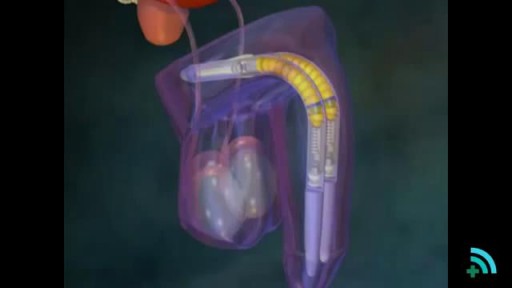
Beating heart or "off pump" coronary artery surgery is the latest revolution in the management coronary disease. It is being embraced world-wide by increasing numbers of surgeons. Many of the advantages are subtle but reduced mortality, stroke, and bleeding as well as earlier discharge are well-established benefits. A cardiac stabiliser is mandatory for this surgery, most are single use only and very expensive, this one is multiple use and is saving many healthcare dollars

Ford Interlocking Suture
Watch that video to know if it is safe to have sex during pregnancy
Duhamel's Operation for Chagasic Megacolon
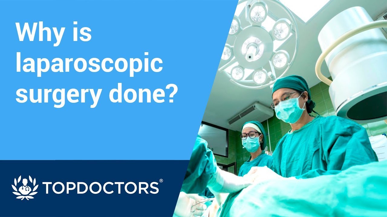
Laparoscopic surgery is minimally-invasive (keyhole) surgery and it is performed through very small incisions, using a camera to guide the surgeon during the procedure. Miss Sarah Mills, a top colorectal surgeon, explains why laparoscopic surgery is performed over alternative methods.
Make an appointment with Miss Sarah Mills here: https://www.topdoctors.co.uk/doctor/sarah-mills

A 55-year-old man presented with recurrent epistaxis. After endoscopic sphenopalatine artery cauterization, the bleeding stopped. The patient was doing well at last follow up.
Transmetatarsal Amputation for Gangrene

Professional Breast Exam
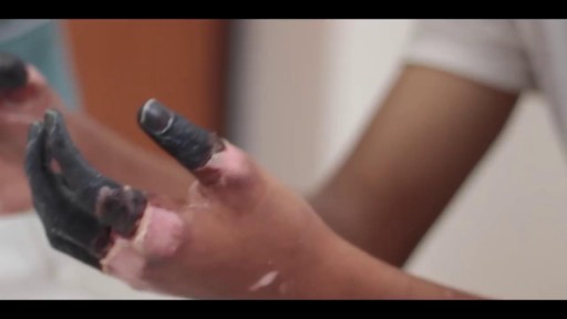
Frostbite is an injury caused by freezing of the skin and underlying tissues. First your skin becomes very cold and red, then numb, hard and pale. Frostbite is most common on the fingers, toes, nose, ears, cheeks and chin. Exposed skin in cold, windy weather is most vulnerable to frostbite. But frostbite can occur on skin covered by gloves or other clothing. Frostnip, the first stage of frostbite, doesn't cause permanent skin damage. You can treat very mild frostbite with first-aid measures, including rewarming your skin. All other frostbite requires medical attention because it can damage skin, tissues, muscle and bones. Possible complications of severe frostbite include infection and nerve damage.
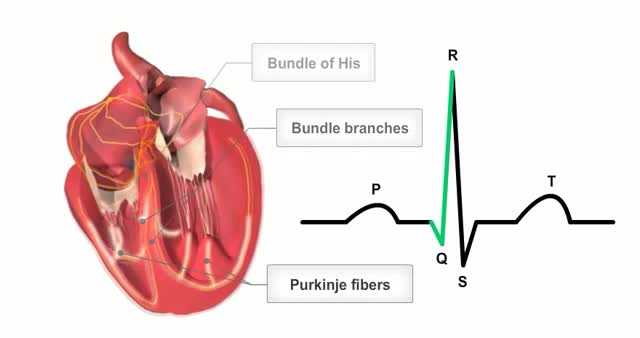
The heart's conductions system controls the generation and propagation of electric signals or action potentials causing the hearts muscles to contract and the heart to pump blood.
USMLE Step 2 CS - Antenatal Visit This is just preview video. To get full access please visit our website : www.usmletutoring.com
SINUS LIFT SURGERY surgical procedure which aims to increase the amount of bone in the posterior maxilla (upper jaw bone), in the area of the premolar and molar teeth, by lifting the lower Schneiderian membrane (sinus membrane) and placing a bone graft.
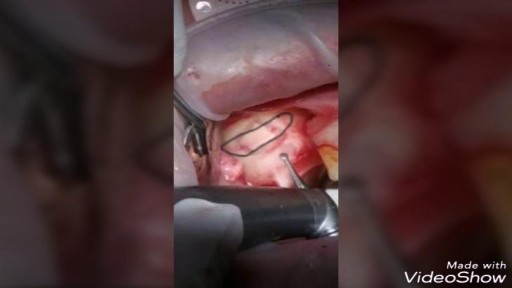
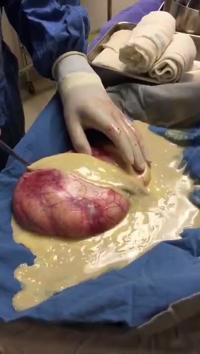
The spleen is one of the most frequently injured intraperitoneal organs, and management of splenic injuries may require splenectomy .. The spleen is an wedge-shaped organ that lies in relation to the ninth and 11th ribs, located in the left hypochondrium and partly in the epigastrium; thus, it is situated between the fundus of the stomach and the diaphragm. The spleen is highly vascular and reddish purple; its size and weight are variable. A normal spleen is not palpable. The spleen's key function is the removal of old red blood cells "RBCs", defective circulating cells, and circulating bacteria. In addition, the spleen helps maintain normal erythrocyte morphology by processing immature erythrocytes, removing their nuclei, and changing the shape of the cellular membrane. Other functions of the spleen include the removal of nuclear remnants of RBCs, denatured hemoglobin, and iron granules ..
Medical Examination of the Lower Limbs
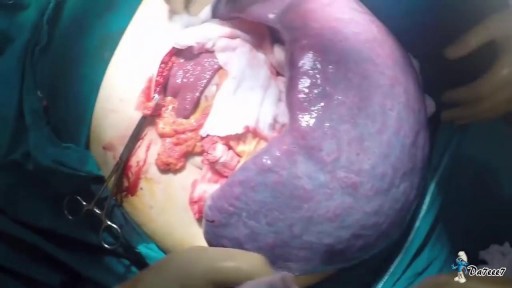
Ovarian teratoma is a type of germ cell tumour. Germ cell tumours are cancers that begin in egg cells in women or sperm cells in men. There are 2 main types of ovarian teratoma. Mature teratoma, which is benign. Immature teratoma, which is cancerous.

I talk about 5 Essential Skills you need as a nurse. These skills are timeless in the fat that you will always need to use them at some level. Of course specific skills are good to have as well but these skills are universal and can help you in other areas of life as well.
NURSING SCHOOL STUDY RESOURCES: https://sellfy.com/nursingschoolstudyNURSING
PHARMACOLOGY: https://sellfy.com/p/fnoy/
INSTAGRAM:https://www.instagram.com/your_mentor_rn/?hl=en
PERSONAL INSTAGRAM: https://www.instagram.com/crosby_steen/
MEDIUM ARTICLES: https://medium.com/@rnacademy1..../7-tips-for-nursing-
AMAZON PRIME STUDENT DISCOUNT: https://amzn.to/2OIleAe
VIDEO GEAR
Camera: G7X Markii - https://amzn.to/2na3OR8
Phone: Galaxy Note 8- https://amzn.to/2nboHM3
Audio: Zoom H4NPro Audio Recorder- https://amzn.to/2vktlf8
Computer: 13 inch Macbook Pro- https://amzn.to/2ndhISw
INSTAGRAM TV https://www.instagram.com/crosby_steen/
Hi Guys! My name is Crosby Steen. I am a Nursing Educator, and ER Travel Nurse. I do videos on daily science based news and travel, with the goal of providing value for you in science based education and travel nursing. Any questions hit me up in the comments or Email below.....
PRIVATE TUTORING OR VIDEO REQUESTS CONTACT:
crosby.steen@gmail.com
MUSIC BY: https://andrewapplepie.com/ and copyrighted by Epidemic Sound
Music by Joakim Karud http://youtube.com/joakimkarud
Music by DJ Quads

Ellis demonstrates how to set up an intravenous piggyback medication (i.e., secondary).
Our Critical Nursing Skills video tutorial series is taught by Ellis Parker MSN, RN-BC, CNE, CHS and intended to help RN and PN nursing students study for your nursing school exams, including the ATI, HESI and NCLEX.
#NCLEX #ClinicalSkills #IVPush #IVpiggyback #HESI #Kaplan #ATI #NursingSchool #NursingStudent #Nurse #RN #PN #Education #LVN #LPN
00:00 What to expect from IV Piggyback
00:32 Ejecting air, saline flush for IV Piggyback
1:11 Saline lock
2:28 Clamping tubing
2:38 Spiking bag
2:50 Hanging bag
3:07 Priming the tubing
3:50 Attaching to pump port
4:04 Unclamping tubing
4:45 Lowering the primary
5:08 Setting the pump
🚨 Reminder: shipping deadlines are looming 👀
🎁 Regular Shipping: Order by Friday, December 15
🚀 Expedited Shipping: Order by Monday, December 18
🔍 Still searching for last-minute gifts? Consider a Level Up RN Gift Card! 💌 It’s not only a thoughtful present but also the perfect way to share treasures like Pharmacology Flashcards OR digital treasures like Flashables Digital Nursing Flashcards & the Level Up RN membership. Give the gift of knowledge this holiday season! 🧠⚡️💖 bit.ly/LevelUpRNGC
🚪 Access our Cram Courses, Quizzes and Videos all in one ad free space with Level Up RN Membership https://bit.ly/LevelUpRNMembership
Want more ways to MASTER Clinical Skills? Check out our flashcards & videos!
👇👇👇👇👇👇👇👇👇👇
👉 https://bit.ly/clinicalnursingskills 👈
☝️👆☝️👆☝️👆☝️👆☝️👆
This is your one-stop-shop for materials to help you LEARN & REVIEW so you can PASS Nursing School.
🤔🤔🤔 DO YOU WANT TO PASS your classes, proctored exams and the NCLEX? 🤔🤔🤔 Our resources are the best you can buy. They are built with a single goal: help you pass with no fluff. Everything you need, and nothing you don’t. Don’t take our word for it, though! Check out our hundreds of ⭐️⭐️⭐️⭐️⭐️ reviews from nurses who passed their exams and the NCLEX with Level Up RN.
🗂️ Our Ultimate Nursing School Survival kit is your number 1 resource to get through nursing school and to pass the NCLEX. Whether you're just starting school or you’re already prepping for the NCLEX, this bundle of flashcards is the best you can buy. It covers all the information you need to know to pass all your exams and it has FREE shipping!
➡️ https://bit.ly/TUNSSK ⬅️
L👀king for EVEN MORE resources to survive Nursing School? Make your Nursing School experience your own! Life’s difficult enough—learning shouldn’t be.
🪅 Games https://nursesquad.com
💻 Digital resources https://bit.ly/NursingStudyCourses
📅 Organizational tools https://bit.ly/OrganizingSchool
✨Want perks? Join our channel!
https://youtube.com/leveluprn/join
🏷 Head to https://leveluprn.com/specials for all our latest deals!🥳️
📧 LOOKING FOR FREE RESOURCES TO HELP WITH YOUR EXAMS? Get exclusive tips, latest video releases and more delivered to your email!
➡️ https://leveluprn.com/signup ⬅️
⚕ 👩 LEVEL UP NURSE SQUAD 👩⚕️
All of the nurses at Level Up RN are here to help! Cathy Parkes started helping her fellow classmates back when she was in nursing school, tutoring so they could pass their exams and graduate. After she got her BSN and started working as an RN at Scripps Encinitas Hospital, she started this YouTube channel to help nursing students around the world. Since then she has built a team of top-notch dedicated nurses and nurse educators who are focused on improving nursing education and supporting career advancement for nurses everywhere. With flashcards, videos, courses, organizational tools and more, we are singularly focused on helping students and nurses Level Up on their exams and nursing careers.
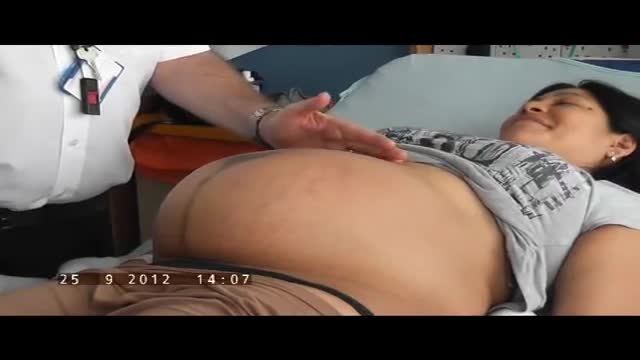
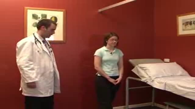
The obstetric examination is distinct from other examinations in that you, the clinician, are trying to assess the health of two individuals – the mother and the fetus – simultaneously. From the initial history, you should be able to judge the health of the pregnancy, any risk factors that need to be addressed, and any concerns from the parents. The history is an opportunity for you to find out how much the parents know about pregnancy, labour and delivery and if they have any preferences to which these events are carried out. A carefully taken history will also direct your attention to specific signs during the examination. As such, it is important that you develop a concise and systematic method of taking the history and carrying out the examination so that you do not miss any important information. This article focuses primarily on the examination. Pregnancy is a sensitive issue, especially for the primigravida’s. Therefore, extra care is needed when you approach a pregnant woman. Always obtain expressed informed consent before examining her and have a chaperone accompany you throughout the examination. A walk-through of what you will be doing is a good way of reassuring the patient and allows the examination to go on smoothly. It is also important to let your patient know that if the examination is too painful, she can stop at any time she wants. Finally, before you begin, you should always wash your hands, especially at an OSCE station.
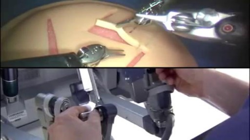
Robotic Surgery Demonstration Using Da Vinci Surgical System
