- Physical Examination
- Surgical Examination
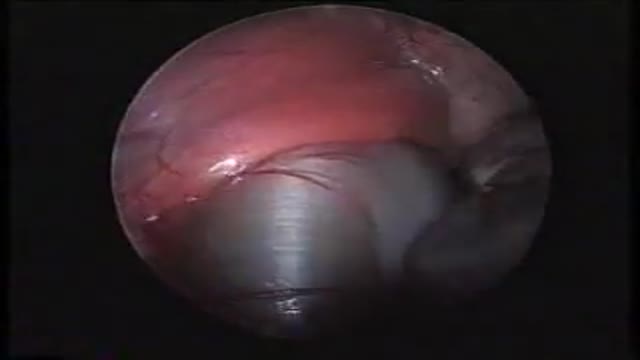
- Ophthalmology
- Clinical Skills
- Orthopedics
- Surgery Videos
- Laparoscopy
- Pediatrics
- Funny Videos
- Cardiothoracic Surgery
- Nursing Videos
- Plastic Surgery
- Otorhinolaryngology
- Histology and Histopathology
- Neurosurgery
- Dermatology
- Pediatric Surgery
- Urology
- Dentistry
- Oncology and Cancers
- Anatomy Videos
- Health and Fitness
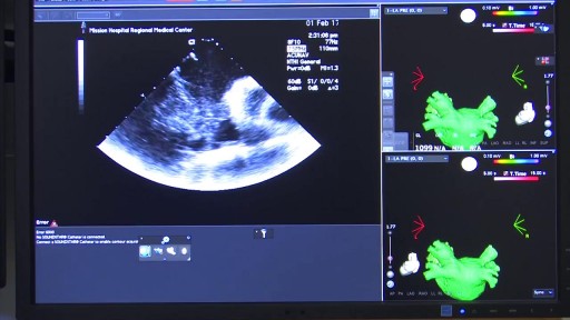
- Radiology
- Anaesthesia
- Physical Therapy
- Pharmacology
- Interventional Radiology
- Cardiology
- Endocrinology
- Gynecology
- Emergency Medicine
- Psychiatry and Psychology
- Childbirth Videos
- General Medical Videos
- Nephrology
- Physiology
- Diet and Food Health
- Diabetes Mellitus
- Neurology
- Women Health
- Osteoporosis
- Gastroenterology
- Pulmonology
- Hematology
- Rheumatology
- Toxicology
- Nuclear Medicine
- Infectious Diseases
- Vascular Disease
- Reproductive Health
- Burns and Wound Healing
- Other
Top videos

Watch that video of Unreal Mutations and Medical Condition
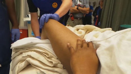
Traumatic Knee Dislocation Reduction-Quick Version
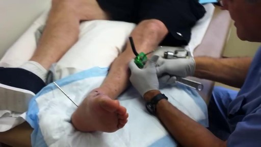
Pulling pins out of the leg


The procedure is used most often to treat a condition called supraventricular tachycardia, or SVT, which occurs because of abnormal conduction fibers in the heart. Catheter ablation is also used to help control other heart rhythm problems such as atrial flutter and atrial fibrillation.

operation on the stomach

Endoscopic Brain Surgery, third Ventriculostomy
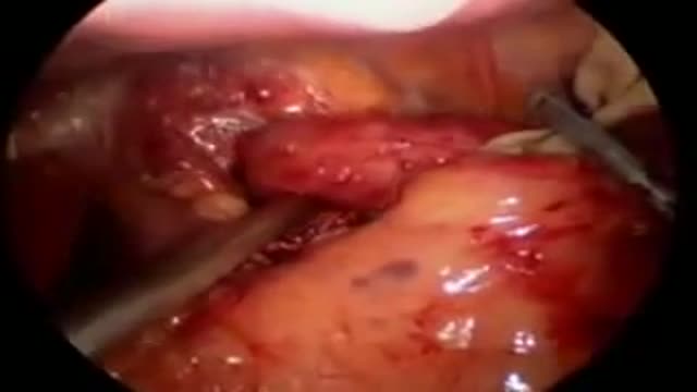
A laparoscopic view of the diaphragmatic hernia

Using a common laser for treatment of eye cancer may benefit some patients by preserving site.

Combined Spinal-Epidural Obstetric Anesthesia
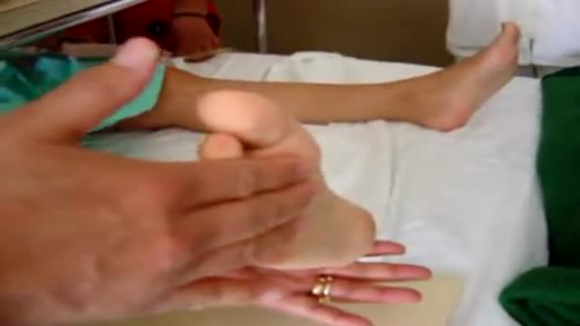
Clonus is tested for in the ankles by rapidly dorsiflexing the relaxed ankle joint.
Faster, more efficient recovery after Orthopedic surgery
Fundoplication HD GERD Surgery 3D Animation
Nissen Laparoscopic Fundoplication Acid Reflux Surgery Stomach
Mechanism of Addiction
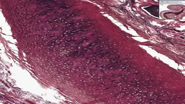
Histology of Elastic Cartilage

Discover how hemodialysis works and the different options available for this dialysis treatment.
Related articles on DaVita.com:
What Is Hemodialysis? (http://www.davita.com/treatmen....t-options/hemodialys
How Does a Dialysis Machine Work? (http://www.davita.com/treatmen....t-options/hemodialys
Peritoneal dialysis is a treatment for kidney failure that uses the lining of your abdomen, or belly, to filter your blood inside your body. ... The process of first draining the used dialysis solution and then replacing it with fresh solution is called an exchange.
The annual incidence of primary intraspinal neoplasm is approximately five per million for females and three per million for males.[9] Spinal intradural extramedullary tumors account for two thirds of all intraspinal neoplasms and include neuromas and meningiomas.[1] Overall, meningiomas account for 25 to 46% of primary spinal neoplasms and are the second most common intradural spine tumor after neuromas.[9] Spinal meningiomas occur less frequently than intracranial ones and account for approximately 7.5 to 12.7% of all meningiomas.[25]
