- Physical Examination
- Surgical Examination
- Ophthalmology
- Clinical Skills
- Orthopedics
- Surgery Videos
- Laparoscopy
- Pediatrics
- Funny Videos
- Cardiothoracic Surgery
- Nursing Videos
- Plastic Surgery
- Otorhinolaryngology
- Histology and Histopathology
- Neurosurgery
- Dermatology
- Pediatric Surgery
- Urology
- Dentistry
- Oncology and Cancers
- Anatomy Videos
- Health and Fitness
- Radiology
- Anaesthesia
- Physical Therapy
- Pharmacology
- Interventional Radiology
- Cardiology
- Endocrinology
- Gynecology
- Emergency Medicine
- Psychiatry and Psychology
- Childbirth Videos
- General Medical Videos
- Nephrology
- Physiology
- Diet and Food Health
- Diabetes Mellitus
- Neurology
- Women Health
- Osteoporosis
- Gastroenterology
- Pulmonology
- Hematology
- Rheumatology
- Toxicology
- Nuclear Medicine
- Infectious Diseases
- Vascular Disease
- Reproductive Health
- Burns and Wound Healing
- Other
Top videos
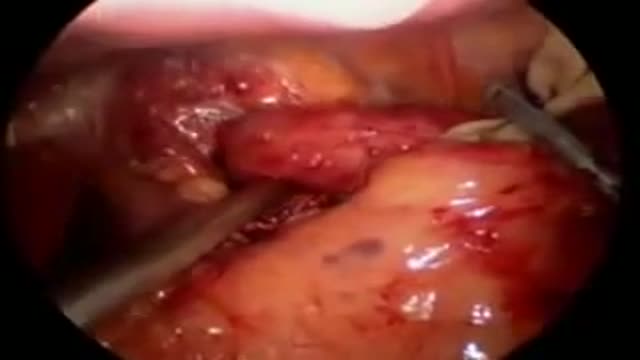
Fundoplication HD GERD Surgery 3D Animation
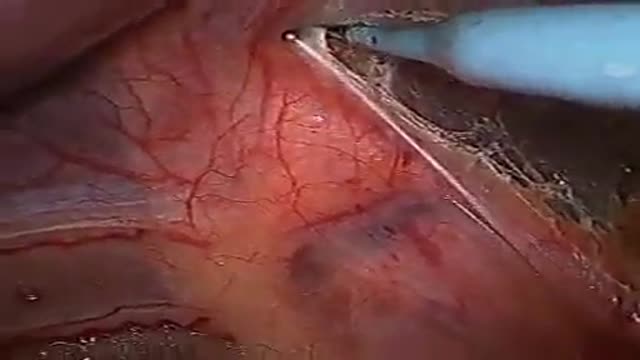
Nissen Laparoscopic Fundoplication Acid Reflux Surgery Stomach
Mechanism of Addiction
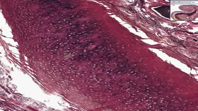
Histology of Elastic Cartilage
Nail biting is a bad habit that can not only make your hands look unsightly, but if you bite your nails badly enough, you may permanently damage your nails, your teeth, or even your gums. Many people deal with this problem, so you're not alone. If you are tired of stubby and bleeding nails try these simple remedies to promote normal and beautiful nail growth.

Can the flu be treated? Yes. There are prescription medications called “antiviral drugs” that can be used to treat flu illness. What are antiviral drugs? Antiviral drugs are prescription medicines (pills, liquid, an inhaled powder, or an intravenous solution) that fight against the flu in your body. Antiviral drugs are not sold over-the-counter. You can only get them if you have a prescription from your doctor or health care provider. Antiviral drugs are different from antibiotics, which fight against bacterial infections. What should I do if I think I have the flu? If you get the flu, antiviral drugs are a treatment option. Check with your doctor promptly if you are at high risk of serious flu complications (see box below for the full list of high risk factors). Flu symptoms can include fever, cough, sore throat, runny or stuffy nose, body aches, headache, chills and fatigue. Your doctor may prescribe antiviral drugs to treat your flu illness. Should I still get a flu vaccine? Yes. Antiviral drugs are not a substitute for getting a flu vaccine. While flu vaccine can vary in how well it works, a flu vaccine is the first and best way to prevent seasonal influenza. Antiviral drugs are a second line of defense to treat the flu (including seasonal flu and variant flu viruses) if you get sick.

Abnormally Large Knee (part 1) - Bizarre ER
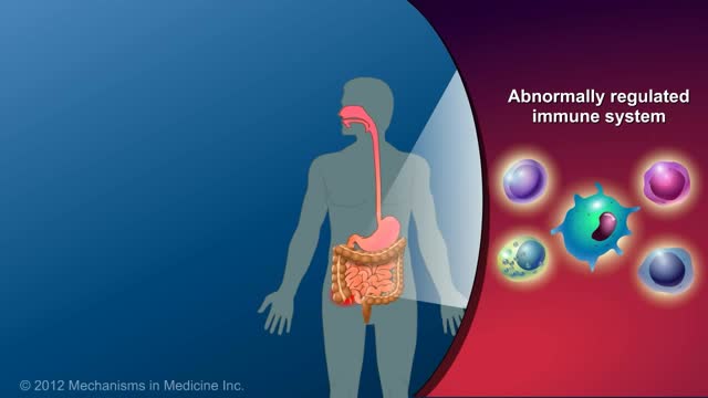
Crohn's disease is an inflammatory bowel disease (IBD). It causes inflammation of the lining of your digestive tract, which can lead to abdominal pain, severe diarrhea, fatigue, weight loss and malnutrition. Inflammation caused by Crohn's disease can involve different areas of the digestive tract in different people. The inflammation caused by Crohn's disease often spreads deep into the layers of affected bowel tissue. Crohn's disease can be both painful and debilitating, and sometimes may lead to life-threatening complications. While there's no known cure for Crohn's disease, therapies can greatly reduce its signs and symptoms and even bring about long-term remission. With treatment, many people with Crohn's disease are able to function well.

Body-Safe Sex Toys
You can decrease your risk of getting Lyme disease with some simple precautions: Cover up. ... Use insect repellents. ... Do your best to tick-proof your yard. ... Check yourself, your children and your pets for ticks. ... Don't assume you're immune. ... Remove a tick as soon as possible with tweezers.
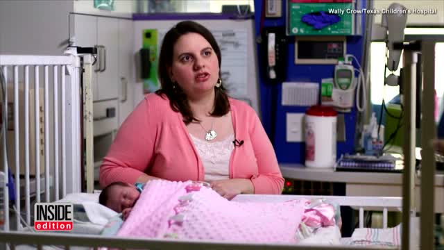
Baby Survives Being Born Twice:
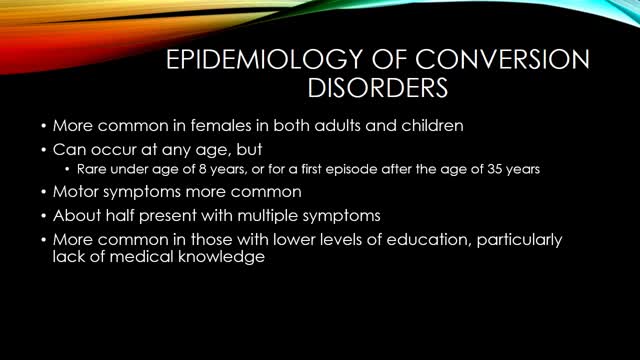
Conversion disorder, also called functional neurological symptom disorder, is a condition in which you show psychological stress in physical ways. The condition was so named to describe a health problem that starts as a mental or emotional crisis — a scary or stressful incident of some kind — and converts to a physical problem.

SEX WITH DIAPHRAGM TO CONTROL UNWANTED PREGNANCY

HAPPY THANKSGIVING
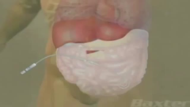
Peritoneal dialysis is a treatment for kidney failure that uses the lining of your abdomen, or belly, to filter your blood inside your body. ... The process of first draining the used dialysis solution and then replacing it with fresh solution is called an exchange.
The annual incidence of primary intraspinal neoplasm is approximately five per million for females and three per million for males.[9] Spinal intradural extramedullary tumors account for two thirds of all intraspinal neoplasms and include neuromas and meningiomas.[1] Overall, meningiomas account for 25 to 46% of primary spinal neoplasms and are the second most common intradural spine tumor after neuromas.[9] Spinal meningiomas occur less frequently than intracranial ones and account for approximately 7.5 to 12.7% of all meningiomas.[25]
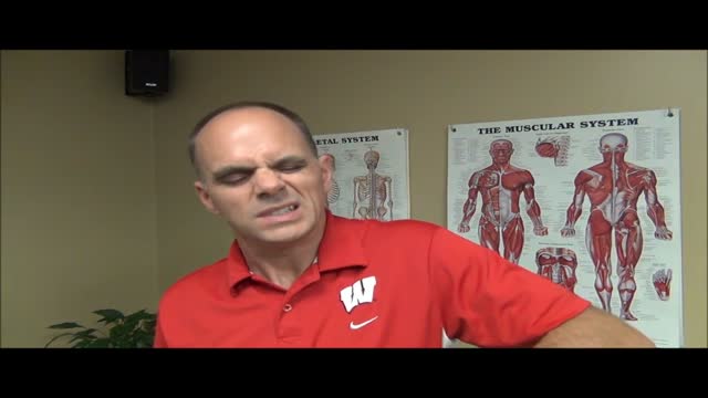
Shoulder pain and exercises Milwaukee WI
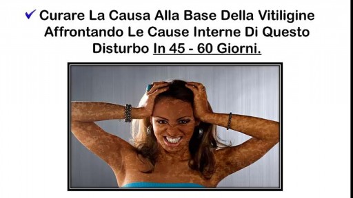
Vitiligine Cause, Vitiligine Bambini, Micropigmentazione Vitiligine, Vitiligine Trucco, Vitiligine --- http://vitiligine-cura.good-info.co --- Un Ricercatore Medico, Nutrizionista, Consulente Di Salute Ed Ex Malato Cronico Di Vitiligine Ti Spiega Come: Curare La Vitiligine E Ripristinare Il Colore Naturale Della Tua Pelle In 7 Giorni! Curare La Causa Alla Base Della Vitiligine Affrontando Le Cause Interne Di Questo Disturbo In 45 - 60 Giorni. Prevenire La Comparsa Di Cicatrici E Segni Gettare Via . Lozioni O Creme "Miracolo" E Sentirti Subito Più Fiducioso! Risparmiare Migliaia Di Euro In Farmaci, Laser E Trattamenti UV, Visite Dal Dottore O Operazioni Chirurgiche! Ripristinare Il Tuo Equilibrio Interno E Fermare I Problemi Di Salute Legati Alla Vitiligine Mantenendoli Alla Larga Per Sempre! Perdere Chili In Eccesso, Sembrare Più Giovane E Riguadagnare L'autostima Ripristinare I Livelli Di Energia E Migliorare La Qualità Della Vita Significativamente... Garantito! Se Vuoi Imparare Come Curare La Vitiligine In Modo Definitivo E Riaquistare La Tua Salute E Benessere, Senza Farmaci, Senza I Tradizionali Trattamenti Per La Vitiligine E Senza Alcun Effetto Collaterale, Allora Questa Sarà La Lettura Più Importante Che Abbia Mai Fatto. Te Lo Garantisco E Ho I Risultati Per Provartelo! http://vitiligine-cura.good-info.co

Case of ITP with persistent very low platelet count despite best medical management