- Physical Examination
- Surgical Examination
- Ophthalmology
- Clinical Skills
- Orthopedics
- Surgery Videos
- Laparoscopy
- Pediatrics
- Funny Videos
- Cardiothoracic Surgery
- Nursing Videos
- Plastic Surgery
- Otorhinolaryngology
- Histology and Histopathology
- Neurosurgery
- Dermatology
- Pediatric Surgery
- Urology
- Dentistry
- Oncology and Cancers
- Anatomy Videos
- Health and Fitness
- Radiology
- Anaesthesia
- Physical Therapy
- Pharmacology
- Interventional Radiology
- Cardiology
- Endocrinology
- Gynecology
- Emergency Medicine
- Psychiatry and Psychology
- Childbirth Videos
- General Medical Videos
- Nephrology
- Physiology
- Diet and Food Health
- Diabetes Mellitus
- Neurology
- Women Health
- Osteoporosis
- Gastroenterology
- Pulmonology
- Hematology
- Rheumatology
- Toxicology
- Nuclear Medicine
- Infectious Diseases
- Vascular Disease
- Reproductive Health
- Burns and Wound Healing
- Other
Top videos
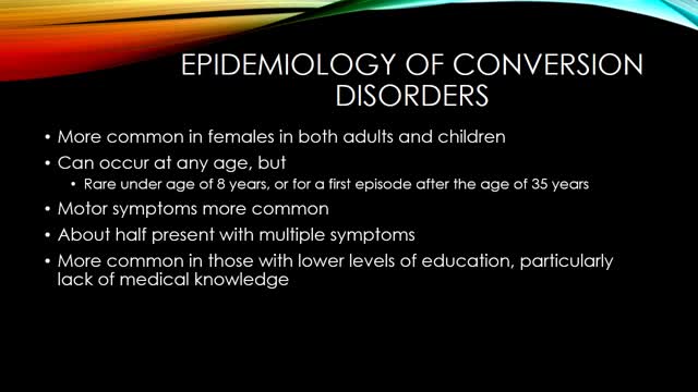
Conversion disorder, also called functional neurological symptom disorder, is a condition in which you show psychological stress in physical ways. The condition was so named to describe a health problem that starts as a mental or emotional crisis — a scary or stressful incident of some kind — and converts to a physical problem.

SEX WITH DIAPHRAGM TO CONTROL UNWANTED PREGNANCY

HAPPY THANKSGIVING
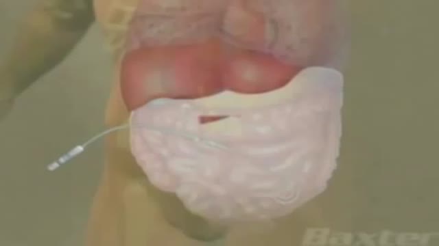
Peritoneal dialysis is a treatment for kidney failure that uses the lining of your abdomen, or belly, to filter your blood inside your body. ... The process of first draining the used dialysis solution and then replacing it with fresh solution is called an exchange.
The annual incidence of primary intraspinal neoplasm is approximately five per million for females and three per million for males.[9] Spinal intradural extramedullary tumors account for two thirds of all intraspinal neoplasms and include neuromas and meningiomas.[1] Overall, meningiomas account for 25 to 46% of primary spinal neoplasms and are the second most common intradural spine tumor after neuromas.[9] Spinal meningiomas occur less frequently than intracranial ones and account for approximately 7.5 to 12.7% of all meningiomas.[25]
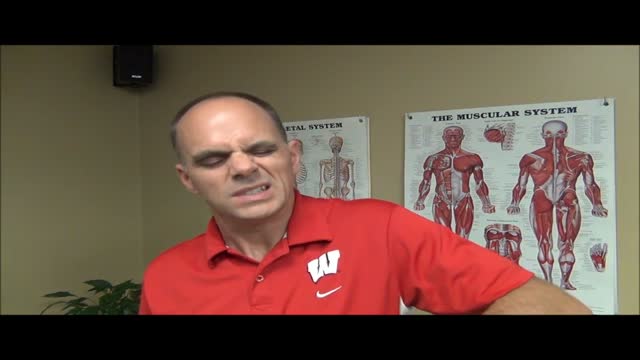
Shoulder pain and exercises Milwaukee WI
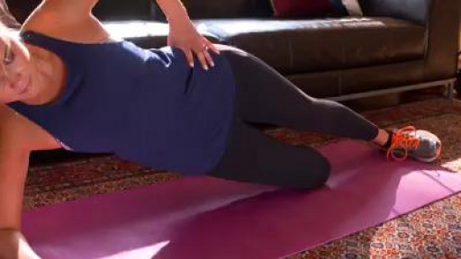
Master perfect plank form and you .ll strengthen your core in no time.
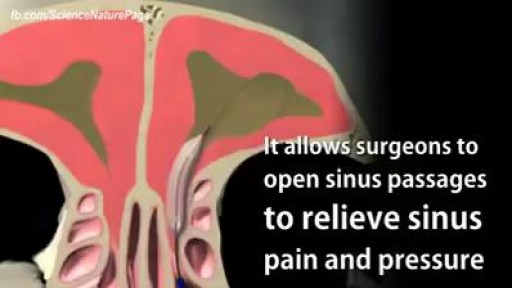
Balloon Sinuplasty for Sinus Infection

how to calm a crying baby everytime
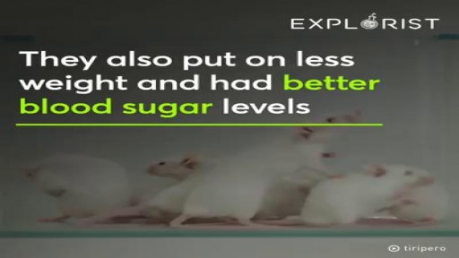
Scientists are working on a pill that could replace exercise.
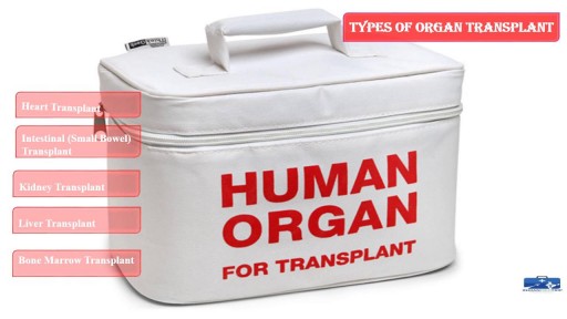
This process involves surgical removing of an #organ or tissue from one person (organ donor) & placing into another person (recipient) body. https://goo.gl/JfoN8y
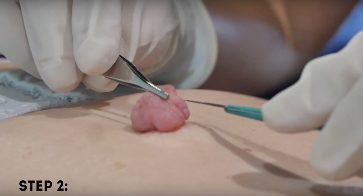
Watch that Huge Skin Tag Removal Procedure

Home Remedies For Acid Reflux, Ginger For Acid Reflux, Heartburn After Gallbladder Removal --- http://heartburn-acid-reflux.info-pro.co --- Stop using Pepto Bismol until you read the following… There is BREAKING scientific news reporting that many of America’s most popular antacids – both the ones you buy at the drug store and the ones you need prescriptions for… Are linked to more than a dozen forms of potentially DEADLY cancers. Click this link now to get the full story and see if you’re at risk. You’ll find out about a “just discovered” alternative to antacids…. Something that can permanently cure even the worst cases of acid reflux in as little few days, and that doesn’t require any pills or medications. Click Here: http://heartburn-acid-reflux.info-pro.co

Come Rimanere Incinta Subito, Probabilità Di Rimanere Incinta A 40 Anni, Primi Sintomi Gravidanza---- http://come-rimanere-incinta.info-pro.co --- Esperta in Medicina Cinese, Specializzata in Trattamenti per la Salute e in Nutrizione, Consulente per la Salute, Dopo Aver Provato in Prima Persona l'Infertilità e Averla Sconfitta ti Insegna Come: Rimanere Incinta in Modo Rapido e del Tutto Naturale in Soli 2 Mesi Dare alla Luce Bambini Sani e Forti Invertire il Problema dell'Infertilità, sia Maschile che Femminile Migliorare la Qualità della Tua Vita...Drasticamente! Scopri Come Ha Vinto la Sua Infertilità e Ha Insegnato a Migliaia di Donne in Tutto il Mondo Come Eliminare Tutti i Problemi dell'Infertilità e Come Rimanere Incinta in Modo Rapido e, Soprattutto, del Tutto Naturale Blog: http://bit.ly/2F3k8xR Se Stai Lottando con Tutte le Tue Forze per Rimanere Incinta e Nonostante Tutto Ancora Non Hai Ottenuto Risultati, Questa Sarà la Lettera Più Importante che Potrai Mai Leggere... Clicca sul link http://come-rimanere-incinta.info-pro.co

baby wrapping
Is Shingles Contagious, What Are Shingles, Herpes Zoster Pictures, Shingles Home Remedies --- http://shingles-cure.good-info.co/ --- If You Are A Newcomer To This Disease, I Hate To Be The Bringer Of Bad News But You Should Know That The List Of Potential Symptoms Is Depressingly Long. These Include The Following: A General Feeling Of Muscle Pain To Begin With, Almost Like Flu A Tingling, Burning Type Sensation In A Specific Area Of The Skin Fever And Headache And Sometimes A Swelling Of The Lymph Nodes A Band Of Spots And Then A Rash At A Specific Part Of Your Body – Very Often The Head Or The Side Of The Trunk Infection Over The Site Of The Rash – Leaving It Prone To Additional Tissue Damage From Bacteria Postherpetic neuralgia leading to chronic nerve pain Ulceration Of The Eye – In Those Cases Where The Shingles Rash Occurs In The Area Of The Eye – Known As Zoster Ophthalmicus. Stress And Depression – Particularly Where The Illness Lingers On For A Long Period Everyone Is Different And Not Everyone Will Experience All Of Those Symptoms. However Even The Most Mild Case Of Shingles Can Be Extremely Debilitating – Something Of Which I Am All Too Aware. Tired Of Fighting A Never Ending Battle Against Shingles? Sick Of Being Told That There´s Nothing You Can Do To Speed Up Recovery? Wherever You Are In Your Fight Against Shingles, I Can Help In this presentation, shows you some unique and rare methods to get rid of shingles naturally in as little as 14 days! This is based on proven techniques used by shingles sufferers without the use of pills and other medication. Get Rid of Shingles will also boost your energy and health dramatically and improve the quality of your life. IMPORTANT NOTE: I can't leave this video up for long, so be sure to watch it from beginning to end while it's still here. REMEMBER: Watch the whole video, as the ending will pleasantly surprise you. click here: http://shingles-cure.good-info.co/

Vitiligine Cause, Vitiligine Bambini, Micropigmentazione Vitiligine, Vitiligine Trucco, Vitiligine --- http://vitiligine-cura.good-info.co --- Un Ricercatore Medico, Nutrizionista, Consulente Di Salute Ed Ex Malato Cronico Di Vitiligine Ti Spiega Come: Curare La Vitiligine E Ripristinare Il Colore Naturale Della Tua Pelle In 7 Giorni! Curare La Causa Alla Base Della Vitiligine Affrontando Le Cause Interne Di Questo Disturbo In 45 - 60 Giorni. Prevenire La Comparsa Di Cicatrici E Segni Gettare Via . Lozioni O Creme "Miracolo" E Sentirti Subito Più Fiducioso! Risparmiare Migliaia Di Euro In Farmaci, Laser E Trattamenti UV, Visite Dal Dottore O Operazioni Chirurgiche! Ripristinare Il Tuo Equilibrio Interno E Fermare I Problemi Di Salute Legati Alla Vitiligine Mantenendoli Alla Larga Per Sempre! Perdere Chili In Eccesso, Sembrare Più Giovane E Riguadagnare L'autostima Ripristinare I Livelli Di Energia E Migliorare La Qualità Della Vita Significativamente... Garantito! Se Vuoi Imparare Come Curare La Vitiligine In Modo Definitivo E Riaquistare La Tua Salute E Benessere, Senza Farmaci, Senza I Tradizionali Trattamenti Per La Vitiligine E Senza Alcun Effetto Collaterale, Allora Questa Sarà La Lettura Più Importante Che Abbia Mai Fatto. Te Lo Garantisco E Ho I Risultati Per Provartelo! http://vitiligine-cura.good-info.co

Case of ITP with persistent very low platelet count despite best medical management
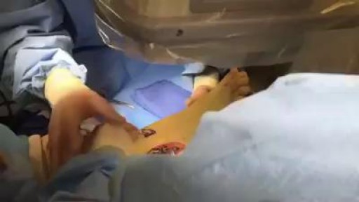
Ankle Fracture Surgery Video
