- Physical Examination
- Surgical Examination
- Ophthalmology
- Clinical Skills
- Orthopedics
- Surgery Videos
- Laparoscopy
- Pediatrics
- Funny Videos
- Cardiothoracic Surgery
- Nursing Videos
- Plastic Surgery
- Otorhinolaryngology
- Histology and Histopathology
- Neurosurgery
- Dermatology
- Pediatric Surgery
- Urology
- Dentistry
- Oncology and Cancers
- Anatomy Videos
- Health and Fitness
- Radiology
- Anaesthesia
- Physical Therapy
- Pharmacology
- Interventional Radiology
- Cardiology
- Endocrinology
- Gynecology
- Emergency Medicine
- Psychiatry and Psychology
- Childbirth Videos
- General Medical Videos
- Nephrology
- Physiology
- Diet and Food Health
- Diabetes Mellitus
- Neurology
- Women Health
- Osteoporosis
- Gastroenterology
- Pulmonology
- Hematology
- Rheumatology
- Toxicology
- Nuclear Medicine
- Infectious Diseases
- Vascular Disease
- Reproductive Health
- Burns and Wound Healing
- Other
Top videos
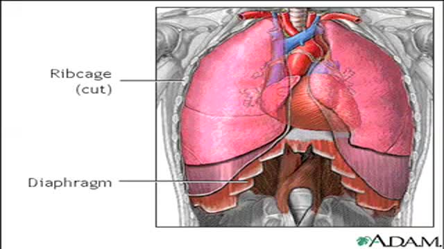
How Do We Breathe ?
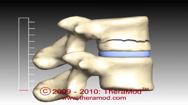
The vertebrae are the bony building blocks of the spine. Between each of the largest parts (bodies) of the vertebrae are the discs. Ligaments are situated around the spine and discs. The spine has seven vertebrae in the neck (cervical vertebrae), 12 vertebrae in the mid-back (thoracic vertebrae), and five vertebrae in the low back (lumbar vertebrae). In addition, in the mid-buttock, beneath the fifth lumbar vertebra, is the sacrum, followed by the tailbone (coccyx).
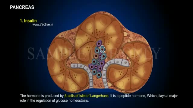
Enzymes, or digestive juices, produced by the pancreas are secreted into the small intestine to further break down food after it has left the stomach. The gland also produces the hormone insulin and secretes it into the bloodstream in order to regulate the body's glucose or sugar level.
The term hypoplastic left heart syndrome (HLHS), initially proposed by Noonan and Nadas, [1] describes a spectrum of cardiac abnormalities characterized by marked hypoplasia of the left ventricle and ascending aorta. This is the same disorder characterized as hypoplasia of the aortic tract complex by Lev. [2] The aortic and mitral valves are atretic, hypoplastic, or stenotic. A patent foramen ovale or an atrial septal defect is usually present. The ventricular septum is usually intact. A large patent ductus arteriosus supplies blood to the systemic circulation. Systemic arterial desaturation may be present because of complete mixing of pulmonary and systemic venous blood in the right atrium. Coarctation of the aorta is also commonly present. See the images below.
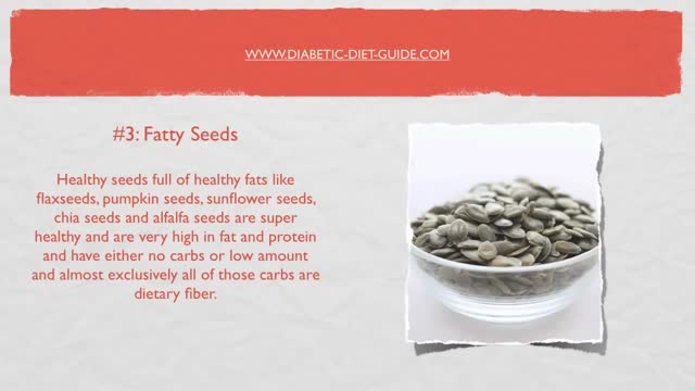
Such foods include carrots, eggplant, cauliflower, green beans, broccoli, peppers, onions, lettuce, zucchini, tomatoes, peanuts and walnuts. These foods are generally safe for you to eat at each meal without spiking your blood sugar.
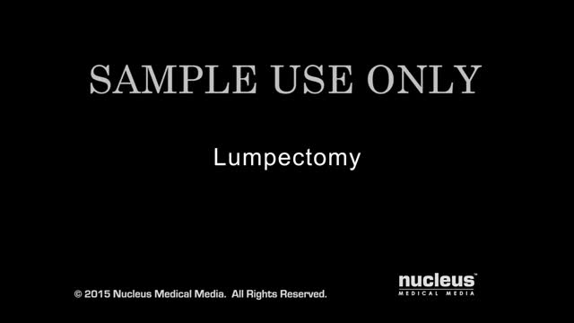
Breast lump removal is surgery to remove a lump that may be breast cancer. Tissue around the lump is also removed. This surgery is called a lumpectomy. When a noncancerous tumor such as a fibroadenoma of the breast is removed, it is often called an excisional breast biopsy, instead of a lumpectomy

1500 mg of sodium amounts to 0.75 teaspoons or 3.75 grams of salt per day, while 2300 mg amounts to one teaspoon or 6 grams of salt per day. Most people today are eating much more than that. The average intake of sodium is about 3400 mg, most of it coming from processed foods.
As a result, the amount of glucose in the blood increases while the cells are starved of energy. Over time, high blood glucose levels damage nerves and blood vessels, leading to complications such as heart disease and stroke, the leading causes of death among people with diabetes.
Verrugas Genitales, Verrugas Del Papiloma Humano, Verrugas En El Cuerpo, Como Eliminar Lunares -- http://sinverrugasylunares.plus101.com --- El Nitrógeno Liquido Es Seguro Para Eliminas Tus Verrugas? Dentro de los tratamientos para las verrugas que la medicina convencional ofrece existe la crioterapia el cual se utiliza nitrógeno líquido. Este tratamiento es de los más comunes que se utilizan para remover las verrugas, este tratamiento debe ser administrado por un profesional especializado. Ya que si se hace sin la supervisión adecuada puede provocar severos daños en la piel. El tratamiento se aplica de la forma siguiente: Se aplica sobre la verruga un poco de nitrógeno líquido ya sea en aerosol o con un algodón, teniendo cuidado de no aplicar a áreas sanas de la piel, esto debido a que el nitrógeno líquido puede afectar severamente a la piel sana, es por eso que debe ser aplicado por un profesional. Para eliminar la verruga serán necesarias varias aplicaciones, al cabo de 2 a 3 semanas la verruga se caerá dejando una costra sobre la piel. Las molestias sobre este tratamiento depende mucho del especialista que lo aplique, existen casos en que a sido doloroso porque el médico a rociado nitrógeno líquido en las partes alrededor de la verruga, lo cual produce un severo daño a la piel, eso sin contar la costra que se forma en la parte donde estuvo la verruga después de que esta se cayera. Generalmente el dolor es mínimo pero también se tiene la opción de aplicar anestesia local sobre la parte afectada. Tu sistema inmunológico es un arma poderosa contra todas las infecciones que existen, pero lamentablemente no le damos el debido mantenimiento q ue necesita para poder luchar contra las infecciones virales. Te invito a conocer esta guía aquí: http://sinverrugasylunares.plus101.com
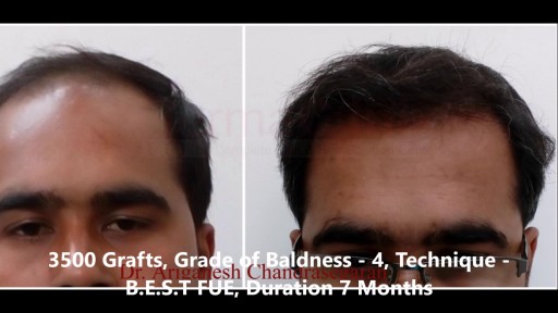
Hair Transplant Results Before and After Photos who undergone Hair Transplant. View our patient's successful results with the FUE, Bio - FUE and B.E.S.T FUE hair transplant technique. Comparable before & after photos! For More Visit Here:- https://www.hairtransplantchennai.org/hair-transplant-results-chennai.php or call us:- +91-8939636222
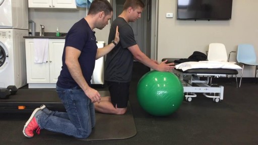
This is the first video of 5, where Mike teamed up with Graham from On Your Marks Fitness and Coaching to show us some exercises to strengthen our muscles, and improve our soccer game! Make sure your feet are planted safely or held by a friend, and keep your back straight, and over your knees. Use the swiss ball to keep you steady, and SQUEEZE those muscles! Check us out on Social Media! Facebook: https://www.facebook.com/striveptandperformance/ Instagram: https://www.instagram.com/striveptandperf/ Twitter: https://twitter.com/StrivePTandPerf Blog: http://www.strivept.ca/blog
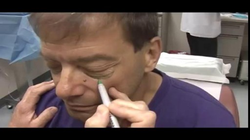
Blepharoplasty
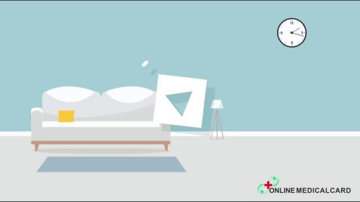
Like a fine whiskey barrel and wine cellar, cannabis also comes at its best when aged in a dark, cool place. Though there is no steadfast expiration date for cannabis, the method you use for preserving the cannabis makes a big difference in maintaining the buds’ freshness and potency. The question is, how do you store cannabis in a way that could extend its longevity while maintaining the vigor and freshness? Experts have described different methods. However, here are some time-proven methods that are easy and inexpensive and require very less equipment. Use air-tight glass containers to store the weed Use clean air-tight glass containers or jars to store cannabis. You can buy glass containers from any ordinary supermarket or hardware store. The tricky part is to make sure you do leave some air in the container while the air stored with cannabis isn’t in detrimental extent. Always leave 1/4 space at the top of the canister or container. Do not fill the containers to the brim with the buds. If you leave no air, then the buds will dry out. If you have too much air, the buds will get damp and moldy. Freeze your cannabis in a convenient temperature The best way to store your buds is in air-tight glass jars, in a cool and dark place under an ideal temperature between 60 and 70 degrees Fahrenheit. If you need to store a high volume of cannabis, you can freeze them after keeping them completely dry for a period of 4 weeks. On this note, you should know that you must not handle frozen buds until it becomes normal in room temperature as trichomes become brittle and can easily break off in freezing temperature. Refrigerate your cannabis (Not Recommended) Even if you use airtight jars, cannabis can grow mold in the fridge. So, you should avoid storing cannabis in the fridge. If you can’t help but doing it, make sure the weed is completely dry and put them in the back where the humidity and temperature don’t fluctuate. Plastic Baggies (Worst method!) Albeit this is very common among people who aren’t expert in handling cannabis, this is the worst of all storage methods. Cannabis gets brittle and dries out in plastic bags. It also loses its natural smell, and the potency deteriorates sharply. So, it should be avoided entirely or can be used for a short-term if there is no better alternative. Here are some things you should know while storing cannabis - Make sure cannabis has been cured for at least 4 weeks before putting them into long-term storage. Without proper curing before storage, the buds can lose their strength and smoothness. - Sunlight can stop the medicinal qualities of cannabis. Your cannabis, if stored correctly, can maintain its medicinal qualities for a few years. Exposure to Sun will turn your cannabis brown, no matter how you have stored it away. - Air-tight, nonporous glass jar are the best way for storing the buds for long term. You can use metal or plastic box/bag, but that could reduce the smell and taste after a while. - Avoid heat and middling temperature in the place where you store your buds. The ideal temperature is 60-70°F (15-21°C) or under 32°F (0°C). Extra heat, cold or middling temperature cause the cannabis potency to decrease. - Keep your cannabis away from any electronic devices or appliances that will expose the cannabis to heat. Keeping cannabis on top of a microwave, or near a laptop or mobile charge is a bad idea. Now, as you know that how to store cannabis properly and make it last for years, enjoy the best form of your weeds even it comes from the previous year. Do write to us in the comments section if you have any questions. Also, don’t forget to hit the subscribe button below. Visit OnlineMedicalCard.com now to get an MMJ recommendation online in less than 10 minutes.
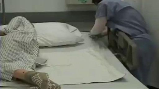
How to change Bed sheet/Bedding of someone Sick or bedridden Elders at home.. Everyone needs it at sometime,

How to Imporve Sexual Health or Stamina Part 2 https://youtu.be/S17bCnwCLuI Dr. Aslam Naveed is a well known sexologist in Pakistan. He has treated more than 1 Lac patients since last 30 years of clinical Practice in sexology, he knows how to help the people facing sexual disorders. Contact: 021-34595050, 03432821919 sexologistpakistan.com facebook.com/menssexcareclinic/ Address: Men's Care Clinic, 2nd floor, The Modern Hospital Opposite Safari Park, University Road. Karachi.
