- Physical Examination
- Surgical Examination
- Ophthalmology
- Clinical Skills
- Orthopedics
- Surgery Videos
- Laparoscopy
- Pediatrics
- Funny Videos
- Cardiothoracic Surgery
- Nursing Videos
- Plastic Surgery
- Otorhinolaryngology
- Histology and Histopathology
- Neurosurgery
- Dermatology
- Pediatric Surgery
- Urology
- Dentistry
- Oncology and Cancers
- Anatomy Videos
- Health and Fitness
- Radiology
- Anaesthesia
- Physical Therapy
- Pharmacology
- Interventional Radiology
- Cardiology
- Endocrinology
- Gynecology
- Emergency Medicine
- Psychiatry and Psychology
- Childbirth Videos
- General Medical Videos
- Nephrology
- Physiology
- Diet and Food Health
- Diabetes Mellitus
- Neurology
- Women Health
- Osteoporosis
- Gastroenterology
- Pulmonology
- Hematology
- Rheumatology
- Toxicology
- Nuclear Medicine
- Infectious Diseases
- Vascular Disease
- Reproductive Health
- Burns and Wound Healing
- Other
Top videos
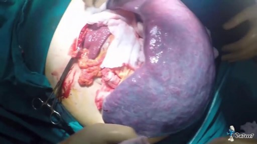
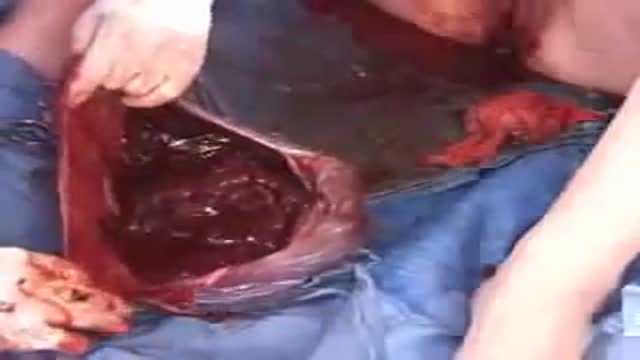
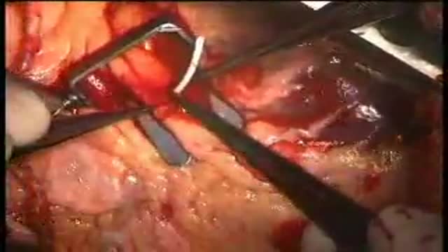
The spleen is one of the most frequently injured intraperitoneal organs, and management of splenic injuries may require splenectomy .. The spleen is an wedge-shaped organ that lies in relation to the ninth and 11th ribs, located in the left hypochondrium and partly in the epigastrium; thus, it is situated between the fundus of the stomach and the diaphragm. The spleen is highly vascular and reddish purple; its size and weight are variable. A normal spleen is not palpable. The spleen's key function is the removal of old red blood cells "RBCs", defective circulating cells, and circulating bacteria. In addition, the spleen helps maintain normal erythrocyte morphology by processing immature erythrocytes, removing their nuclei, and changing the shape of the cellular membrane. Other functions of the spleen include the removal of nuclear remnants of RBCs, denatured hemoglobin, and iron granules ..
Polymyalgia rheumatica is an inflammatory disorder that causes muscle pain and stiffness, especially in the shoulders. Symptoms of polymyalgia rheumatica (pol-e-my-AL-juh rue-MAT-ih-kuh) usually begin quickly and are worse in the morning. Most people who develop polymyalgia rheumatica are older than 65. It rarely affects people under 50. You may receive symptom relief by taking anti-inflammatory drugs called corticosteroids. But relapses are common, and you'll need to visit your doctor regularly to watch for serious side effects of these drugs. Polymyalgia rheumatica is related to another inflammatory disorder called giant cell arteritis, which can cause headaches, vision difficulties, jaw pain and scalp tenderness. It's possible to have both of these conditions together.
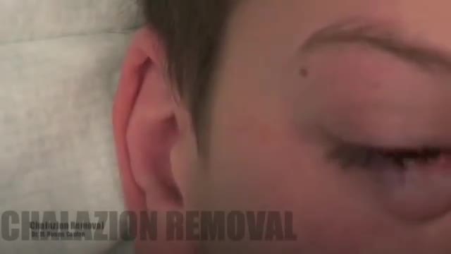
Chalazions are extremely common, and having a sound surgical technique to drain a chalazion is a fundamental in general ophthalmology and oculoplastic surgery. I believe one of the biggest downfalls in treating chalazions is inadequate local anesthetic. Please that both the outer and inner surface to the eyelid need to receive local anesthesia to make the patient totally comfortable. It is important to be careful in delivering the local anesthetic and making sure you have control of the head position, and the position of your needle is bent to minimize any possibility of contact with the globe.
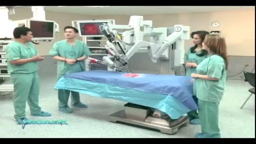
Understand how this world-class surgery platform operates a minimally invasive robotic surgery during a medical procedure for prostate cancer.
Robotic Simple Prostatectomy

Vial medication administration nursing skill. Learn techniques to withdraw medication from a vial using a syringe with a needle.
Medications can come in different forms, such as ampules, vials, tablets, capsules, and so forth. When withdrawing medication from a vial, there are a few things you'll want to know as a nursing student or nurse.
First, there are different needles that can be attached to the syringe. You can use a traditional needle with a beveled tip; you can use a blunt-tip needle to reduce the risk of needle sticks; or you can use a filter needle, which is sometimes required or recommended when drawing medication from a vial, particularly in cases of reconstituted medication.
When withdrawing from a vial, you'll want to do these things (assuming they fit with the protocols and manufacturer's instructions):
NOTE: Some medications or vaccines may require a different technique, so always consult with the manufacturer's instructions.
-gather your supplies
-perform hand hygiene
-clean the vial's top with alcohol prep
-attach the appropriate needle
-stick the needle using a technique to prevent coring of the rubber on the vial (start with 45 degree angle, and as you puncture the vial, rotate the needle to a 90 degree angle in one smooth motion).
-push air into the vial equal to the amount of medication you plan to draw
-invert the vial to withdraw medication
-remove air bubbles
-and much more
See more Nursing Skills: https://www.youtube.com/playli....st?list=PLQrdx7rRsKf
Notes: https://www.registerednursern.....com/how-to-withdraw-
Website: https://www.registerednursern.com/
More Videos: https://www.youtube.com/watch?v=R2XMro13dD0&list=UUPyMN8DzkFl2__xnTEiGZ1w
Nursing Gear: https://teespring.com/stores/registerednursern
Instagram: https://www.instagram.com/registerednursern_com/
Facebook: https://www.facebook.com/RegisteredNurseRNs
Twitter: https://twitter.com/NursesRN
Popular Playlists:
NCLEX Reviews: https://www.youtube.com/playli....st?list=PLQrdx7rRsKf
Fluid & Electrolytes: https://www.youtube.com/playli....st?list=PLQrdx7rRsKf

In order to be able to look at tissues under a microscope, we need to first stain them with the right technique. Learn the main staining techniques used in histology today on our full video: https://khub.me/aux9w
Oh, are you struggling with learning anatomy? We created the ★ Ultimate Anatomy Study Guide ★ to help you kick some gluteus maximus in any topic. Completely free. Download yours today: https://khub.me/e0th1
As you probably know, histology is the study of the microscopic anatomy of cells and tissues. So we use staining methods to visualize and distinguish the different parts of cells and tissues since cells and their structures are usually transparent or colorless. The types of dyes used to color cells and their components can either be specific to particular structures, chemical groups or even molecules, and it can also be non-specific in which case most of the cell is stained in the same way.
When staining tissue samples, dyes that are used are either acidic or basic or a combination of the two. And why is that, you might be asking. Well, cellular structures such as nucleic acids or proteins have charged groups which are known as phosphate groups or carboxyl groups, just to name a couple. The dyes used in histology are colored organic compounds which also have a charge. Acidic dyes carry a negative charge and so they bind to positively-charged cell structures.
In the full version of this tutorial, we will cover some of the most common types of dyes used in histological staining of cells and their structures:
- basic dyes vs acidic dyes vs neutral dyes;
- hematoxylin and eosin;
- PAS - staining;
- Golgi method;
- Toluidine blue;
- Masson's trichrome;
- Osmium tetroxide;
To master this topic, click on the link and carry on watching the full video (available to Premium members): https://khub.me/aux9w !
Want to test your knowledge on the different types of cells and tissues? Take this quiz: https://khub.me/3g19f
Read more on how to interpret different histological sections on this complete article which goes through the different stains used in histology https://khub.me/saimh
For more engaging video tutorials, interactive quizzes, articles and an atlas of Human anatomy and histology, go to https://khub.me/pkvz2
This video shows the delivery of the placenta after delivery of the fetus
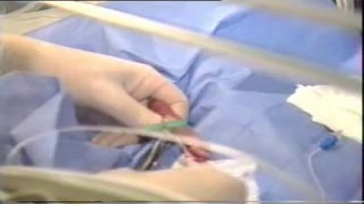
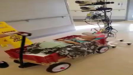
Catheters and Long Lines are introduced in Neonates to administer fluid and Total Parentral Nutrition. The proceedure is not easy to perform and is prone to get infections.
Strict Aseptic technique is mandatory
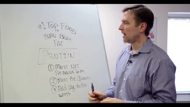
Overweight does not necessarily equal unhealthy. There are actually plenty of overweight people who are in excellent health (1). Conversely, many normal weight people have the metabolic problems associated with obesity (2). That’s because the fat under the skin is actually not that big of a problem (at least not from a health standpoint, it’s more of a cosmetic problem). It’s the fat in the abdominal cavity, the belly fat, that causes the biggest issues (3). If you have a lot of excess fat around your waistline, even if you’re not very heavy, then you should take some steps to get rid of it. Belly fat is usually estimated by measuring the circumference around your waist. This can easily be done at home with a simple tape measure. Anything above 40 inches (102 cm) in men and 35 inches (88 cm) in women, is known as abdominal obesity. There are actually a few proven strategies that have been shown to target the fat in the belly area more than other areas of the body.
Beating heart or "off pump" coronary artery surgery is the latest revolution in the management coronary disease. It is being embraced world-wide by increasing numbers of surgeons. Many of the advantages are subtle but reduced mortality, stroke, and bleeding as well as earlier discharge are well-established benefits. A cardiac stabiliser is mandatory for this surgery, most are single use only and very expensive, this one is multiple use and is saving many healthcare dollars

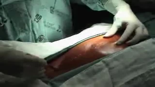
Both lower extremities must be evaluated to determine the presence or extent of any disease and to ascertain the pulse status of the patient. The feet are examined for signs of peripheral vascular disease and the anterior and posterior tibial pulses are palpated. Because an intact arch can supply retrograde flow to the major vessels of the foot, it can be helpful to put pressure on the anterior tibial artery when detecting the presence of a posterior tibial pulse and visa-versa. This "modified Allen's test" may detect proximal vessel obstruction masked by an intact foot arch. The need for preoperative angiography in young, healthy patients with a normal physical examination has been hotly debated. Our tendency has been to obtain preoperative angiograms as a guide. Although rare, we have seen lower extremities with a dominant peroneal artery nourishing the foot and distal anterior and posterior vessels, contraindicating sacrifice of the peroneal artery. MRI or CT angiography can also be used in many circumstances.
Dont worry sister!
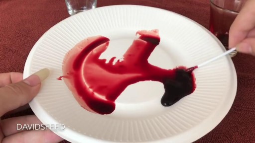
A little venom is drawn into a syringe. ... The quick coagulation or blood clotting caused by the Russell's viper venom is of particular interest to scientists — there's a lot of research into how it might be used in medicine. But this effect is only present in healthy blood.
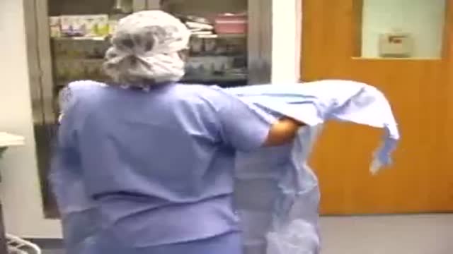
Wearing Surgical Gown Without Help
Duhamel's Operation for Chagasic Megacolon
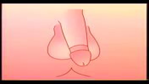
Watch that video to know if Does Circumcision Affect Male Orgasm?

#abdomenliposuction #laserskintightening #drprashantyadav #cosmeticsurgery #plasticsurgery #dezireclinicindia #weightloss #shorts #360degreeabdomenliposuction #lowerbackliposuction
Weight Loss After 360° Abdomen liposuction result, Abdomen Liposuction, lower back liposuction, 360 degree abdomen liposuction
☎️ For more info:
WhatsApp Your Details to know the Cost
Delhi - 8956880644, 9717470550, Pune - 9222122122, Bangalore- 8971224700, Gurugram - 9272007896, Ahmedabad - 9711162746
Why choose Dezire Clinic For Your Cosmetic and plastic surgery treatment ?
Dezire Clinic is a top searched clinic surgical and nonsurgical cosmetic procedure in India when comes to “Cosmetic, Skin ,Laser and Hair transplantation”.
Like and Share the video if you find it useful. Do not forget to Subscribe to our channel to get more updates.
Subscribe on YouTube https://youtube.com/dezireclin....ic?sub_confirmation=
https://youtube.com/dezireplas....ticsurgerycenter?sub
🎦 https://www.youtube.com/dezireclinic
🎦 https://www.youtube.com/DezirePlasticSurgeryCenter
👍🏻 https://www.facebook.com/drprashantmch/
👍🏻 https://www.facebook.com/dezireclinic
📸 https://www.instagram.com/drprashantdezireclinic/
📸 https://www.instagram.com/dezireclinics/
🐥 https://twitter.com/drprashantmch
👍🏻 https://www.linkedin.com/in/drprashantyadav/
🌐 Website: https://www.dezireclinic.in/
📧 dezireclinicindia@gmail.com
📧 info@dezireclinic.in
Dr. Prashant Yadav (M.S., M.Ch. Plastic Surgery ) & Founder of Dezire Clinic
Disclaimer: The content of this channel is for informational and educational purposes only. This content should not be considered a substitute for advice provided by a certified plastic or cosmetic surgeon. Patients must be properly diagnosed by a healthcare professional on an individual basis in order to achieve the desired results. There is no guarantee of getting the results and outcomes shown in videos, as the results can vary at the end. We will not be held liable for any harm caused by someone misusing our name.
#plasticsurgery #cosmeticsurgery #dezireclinic #drprashantyadav
Laparoscopic Uterosacral Colpoplexy HD

#anatomy #histology #bytesizemed
✨If you would like my help studying about cartilage, you can check out my long-form video linked at the bottom of the screen.
💫 For more videos like this, subscribe to my channel, Byte Size Med.
📚Factual References & for Further Reading:
- DiFiore's Atlas of Histology
- Junqueira's Basic Histology
- Gartner's Concise Histology
- Openstax Anatomy and Physiology
https://openstax.org/details/b....ooks/anatomy-and-phy
- Openstax Biology
https://openstax.org/details/books/biology-2e
(The last two are links to open-source references. They are NOT affiliate links)
🌤 Note:
These are just a collection of my notes. So use them the way you would use borrowed notes from a friend. 📝
The images in this video are hand-drawn for illustration and explanation only.✍️ Hence, they may not be anatomically accurate. I am just one person making these videos. If there are any errors, that is unintentional. I try super hard to avoid them. Please let me know if you find any, so it gets clarified for other viewers. Science constantly evolves and changes. New discoveries are made everyday. So some of the information in these videos may become outdated. If you notice that, please let me know so I can update them.
⚡️Disclaimer:
These videos are NOT a substitute for a medical textbook. Textbooks are written by experts (which I do not claim to be), edited, proofread and referenced. Please use them.
The information has been sourced from multiple references as mentioned above. I draw all the pictures myself. But if I have inadvertently infringed on any copyright, that is completely unintentional. I only make these videos to impart education. If I have accidentally violated copyright in any way, do let me know so I can make the necessary changes or give credit to anyone who is owed the same.
These videos are NOT intended for patient education. They are NOT a substitute for diagnosis and treatment by a licensed medical professional. Always seek the advice of a qualified health care provider for any questions you may have regarding any medical condition, so that they can address your individual needs.
🔅They are ONLY meant to help students of medicine and health sciences with studying, and should be used for just that purpose and absolutely nothing else.
Byte Size Med. All Rights Reserved.
