- Physical Examination
- Surgical Examination
- Ophthalmology
- Clinical Skills
- Orthopedics
- Surgery Videos
- Laparoscopy
- Pediatrics
- Funny Videos
- Cardiothoracic Surgery
- Nursing Videos
- Plastic Surgery
- Otorhinolaryngology
- Histology and Histopathology
- Neurosurgery
- Dermatology
- Pediatric Surgery
- Urology
- Dentistry
- Oncology and Cancers
- Anatomy Videos
- Health and Fitness
- Radiology
- Anaesthesia
- Physical Therapy
- Pharmacology
- Interventional Radiology
- Cardiology
- Endocrinology
- Gynecology
- Emergency Medicine
- Psychiatry and Psychology
- Childbirth Videos
- General Medical Videos
- Nephrology
- Physiology
- Diet and Food Health
- Diabetes Mellitus
- Neurology
- Women Health
- Osteoporosis
- Gastroenterology
- Pulmonology
- Hematology
- Rheumatology
- Toxicology
- Nuclear Medicine
- Infectious Diseases
- Vascular Disease
- Reproductive Health
- Burns and Wound Healing
- Other
Top videos
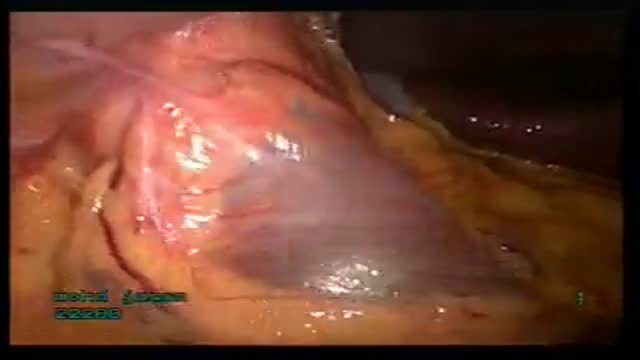
Laparoscopic Abdominal Wall Hernia Repair in Qatar by Dr. Emadi

Laparoscopic Gastric Banding in Qatar by Dr. Emadi
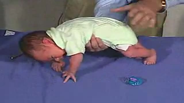
Galant Reflex

Short Leg Cast
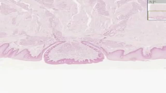
Histology of Tongue Circumvallate Papilla
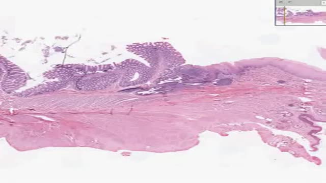
Histology of Rectoanal Junction
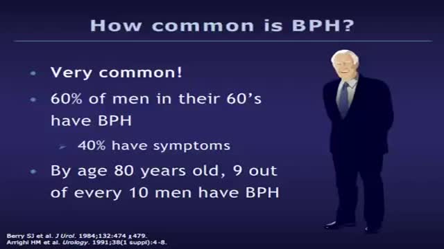
Benign prostatic hyperplasia (BPH), also known as benign prostatic hypertrophy, is a histologic diagnosis characterized by proliferation of the cellular elements of the prostate. Cellular accumulation and gland enlargement may result from epithelial and stromal proliferation, impaired preprogrammed cell death (apoptosis), or both. BPH involves the stromal and epithelial elements of the prostate arising in the periurethral and transition zones of the gland (see Pathophysiology). The hyperplasia presumably results in enlargement of the prostate that may restrict the flow of urine from the bladder. BPH is considered a normal part of the aging process in men and is hormonally dependent on testosterone and dihydrotestosterone (DHT) production. An estimated 50% of men demonstrate histopathologic BPH by age 60 years. This number increases to 90% by age 85 years. The voiding dysfunction that results from prostate gland enlargement and bladder outlet obstruction (BOO) is termed lower urinary tract symptoms (LUTS). It has also been commonly referred to as prostatism, although this term has decreased in popularity. These entities overlap; not all men with BPH have LUTS, and likewise, not all men with LUTS have BPH. Approximately half of men diagnosed with histopathologic BPH demonstrate moderate-to-severe LUTS. Clinical manifestations of LUTS include urinary frequency, urgency, nocturia (awakening at night to urinate), decreased or intermittent force of stream, or a sensation of incomplete emptying. Complications occur less commonly but may include acute urinary retention (AUR), impaired bladder emptying, the need for corrective surgery, renal failure, recurrent urinary tract infections, bladder stones, or gross hematuria. (See Presentation.) Prostate volume may increase over time in men with BPH. In addition, peak urinary flow, voided volume, and symptoms may worsen over time in men with untreated BPH (see Workup). The risk of AUR and the need for corrective surgery increases with age.

A dream is a succession of images, ideas, emotions, and sensations that usually occurs involuntarily in the mind during certain stages of sleep.[1] The content and purpose of dreams are not definitively understood, though they have been a topic of scientific speculation, as well as a subject of philosophical and religious interest, throughout recorded history. The scientific study of dreams is called oneirology
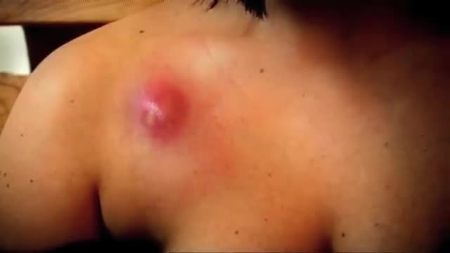
An abscess is a tender mass generally surrounded by a colored area from pink to deep red. Abscesses are often easy to feel by touching. The middle of an abscess is full of pus and debris. Painful and warm to touch, abscesses can show up any place on your body. The most common sites are in your armpits (axillae), areas around your anus and vagina(Bartholin gland abscess), the base of your spine (pilonidal abscess), around a tooth (dental abscess), and in your groin. Inflammation around a hair follicle can also lead to the formation of an abscess, which is called a boil (furuncle). Unlike other infections, antibiotics alone will not usually cure an abscess. In general an abscess must open and drain in order for it to improve. Sometimes draining occurs on its own, but generally it must be opened by a doctor in a procedure called incision and drainage (I&D).

People with panic disorder have sudden and repeated attacks of fear that last for several minutes or longer. These are called panic attacks. Panic attacks are characterized by a fear of disaster or of losing control even when there is no real danger. A person may also have a strong physical reaction during a panic attack. It may feel like having a heart attack. Panic attacks can occur at any time, and many people with panic disorder worry about and dread the possibility of having another attack.
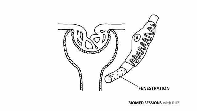
Glomerular filtration is the first step in making urine. It is the process that your kidneys use to filter excess fluid and waste products out of the blood into the urine collecting tubules of the kidney, so they may be eliminated from your body.
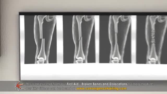
A broken bone requires emergency medical care. Your child might have a broken (fractured) bone if he or she heard or felt a bone snap, has difficulty moving the injured part, or if the injured part moves in an unnatural way or is very painful to the touch. A sprain occurs when the ligaments, which hold bones together, are overstretched and partially torn. A strain is when a muscle or tendon is overstretched or torn. Sprains and strains generally cause swelling and pain, and there may be bruises around the injured area. Most sprains and strains, after proper medical evaluation, can be treated at home.
Alzheimer's worsens over time. Alzheimer's is a progressive disease, where dementia symptoms gradually worsen over a number of years. In its early stages, memory loss is mild, but with late-stage Alzheimer's, individuals lose the ability to carry on a conversation and respond to their environment. Alzheimer's is the sixth leading cause of death in the United States. Those with Alzheimer's live an average of eight years after their symptoms become noticeable to others, but survival can range from four to 20 years, depending on age and other health conditions. Alzheimer's has no current cure, but treatments for symptoms are available and research continues. Although current Alzheimer's treatments cannot stop Alzheimer's from progressing, they can temporarily slow the worsening of dementia symptoms and improve quality of life for those with Alzheimer's and their caregivers. Today, there is a worldwide effort under way to find better ways to treat the disease, delay its onset, and prevent it from developing. Alzheimer's has no current cure, but treatments for symptoms are available and research continues. Although current Alzheimer's treatments cannot stop Alzheimer's from progressing, they can temporarily slow the worsening of dementia symptoms and improve quality of life for those with Alzheimer's and their caregivers. Today, there is a worldwide effort under way to find better ways to treat the disease, delay its onset, and prevent it from developing.

The DASH diet is a lifelong approach to healthy eating that's designed to help treat or prevent high blood pressure (hypertension). The DASH diet encourages you to reduce the sodium in your diet and eat a variety of foods rich in nutrients that help lower blood pressure, such as potassium, calcium and magnesium.

What Happens When You're In a Coma?
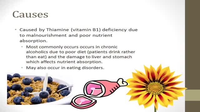
-Korsakoff's syndrome is a common and preventable sequel of Wernicke's encephalopathy. Thiamine, if given during the stage of Wernicke's encephalopathy, can prevent the onset of Korsakoff's psychosis. The administration of glucose prior to thiamine can precipitate Korsakoff's syndrome, as seen in this case. In such patients, brain MRI frequently shows abnormal enhancement of the mammillary bodies & thallamus

Panic attacks are discrete periods of intense fear or discomfort. Symptoms may include palpitations, sweating, trembling, shortness of breath, a choking sensation, chest pain, nausea, dizziness, paresthesias, and a fear of dying or losing control
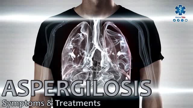
Aspergillosis is the name given to a wide variety of diseases caused by infection by fungi of the genus Aspergillus. The majority of cases occur in people with underlying illnesses such as tuberculosis or chronic obstructive pulmonary disease (COPD), but with otherwise healthy immune systems.
Curious about physiotherapy or wanting to know how to properly perform an exercise? Check us out on Social Media! Facebook: https://www.facebook.com/striveptandperformance/ Instagram: https://www.instagram.com/striveptandperf/ Twitter: https://twitter.com/StrivePTandPerf Blog: http://www.strivept.ca/blog
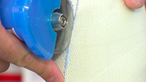
How to Prepare, Apply & Remove a Total Contact Cast
