- Physical Examination
- Surgical Examination
- Ophthalmology
- Clinical Skills
- Orthopedics
- Surgery Videos
- Laparoscopy
- Pediatrics
- Funny Videos
- Cardiothoracic Surgery
- Nursing Videos
- Plastic Surgery
- Otorhinolaryngology
- Histology and Histopathology
- Neurosurgery
- Dermatology
- Pediatric Surgery
- Urology
- Dentistry
- Oncology and Cancers
- Anatomy Videos
- Health and Fitness
- Radiology
- Anaesthesia
- Physical Therapy
- Pharmacology
- Interventional Radiology
- Cardiology
- Endocrinology
- Gynecology
- Emergency Medicine
- Psychiatry and Psychology
- Childbirth Videos
- General Medical Videos
- Nephrology
- Physiology
- Diet and Food Health
- Diabetes Mellitus
- Neurology
- Women Health
- Osteoporosis
- Gastroenterology
- Pulmonology
- Hematology
- Rheumatology
- Toxicology
- Nuclear Medicine
- Infectious Diseases
- Vascular Disease
- Reproductive Health
- Burns and Wound Healing
- Other
Top videos
Dieta Alcalina, Lista Alimentos Alcalinos, Agua Alcalina Beneficios, Consejos Para Adelgazar---- http://dieta-alcalina-alimentos.good-info.co --- Entendiendo Como Funciona Una Dieta. Alcalina Las dietas Alcalinas son una forma popular para las personas que quieren logran una salud óptima. Sin embargo, muchas personas en verdad no entienden como esta dieta funciona. El concepto es realmente muy simple – la dieta se centra en volver a ganar el equilibrio que se perdió cuando el hombre empezó a tener una alimentación más "domesticada" en la revolución industrial y grandes almacenamientos de alimentos. Cuando comenzó a primar el beneficios económico a la salud de las personas. En vez de centrarse en la comida alta en azúcar, grasa y colesterol, una dieta alcalina consiste principalmente en frutas y vegetales frescos, cereales integrales, fuentes de proteínas saludables, como soya, frijoles, legumbres y aceites saludables como la canola, oliva y la semilla de lino. Estos alimentos pueden ser alcalinos o ácidos en su estado natural, pero todos ellos producen lo que se denomina “cenizas alcalinas” una vez que son digeridos y metabolizados por el cuerpo. Cuando el pH del cuerpo se mantiene a un nivel bajo de alcalinidad, todo el sistema puede trabajar más eficientemente. Descubre como la dieta alcalina funciona & por qué los alimentos alcalinos son altamente recomendados para tu salud. Haz clic aquí http://dieta-alcalina-alimentos.good-info.co
Polio virus

Pictures Of Shingles, Images Of Shingles, Cause Of Shingles, Can You Catch Shingles, Cause Shingles --- http://shingles-cure.good-info.co/ ---- Home Remedy For Shingles Treatment. There are surprisingly diverse amounts of treatments that can be used for viruses, especially one in particular known as Shingles. Shingles is the type of virus that is annoyingly unpredictable. Some people get it while others do not, and while it predominant after a certain age, most people can go their whole lives without ever seeing a hint of it. Yet, we all have it living inside of us. The first step to treating the virus correctly is to understand what can set it off. The first pre-requisite is to have had chicken-pox before. Most things manage to squeeze through an immune system if it is weak enough and the shingles virus is no different. In some cases that can kill someone with a weak enough system The shingles virus can also ‘wake up’ if sufficient levels of stress agitate a person’s immune system. As mentioned before, the virus is unpredictable, and even if you meet all the requirements you could go through your entire life without one outbreak. The second step to treating the virus is catching it in time. 72 hours after you first begin to notice symptoms of shingles is pushing your luck so within that time frame some type of treatment should be instigated. For the most part, for a disease like shingles, the main source of relief comes from skin creams and pain killers. Neither of which is a cure for the pain, but which do happen to fall under the heading of ‘home remedy’ Wet rags can be used to soothe the inflamed and tender skin, and a binding with a substance known as aluminum acetate can protect the infected area without causing further irritation. Calamine lotion is a fail safe for almost any sort of skin irritation so it would be smart to go through the aisles at your local grocery store to find products similar. Then of course there are the most obvious measures that need to be taken during your do it yourself treatment sessions. Stay out of the sun since the heat and UV rays can cause unbearable pain against already brutalized skin. Also, keep yourself from scratching at the area since, like the chicken-pox, you will only end up making it worse. If you do not believe me then consider the unlucky individuals who do not have the rash on a small area, but rather all over their bodies. Anti-itch cream would probably be more than appreciated at that point. Should the time come where the virus is making repeated appearances, then it may be time to throw in the towel and head for the hospital. The reason is that after the first outbreak, it isn’t unusual that the virus that was originally stored in the roots of your nerves ‘burned’ itself out. However, continual outbreaks are indications of something deeper that can only grow worse as the attacks continue. Some victims of the virus have described the sensation as having your flesh eaten from the inside out. Once the pain reaches that level, stubbornness should be put on the backburner. If you are forced to go to your local doctor, you may be provided with some of the medicines that have recently been created to help fight off shingles. They are by no means brand names, but neither are they over the counter drugs either. After a few more years to study their effectiveness, there is no reason why this new medication cannot ease the pain caused by shingles, and even, eventually cure it. In this presentation, shows you some unique and rare methods to get rid of shingles naturally in as little as 14 days! This is based on proven techniques used by shingles sufferers without the use of pills and other medication. Get Rid of Shingles will also boost your energy and health dramatically and improve the quality of your life. IMPORTANT NOTE: I can't leave this video up for long, so be sure to watch it from beginning to end while it's still here. REMEMBER: Watch the whole video, as the ending will pleasantly surprise you. click here: http://shingles-cure.good-info.co/
Curious about physiotherapy or wanting to know how to properly perform an exercise? Check us out on Social Media! Facebook: https://www.facebook.com/striveptandperformance/ Instagram: https://www.instagram.com/striveptandperf/ Twitter: https://twitter.com/StrivePTandPerf Blog: http://www.strivept.ca/blog
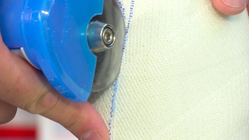
How to Prepare, Apply & Remove a Total Contact Cast


How to Know If You Have a Serious Knee Injury or Problem
Youtube Channel: https://www.youtube.com/user/physicaltherapyvideo
Website: https://bobandbrad.com/
Bob & Brad discuss how to know if you have a serious knee injury. They show you what to look for and what you should do.
This Week's Giveaway:
This month we are giving away a grand prize of a Sleepovation mattress and two pillows!
BONUS: 2 runners-up will receive a Sleepovation pillow!
December Giveaway link: https://shrsl.com/2ob0e
Purchase Mattress: http://shrsl.com/1n2e2
Purchase Pillow: http://shrsl.com/1xch4
Discount: Make sure to use the discount code FAMOUSPT to receive 15% off of your purchase of a mattress or use FAMOUSPTPIL for 25% off of their pillows! This is the biggest discount of the year!
Sleepovation will reimburse winners of the giveaway if they have already purchased, so no need to wait to buy Bob & Brad's favorite mattress and pillow!
Our videos offer the best "get fit , stay healthy, and pain-free" information directed toward people 0 to 101 years old. Physical Therapists Bob Schrupp and Brad Heineck have over 50 years of combined. We try to add a twist of our humor into each video in our quest to be the "Most Famous Physical Therapists on the Internet" In our opinion of course!!! Subscribe to us now and join the fun. Not only will these videos provide outstanding health information on treating yourself at home, we also do product reviews.
For our favorite products on Amazon click on this link: https://www.amazon.com/shop/physicaltherapyvideo
Visit us on our other social media platforms:
Website: https://bobandbrad.com/
Facebook: https://www.facebook.com/BobandBrad/
Instagram: https://www.instagram.com/officialbobandbrad/
Twitter: https://twitter.com/ptfamous
Bob and Brad’s Products:
Grip and Forearm Strengthener: https://store.bobandbrad.com
Wall Anchor: https://store.bobandbrad.com
Booyah Stik: https://store.bobandbrad.com
Knee Glide: https://store.bobandbrad.com
Fit Glide: https://store.bobandbrad.com
Massage Gun: https://amzn.to/36pMekg
Hanging Handles: https://amzn.to/2RXLVFF
Resistance Bands: https://amzn.to/36uqnbr
Pull Up Bands: https://amzn.to/3qmI4Rv
If you order from the Bob and Brad Store Links, you will receive 15% off your purchase.
Check out our shirts, mugs, bags and more in our Bob and Brad merchandise shop here: https://shop.spreadshirt.com/bob-brad
Bob & Brad Amazon Store: https://amzn.to/2RTSLLh
Other Products We Love: https://www.amazon.com/shop/physicaltherapyvideo?listId=3581Z1XUVFAFY
Check out The Bob & Brad Crew on YouTube by clicking here: https://www.youtube.com/c/thebobbradcrew
Want to help translate our videos? We would so love the help! http://www.youtube.com/timedtext_cs_panel?c=UCmTe0LsfEbpkDpgrxKAWbRA&tab=2
Medical Disclaimer All information, content, and material of this website is for informational purposes only and are not intended to serve as a substitute for the consultation, diagnosis, and/or medical treatment of a qualified physician or healthcare provider.
Affiliate disclaimer: Keep in mind that we may receive commissions when you click our links and make purchases. However, this does not impact our reviews and comparisons. We are highly selective in our products and try our best to keep things fair and balanced in order to help you make the best choice for you.

Shoulder Exam
I think that the most daunting aspect of the shoulder exam is appreciating the functional anatomy of this incredibly mobile joint. The primary benefit of the ball and socket arrangement is that it allows the hand to be positioned precisely in space, maximizing our ability to function. In terms of functionality, the shoulder might be best described as having a golf ball-on-a-tee design.
Location Of The Muscle Groups Is Approximated In The Pictures Above.
Start by looking at the normal (or more normal) side. Note any scars, obvious asymmetry, discoloration, swelling, or muscle asymmetry.
Palpation
Gently palpate around the shoulder, touching each of the landmarks noted above. Make note of pain.
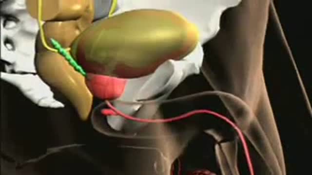
Anatomy of the Prostate
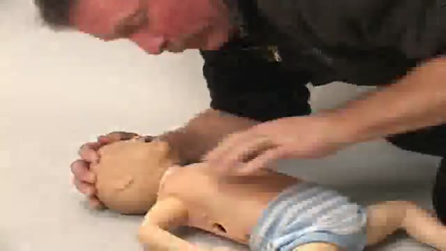
Infant CPR Video Demonstration

Mesenteric artery illuminated with luciferase

Phlebotomy Procedure

Rhinoplasty Surgery Video
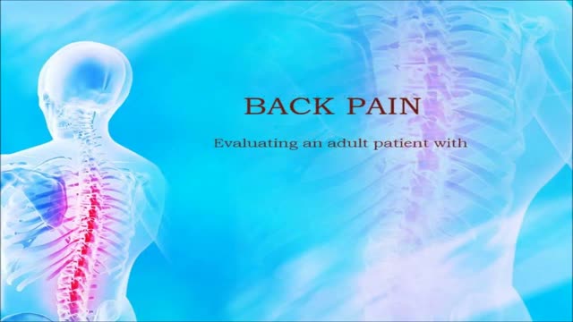
USMLE Step 2 CS - Back Pain - This is just preview video. To get full access please visit our website : www.usmletutoring.com

WORLD`S BEST IMMEDIATE ZIRCONIA DENTAL IMPLANT SOLUTION video

A video from the American Academy of Family Physicians

Histology of Hyaline Cartilage

Blood type (or blood group) is determined, in part, by the ABO blood group antigens present on red blood cells. A blood type (also called a blood group) is a classification of blood based on the presence or absence of inherited antigenic substances on the surface of red blood cells (RBCs).

Ehlers-Danlos syndrome is a group of disorders that affect the connective tissues that support the skin, bones, blood vessels, and many other organs and tissues. Defects in connective tissues cause the signs and symptoms of Ehlers-Danlos syndrome, which vary from mildly loose joints to life-threatening complications. Previously, there were more than 10 recognized types of Ehlers-Danlos syndrome, differentiated by Roman numerals. In 1997, researchers proposed a simpler classification that reduced the number of major types to six and gave them descriptive names: the classical type (formerly types I and II), the hypermobility type (formerly type III), the vascular type (formerly type IV), the kyphoscoliosis type (formerly type VIA), the arthrochalasia type (formerly types VIIA and VIIB), and the dermatosparaxis type (formerly type VIIC). This six-type classification, known as the Villefranche nomenclature, is still commonly used. The types are distinguished by their signs and symptoms, their underlying genetic causes, and their patterns of inheritance. Since 1997, several additional forms of the condition have been described. These additional forms appear to be rare, affecting a small number of families, and most have not been well characterized.

An ICD is a battery-powered device placed under the skin that keeps track of your heart rate. Thin wires connect the ICD to your heart. If an abnormal heart rhythm is detected the device will deliver an electric shock to restore a normal heartbeat if your heart is beating chaotically and much too fast.
