- Physical Examination
- Surgical Examination
- Ophthalmology
- Clinical Skills
- Orthopedics
- Surgery Videos
- Laparoscopy
- Pediatrics
- Funny Videos
- Cardiothoracic Surgery
- Nursing Videos
- Plastic Surgery
- Otorhinolaryngology
- Histology and Histopathology
- Neurosurgery
- Dermatology
- Pediatric Surgery
- Urology
- Dentistry
- Oncology and Cancers
- Anatomy Videos
- Health and Fitness
- Radiology
- Anaesthesia
- Physical Therapy
- Pharmacology
- Interventional Radiology
- Cardiology
- Endocrinology
- Gynecology
- Emergency Medicine
- Psychiatry and Psychology
- Childbirth Videos
- General Medical Videos
- Nephrology
- Physiology
- Diet and Food Health
- Diabetes Mellitus
- Neurology
- Women Health
- Osteoporosis
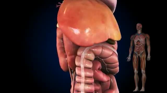
- Gastroenterology
- Pulmonology
- Hematology
- Rheumatology
- Toxicology
- Nuclear Medicine
- Infectious Diseases
- Vascular Disease
- Reproductive Health
- Burns and Wound Healing
- Other
Top videos
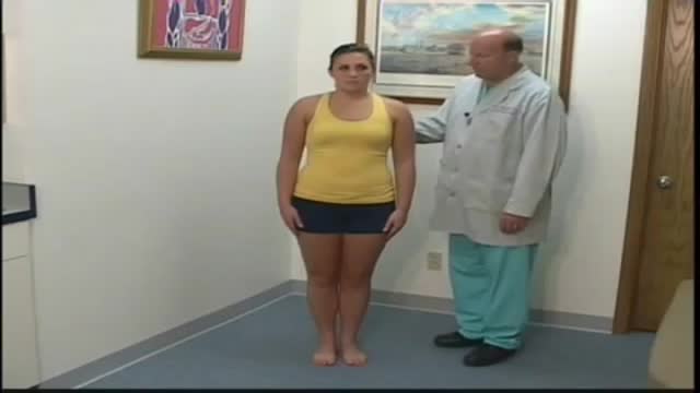
A patient who has a problem with proprioception can still maintain balance by using vestibular function and vision. In the Romberg test, the standing patient is asked to close his or her eyes. A loss of balance is interpreted as a positive Romberg's test.
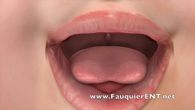
Tongue-tie (ankyloglossia) is a condition present at birth that restricts the tongue's range of motion. With tongue-tie, an unusually short, thick or tight band of tissue (lingual frenulum) tethers the bottom of the tongue's tip to the floor of the mouth
Questo Video 3D illustra la tecnica della Microlipocavitazione: sistema chirurgico ad ultrasuoni per ottenere l'emulsione del grasso in eccesso da eliminare. La Microlipocavitazione è una tecnica di chirurgia ambulatoriale, che richiede una modesta anestesia locale con un recupero delle proprie attività pressoché immediato.

Head Cyst watch to see more

This device gives you super strength to lift heavy items.
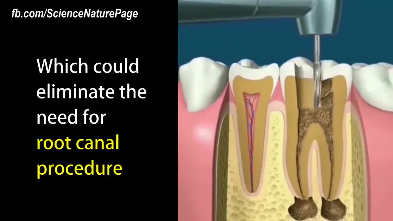
New dental fillings could allow your teeth to heal themselves.
Medications to treat high blood pressure Thiazide diuretics. ... Beta blockers. ... Angiotensin-converting enzyme (ACE) inhibitors. ... Angiotensin II receptor blockers (ARBs). ... Calcium channel blockers. ... Renin inhibitors.

Problems with Constipation? with medical students
In this video we give examples of five proven techniques for popping. Viewer discretion is advised as this may not be something that all people want to see. Popping isn't for everyone!
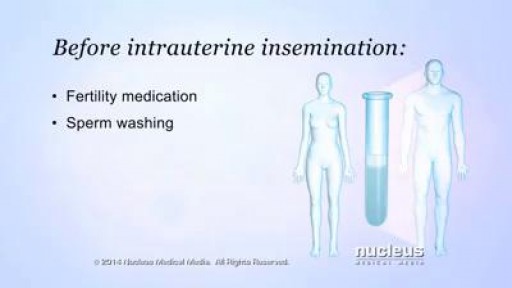
Intrauterine insemination (IUI) is a fertility treatment that involves placing sperm inside a woman's uterus to facilitate fertilization. The goal of IUI is to increase the number of sperm that reach the fallopian tubes and subsequently increase the chance of fertilization
This video was taken 2 weeks after this lovely patient had a Endoscopic Brow Lift, Face and Neck lift, and Fat Grafting. Know more about Handal Plastic Surgery Call us at (561) 912-9888 for more info!

Heart And Stroke, Good Health, Heart Attack And Stroke, Ways To Be Healthy, Tips On Healthy Living.-- http://grow-younger-blood.good-info.co -- Clicking the link below, you'll learn three easy exercises so effective that even if you have suffered from life-threatening hypertension for years, you can bring it down to 120/80 – as soon as TODAY! Plus, they take very little time – just 9 minutes. Lowering high blood pressure helps prevent… - stroke - heart attack - kidney failure - impotence …just to name a few conditions – caused by high blood pressure – you will avoid using these exercises! Bringing your blood pressure below 120/80 eliminates any need for blood pressure medications – so you’ll never have to suffer the horrendous side effects again. Every single brand of blood pressure drug creates serious side effects. The sad fact is, so do herbal medications. And the cost of buying pills these days is horrendous. The Solution Is Simple! Try these easy exercises now and naturally drop your blood pressure as soon as today. There are NO side effects and NO extra medical costs. To learn more and test the exercises for yourself, click here... http://grow-younger-blood.good-info.co

Come Rimanere Incinta Velocemente, Per Restare Incinta, Rimanere Incinta A 45 Anni, In Gravidanza--- http://come-rimanere-incinta.info-pro.co -- Farti rimanere incinta rapidamente e allo stesso tempo invertire l'infertilità. E' un dato di fatto. Il 92% delle donne che usano trattamenti convenzionali per aumentare le loro probabilità di concepire non riescono a rimanere incinta e, a volte, la loro situazione peggiora anzichè migliorare. Ora tu puoi decidere di far parte del 8% delle donne che sono guarite dall'infertilità per sempre, imparando a lavorare sinergicamente con il tuo corpo. Contrariamente agli approcci convenzionali, lavorando con il tuo corpo, eliminando la causa principale e specifica della tua infertilità (come: cisti ovariche, fibromi uterini, endometriosi, livelli di follitropina alti, sindrome dell'ovaio policistico, ecc), migliorando contemporaneamente la tua mentalità, il tuo stato emotivo e biologico-riproduttivo, rimarrai velocemente incinta e darai alla luce un bimbo sano e forte, indipendentemente dalla tua età, dal numero di tentativi andati male o dalla gravità della tua situazione. Farti rimanere incinta olisticamente. E' un dato di fatto, non potrai mai rimanere incinta naturalmente e curare la tua infertilità affrontando solo uno dei tanti fattori responsabili dell'infertilità. Ad esempio, se hai già provato trattamenti come le pillole ormonali, posizioni sessuali o diete differenti, e non hai ottenuto nessun risultato probabilmente è perchè ti sei concentrata solo su un aspetto della tua condizione. Il mio sistema non ti insegnerà solo l'unico modo per rimanere incinta naturalmente, ma imparerai anche l'unico modo per invertire la tua infertilità per sempre, in modo olistico. Questo Rivoluzionario Sistema E' Talmente Unico ed Efficace che Ha il Potere di... Clicca sul link http://come-rimanere-incinta.info-pro.co
Dieta Iposodica Per Acufeni, Agopuntura Efficacia Contro Acufeni, Ronzio Orecchie Nel Silenzio---- http://acufeni-cura.plus101.com/ --- I sintomi dell'acufene, I sintomi dell'acufene possono causare molti fastidi e problemi a chi ne soffre. Inoltre anche i sintomi possono causare confusione. Prendiamo un esempio per capire meglio. Stai conducendo una vita perfetta, vai al lavoro e torni a casa ogni giorno, ma improvvisamente incominci a sentire strani rumori all'orecchio. Ovviamente credi che ci sia una sorgente di tali rumori. Stranamente nessun altro sembra sentirli. Puoi spaventarti e pensare che quei rumori provengano dal tuo corpo, così vai dal dottore. Dopo qualche esame il dottore conferma che è tutto a posto. Il problema è che tu i rumori continui a sentirli. Incominciano a darti sui nervi e ti influenzano nel lavoro. E ancora peggio, i rumori sembrano farsi più forti la notte, privandoti del tuo prezioso sonno. Si tratta di acufene. I rumori che senti sono i sintomi principali e possono essere di vario tipo. Puoi sentire strani scricchilii, ronzii, brusii, fischi o sibili all'orecchio. Non è mai lo stesso per tutti. Alcuni li sentono di tanto in tanto, altri per tutto il tempo. alcuni raccontano di forti attacchi. Ed altri sono stremati da trapanamenti costanti. Ma una cosa è certa - molte persone negli USA e altrove hanno gli acufeni. Quindi non sei davvero l'unico in queste condizioni. La maggior parte dei sintomi dell'acufene non sono altro che rumori fantasma Molte persone sono confuse se non riescono ad indivuare la fonte del rumore alle orecchie. Alcuni si spaventano. Spesso queste persone sono messe in ridicolo da chi non sente quei rumori. Ma per chi soffre di acufeni i suoni sono assolutamente reali. Di fatto sono rumori fantasma, una percezione delle orecchie. Vi sono eccezioni. In almeno un caso il rumore può essere reale. In altre parole c'è una effettiva fonte del rumore che senti. Questo è l'acufene pulsatile. In questo caso sei in grado di sentire il battito del tuo cuore e ciò ti può far impazzire perchè lo senti in continuazione. Vi è un'altra differenza. C'è sicuramente più di una persona che sente questi rumori e questo è il tuo dottore. Avrà bisogno di uno strumento di ascolto per sentirli. Prendi nota - non è come sentire il cuore con uno stetoscopio. in questo caso il dottore utilizza uno strumento di ascolto per sentire il rumore all'orecchio, non nel petto. Che cosa causa l'acufene pulsatile? Potresti sentire il tuo cuore in caso di pressione alta, danni alle arterie e anche cambiamenti della circolazione del sangue. A volte, una grande dose di stress, ansia o depressione può causare questa situazione. Non Importa Quale Sia La Causa, Tipo O Gravità Del Tuo Acufene, Puoi Iniziare Ad Utilizzare Questo Efficare Sistema PROPRIO ORA Ed Ottenere Un Sollivo E Una Liberazione Permanente ISTANTANEA Dai Tuoi Acufeni! inserisci ora: http://acufeni-cura.plus101.com/

Watch that video of The Most Terrible Skin Jiggers Removal
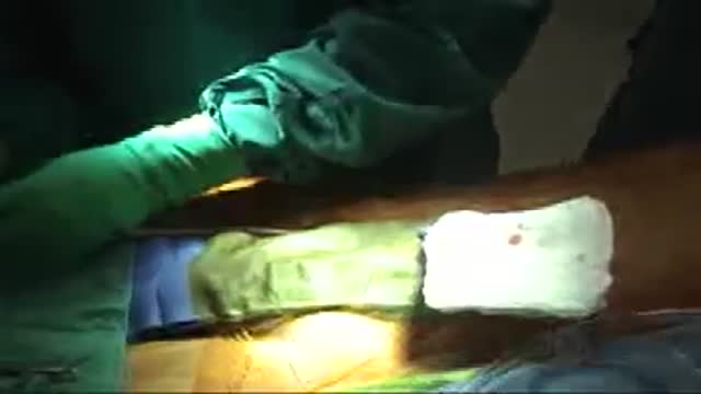
Mitral Valve Chorda Repair
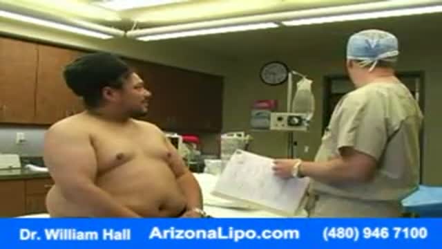
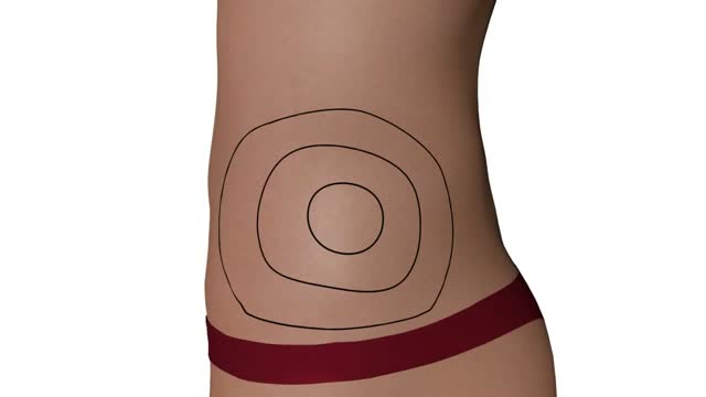
Abdomen Waist Liposuction for Weight Loss
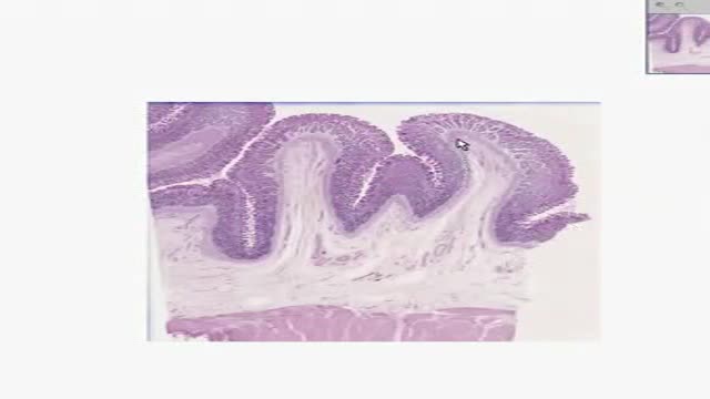
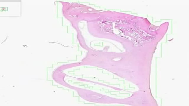
Histology of Gastric Fundus
Histology of Inner Ear 2
Possible complications include: Cardiovascular disease. ... Nerve damage (neuropathy). ... Kidney damage (nephropathy). ... Eye damage (retinopathy). ... Foot damage. ... Skin conditions. ... Hearing impairment. ... Alzheimer's disease.
