- Physical Examination
- Surgical Examination
- Ophthalmology
- Clinical Skills
- Orthopedics
- Surgery Videos
- Laparoscopy
- Pediatrics
- Funny Videos
- Cardiothoracic Surgery
- Nursing Videos
- Plastic Surgery
- Otorhinolaryngology
- Histology and Histopathology
- Neurosurgery
- Dermatology
- Pediatric Surgery
- Urology
- Dentistry
- Oncology and Cancers
- Anatomy Videos
- Health and Fitness
- Radiology
- Anaesthesia
- Physical Therapy
- Pharmacology
- Interventional Radiology
- Cardiology
- Endocrinology
- Gynecology
- Emergency Medicine
- Psychiatry and Psychology
- Childbirth Videos
- General Medical Videos
- Nephrology
- Physiology
- Diet and Food Health
- Diabetes Mellitus
- Neurology
- Women Health
- Osteoporosis
- Gastroenterology
- Pulmonology
- Hematology
- Rheumatology
- Toxicology
- Nuclear Medicine
- Infectious Diseases
- Vascular Disease
- Reproductive Health
- Burns and Wound Healing
- Other
Top videos
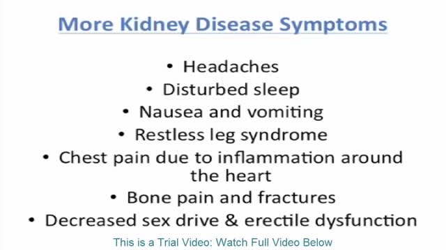
Signs and symptoms of chronic kidney disease develop over time if kidney damage progresses slowly. Signs and symptoms of kidney disease may include: Nausea Vomiting Loss of appetite Fatigue and weakness Sleep problems Changes in how much you urinate Decreased mental sharpness Muscle twitches and cramps Swelling of feet and ankles Persistent itching Chest pain, if fluid builds up around the lining of the heart Shortness of breath, if fluid builds up in the lungs High blood pressure (hypertension) that's difficult to control Signs and symptoms of kidney disease are often nonspecific, meaning they can also be caused by other illnesses. Because your kidneys are highly adaptable and able to compensate for lost function, signs and symptoms may not appear until irreversible damage has occurred.

Remove Acne Marks
Malaria is a serious and sometimes fatal disease caused by a parasite that commonly infects a certain type of mosquito which feeds on humans. People who get malaria are typically very sick with high fevers, shaking chills, and flu-like illness.
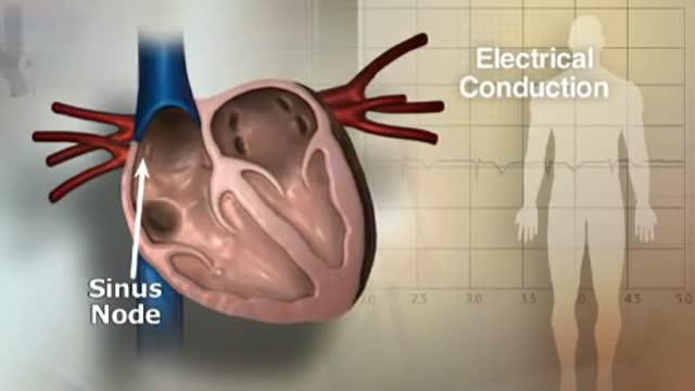
If you or someone you love has atrial fibrillation, learn more about what AFib is, why treatment can save lives, and what you can do to reach your goals, lower your risks and live a healthy life.
During in vitro fertilization (IVF), eggs and sperm are brought together in a laboratory glass dish to allow the sperm to fertilize an egg. With IVF, you can use any combination of your own eggs and sperm and donor eggs and sperm. After IVF, one or more fertilized eggs are placed in the uterus .
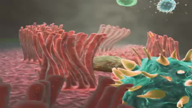
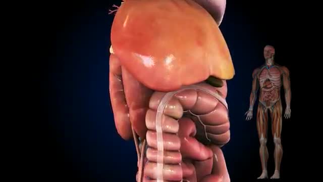
Cells may have slender extensions of the cell membrane to form cilia or the smaller extensions called microvilli. The microscopic microvilli effectively increase the surface area of the cell and are useful for absorption and secretion functions. A dramatic example is the human small intestine. The tissue has small fingerlike extensions called villi which are collections of cells, and those cells have many microvilli to even further increase the available surface area for the digestion process. According to Audesirk & Audesirk, this can give an effective surface area of about 250 square meters for absorption.
Possible complications include: Cardiovascular disease. ... Nerve damage (neuropathy). ... Kidney damage (nephropathy). ... Eye damage (retinopathy). ... Foot damage. ... Skin conditions. ... Hearing impairment. ... Alzheimer's disease.

Warfarin is an anticoagulant medication - it is used to slow down the blood-clotting process. Anticoagulants are used to prevent blood clots which may cause vein blockages, heart attack and stroke. Warfarin is known under the brand names Warfant, Jantoven, Coumadin, Lawarin, Marevan, and Waran.
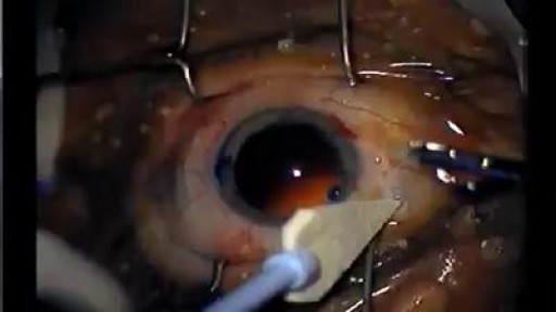
A cataract is a cloudiness of the normally transparent lens that is situated behind the iris. The lens focuses light rays on the retina at the back of the eye to produce a sharp image of what we see. When the lens becomes cloudy, the light rays cannot pass easily through it, and the image becomes blurry. It would be equivalent to having the lens in your camera becoming murky.
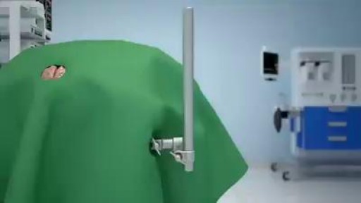
A craniotomy involves making an incision in the scalp and creating a hole known as a bone flap in the skull. The hole and incision are made near the area of the brain being treated. During open brain surgery, your surgeon may opt to: remove tumors. clip off an aneurysm

http://eliminar-seu-diabetes.good-info.co/ Tipos De Diabetes, Diabets, Alimentação Para Diabéticos, Diabetes Tipo Ii, Yacon Diabetes, https://youtu.be/iDK8jKuR_VQ É provável que se sinta identificado com alguma destas situações. Tem medo de uma complicação a longo prazo, como a perda da visão, a amputação dos dedos dos pés, de extremidades ou inclusive da morte? Quer terminar com as injeções diárias de insulina e as picadas nos dedos? Enfrenta diariamente o fato de que tem 80% de probabilidades de morrer com doenças cardíacas ou derrame cerebral? Sofre de excesso de peso que não pode eliminar, causada por seus medicamentos? Quer deixar de se sentir culpado por ter dietas especiais que complicam a organização da sua família? Está oprimido pelo cuidado e controle que diariamente esta doença precisa? Quer deixar de sofrer os terríveis efeitos secundários que provocam os medicamentos para o Diabetes? Sei o que se sente. pensar que não existe esperança, que não tem cura e que estamos condenados a viver doentes. Mas hoje Tenho Excelentes Notícias para lhe dar e posso garantir que o que você vai ler neste site será o mais importante que tenha lido em toda a sua vida.
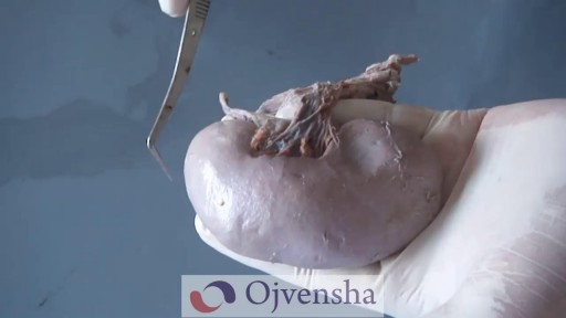
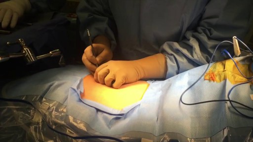
seroma 3 years after surgery
Each kidney contains around 1 million individual nephrons, the kidneys' microscopic functional units that filter blood to produce urine. The nephron is made of 2 main parts: the renal corpuscle and the renal tubule.

Suppurativa Hidradenitis, Hidradenitis Suppurativa Support, Hidradenitis Suppurativa Cures.--- http://hidradenitis-suppurativa-cure.good-info.co --- Causes of hidradenitis suppurativa Now that we know the symptoms, we must look deeper into the causes so as to get to the root of the problem Caused when hair follicles are blocked or inflamed When hair follicles are blocked due to smoking When hair follicles are blocked due to excess weight Hormonal fluctuations May be caused due to hyper active immune system Causes are sometimes also genetic Severe effects of hidradenitis suppurativa Smells, scars, itches and pains Fear of isolation due to stigmatization Overtime leading to depression Debility and strike on self esteem More information in. http://hidradenitis-suppurativa-cure.good-info.co

Anti Anxiety Medication, Is Depression A Mental Illness, How To Overcome Depression, Depression Cure--- http://depression-help.info-pro.co --- Symptoms of Depression, Signs and symptoms of depression include: Feelings of helplessness and hopelessness. A bleak outlook—nothing will ever get better, and there’s nothing you can do to improve your situation. Loss of interest in daily activities. No interest in former hobbies, pastimes, social activities, or sex. You’ve lost your ability to feel joy and pleasure. Appetite or weight changes. Significant weight loss or weight gain—a change of more than 5% of body weight in a month. Sleep changes. Either insomnia, especially waking in the early hours of the morning, or oversleeping. Anger or irritability. Feeling agitated, restless, or even violent. Your tolerance level is low, your temper short, and everything and everyone gets on your nerves. Loss of energy. Feeling fatigued, sluggish, and physically drained. Your whole body may feel heavy, and even small tasks are exhausting or take longer to complete. Self-loathing. Strong feelings of worthlessness or guilt. You harshly criticize yourself for perceived faults and mistakes. Reckless behavior. You engage in escapist behavior such as substance abuse, compulsive gambling, reckless driving, or dangerous sports. Concentration problems. Trouble focusing, making decisions, or remembering things. Unexplained aches and pains. An increase in physical complaints such as headaches, back pain, aching muscles, and stomach pain. DESTROY Depression The Natural Way! Click Here http://depression-help.info-pro.co
Home Remedies For Gas And Bloating, Get Rid Of Flatulence, Painful Flatulence, Severe Flatulence ----- http://flatulence-cure.plus101.com --- Reducing Flatulence Can Be Simple. No-one wants to gain a reputation as the person you have to open a window around. Breaking wind is perfectly normal, even desirable, but there is a difference between normal everyday wind and problem flatulence. If you can't hold it in even when you're trying hard, it may be that you have a decision to make - and reducing flatulence is more achievable, and more desirable, than stopping it altogether. What cannot be denied is that by following the right steps, you can make sure that reducing flatulence is within your reach whenever you so wish. Reducing Flatulence The Simple Way There are some very simple tips you can follow in order to ensure that flatulence is less of a problem. People who suffer from flatulence on a regular basis can make changes in their life which are ideal for reducing flatulence. They include: 1. Changing your diet. It is a commonly-used tip, but this is not without reason. The fact of the matter is that your diet is sure to affect the level of flatulence - for better or worse. 2. Taking enzyme supplements. These help your body produce more enzymes - helping the digestive system as well as aiding you in healing from injuries and infections. 3. Taking probiotics. Most commonly in the form of a yogurt or yogurt drink, these promote the growth of friendly bacteria, a major step in reducing flatulence. 4. Getting more exercise. A walk around the block may be all it takes to get your digestion running smoothly and ensure that you are less prone to flatulence. flatulence. You can discover how a former chronic gas sufferer is revealing the only holistic system to show you how to elimite your flatulence and bloating problems... FOREVER! And you can see how this approach has worked for hundreds of others just like yourself. Check it out now: http://flatulence-cure.plus101.com
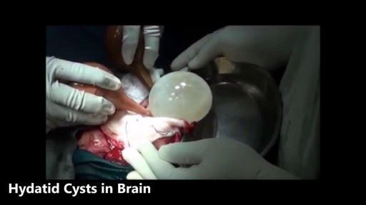
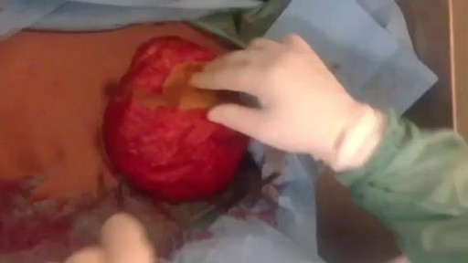
Watch that video of the Worst Brain & Liver Cysts Removal\
Como Curar Boqueras, Remedio Casero Para Boqueras, Porque Se Producen Las Boqueras, Boqueras ---- http://queilitis-angular.good-info.co --- ¿Qué Es Y Cómo Se Trata La Queilitis Angular? Perleche, queilosis, estomatitis, boqueras, son otras denominaciones con las que se conoce a la queilitis angular. Las comisuras de los labios presentan lesiones inflamatorias. Las grietas verticales a nivel de la piel pueden profundizarse y provocar ulceraciones, llagas, sangrados, infecciones, descamaciones, costras. Con ello, sobrevendrán las dificultades para hablar, para sonreír, para ingerir los alimentos y las bebidas. La queilitis angular no discrimina. Puede afectar tanto a los bebés como a los niños, a los adultos o a los ancianos. A menudo las causas derivan de una mala alimentación, carente de los nutrientes esenciales para el organismo. También las deficiencias nutricionales pueden deberse a la incapacidad orgánica para absorber los nutrientes, como sucede con la enfermedad celíaca. O las causas pueden provenir de estímulos que afectan una piel hipersensible, como ciertas alergias. O se puede producir por ciertos medicamentos. O incluso por prótesis dentarias mal ajustadas. Y la queilitis puede agravarse en una persona que padece micosis como la Cándida albicans. Lo cierto es que la boca es una zona húmeda, condición que dificulta la cura e incluso aumenta las manifestaciones nocivas en la piel y en la membrana que tapiza la cavidad interior de la boca. La humedad constante podrá ser caldo de cultivo para hongos y bacterias. Como muchas afecciones, la queilitis angular suele tener su origen en una mala alimentación. La hipovitaminosis o escasa provisión de vitamina A está considerada como posible desencadenante de la afección. Asimismo, es atribuible a la falta de minerales como el zinc, el hierro y la riboflavina (vitamina B2). Una vez que la queilitis angular está en proceso, la falta de nutrientes se agudiza. Sucede que a medida que la dolencia evoluciona se hace cada vez más difícil la ingesta de alimentos y la hidratación. Recordemos que beber suficiente cantidad de agua es esencial para el organismo. ¿Qué podemos hacer YA mismo? Hoy Existe Un Novedoso Tratamiento, Totalmente Natural Y Muy Simple, Con El Que Se Puede Eliminar La Queilitis Angular O Boqueras En Tan Solo 7 Días (O Menos). Este Revolucionario Sistema Ataca La Verdadera Causa De La Enfermedad Y No Solo Los Síntomas, Asegurando Resultados A Largo Plazo. Si Usted Desea Eliminar Para Siempre Esas Grietas Dolorosas Y La Vergüenza Que Causa Esta Afección, Puede Conocer Este Método De Resultados Comprobados Haciendo Clic En El Siguiente Enlace: http://queilitis-angular.good-info.co

A doctor explains the purpose of dialysis and explains the difference between hemodialysis and peritoneal dialysis. Learn more at https://www.niddk.nih.gov/heal....th-information/kidne
