- Physical Examination
- Surgical Examination
- Ophthalmology
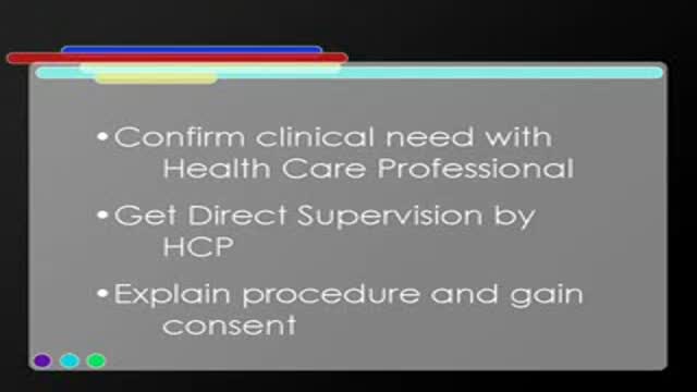
- Clinical Skills
- Orthopedics
- Surgery Videos
- Laparoscopy
- Pediatrics
- Funny Videos
- Cardiothoracic Surgery
- Nursing Videos
- Plastic Surgery
- Otorhinolaryngology
- Histology and Histopathology
- Neurosurgery
- Dermatology
- Pediatric Surgery
- Urology
- Dentistry
- Oncology and Cancers
- Anatomy Videos
- Health and Fitness
- Radiology
- Anaesthesia
- Physical Therapy
- Pharmacology
- Interventional Radiology
- Cardiology
- Endocrinology
- Gynecology
- Emergency Medicine
- Psychiatry and Psychology
- Childbirth Videos
- General Medical Videos
- Nephrology
- Physiology
- Diet and Food Health
- Diabetes Mellitus
- Neurology
- Women Health
- Osteoporosis
- Gastroenterology
- Pulmonology
- Hematology
- Rheumatology
- Toxicology
- Nuclear Medicine
- Infectious Diseases
- Vascular Disease
- Reproductive Health
- Burns and Wound Healing
- Other
Top videos
Cardiac tamponade is a medical emergency that requires urgent drainage of the pericardial fluid. Preferably, patients should be monitored in an intensive care unit. All patients should receive the following: Oxygen Volume expansion with blood, plasma, dextran, or isotonic sodium chloride solution, as necessary, to maintain adequate intravascular volume - Sagristà-Sauleda et al noted significant increase in cardiac output after volume expansion [24] (see the Cardiac Output calculator) Bed rest with leg elevation - This may help increase venous return Positive-pressure mechanical ventilation should be avoided because it may decrease venous return and aggravate signs and symptoms of tamponade. Inpatient care After pericardiocentesis, leave the intrapericardial catheter in place after securing it to the skin using sterile procedure and attaching it to a closed drainage system via a 3-way stopcock. Periodically check for reaccumulation of fluid, and drain as needed. The catheter can be left in place for 1-2 days and can be used for pericardiocentesis. Serial fluid cell counts can be useful for helping to discover an impending bacterial catheter infection, which could be catastrophic. If the white blood cell (WBC) count rises significantly, the pericardial catheter must be removed immediately. A Swan-Ganz catheter can be left in place for continuous monitoring of hemodynamics and to assess the effect of reaccumulation of pericardial fluid. A repeat echocardiogram and a repeat chest radiograph should be performed within 24 hours.
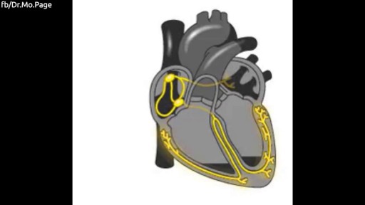
The human heart explained in 1 minute Video by Dr. Mo
http://tipps-gegen-cellulite.good-info.co --- Cellulite Behandeln, Hilfe Gegen Cellulite, Ultraschall Gegen Cellulite, Cellulite Reduzieren. Was Verursacht Cellulite? Wenn man die ursächlichen Faktoren der Cellulite suchen, findet man viele verschiedene Gründe. Websites geben oft falsche Informationen zu diesem Thema. Sie argumentieren, dass die richtige Ursachen schlechte Durchblutung, Flüssigkeitsretention und spezielle Arten von Fetten, usw. Sind, wenn das wirklich nicht wahr ist. Es gibt keine einzelne Ursache der Cellulite. Ihr Erscheinen kann sich auf verschiedene Faktoren zurückzuführen sein. Cellulite ist wie jede andere Art von Fett, es hat keine Besonderheiten und es ist nicht durch Flüssigkeitsretention oder schlechte Durchblutung verursacht. Wissentschaftliche Tests haben bewiesen, dass es nur gewöhnliche Fett ist. Diese vorstehende Fett ist auf der Bindgewebe unter der Haut und das ist der Grund, warum es wie eine Orangenhaut aussieht. Frauen haben Cellulite da das meiste Fett im unteren Teil des Körpers gespeichert ist, wo die Haut dünner ist. Andererseits Männer speichern die meisten Fett im Bauchbereich, wo die Haut dicker ist. Hormonelle Veränderungen sind einem der wichtigsten Faktoren, die das Erscheinungsbild der Cellulite beeinflussen. Diese treten in der Phase der Schwangerschaft oder Antibabypillen Verbrauch. Das Erste System Mit Allem, Was Notwendig lst, Um Cellulite Definitiv Loszuwerden Klicken Sie hier, um mehr zu erfahren http://tipps-gegen-cellulite.good-info.co

Roux-en-Y Gastric Bypass Surgery
Dr. Jawad has been performing Bariatric Surgery in Central Florida since 1984, and Laparoscopic Bariatric Surgery since 1999, having completed over 2000 Bariatric Surgical Cases safely, and with great success. Here you can watch Dr. Jawad performing a Laparoscopic Roux-En-Y Gastric Bypass surgery, with audio commentary describing the procedure.
varicose vein surgery stab and avulsion technique
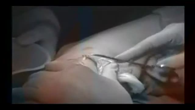
Structure of the Cannula and Cannulation

With a portable pump controlled by a wireless handheld device that automatically delivers insulin.
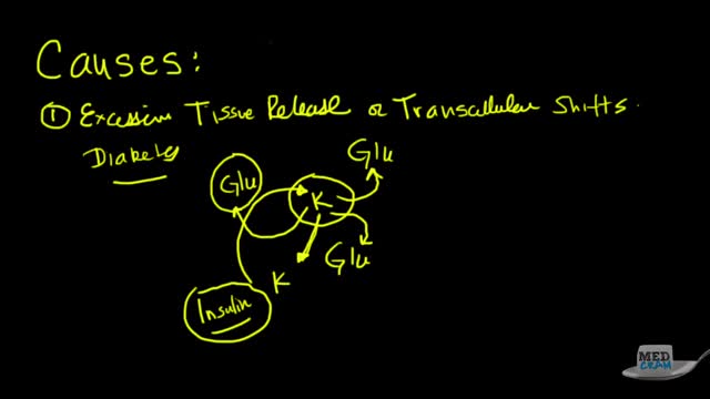
Hyperkalemia is defined as a serum potassium concentration higher than the upper limit of the normal range; the range in infants and children is age-dependent, whereas the range for adults is approximately 3.5-5.5 mEq/L. The upper limit may be considerably higher in young or premature infants, as high as 6.5 mEq/L.[5] Degrees of hyperkalemia are defined as follows[6] : 5.5-6.0 mEq/L – Mild 6.1-7.0 mEq/L – Moderate ≥7.0 mEq/L – Severe levels higher than 7 mEq/L can lead to significant hemodynamic and neurologic consequences. levels exceeding 8.5 mEq/L can cause respiratory paralysis or cardiac arrest and can quickly be fatal. Because of a paucity of distinctive signs and symptoms, hyperkalemia can be difficult to diagnose. Indeed, it is frequently discovered as an incidental laboratory finding. The physician must be quick to consider hyperkalemia in patients who are at risk for this disease process. (See Etiology.) However, any single laboratory study demonstrating hyperkalemia must be repeated to confirm the diagnosis, especially if the patient has no changes on electrocardiography (ECG). Because hyperkalemia can lead to sudden death from cardiac arrhythmias, any suggestion of hyperkalemia requires an immediate ECG to ascertain whether ECG signs of electrolyte imbalance are present (see Workup). Continuous ECG monitoring is essential if hyperkalemia is confirmed. Other testing is directed toward uncovering the condition or conditions that led to the hyperkalemia (see Workup). The aggressiveness of therapy for hyperkalemia is directly related to the rapidity with which the condition has developed, the absolute level of serum potassium, and the evidence of toxicity. The faster the rise of the potassium level, the higher it has reached, and the greater the evidence of cardiotoxicity, the more aggressive therapy should be. In severe cases, treatment focuses on immediate stabilization of the myocardial cell membrane, rapid shifting of potassium to the intracellular space, and total body potassium elimination. In addition, all sources of exogenous potassium should be immediately discontinued. (See Treatment.)
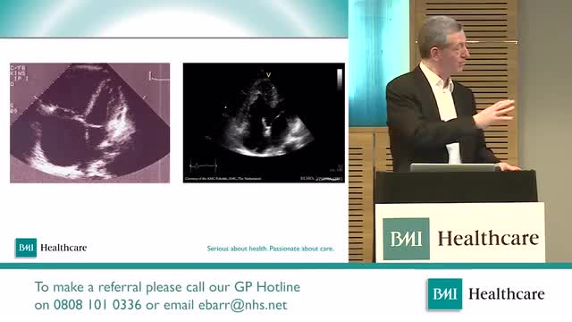
An increased prevalence of cardiovascular disease (CVD) has been found in women of childbearing age,[1] with the presence of CVD in pregnant women posing a difficult clinical scenario in which the responsibility of the treating physician extends to the unborn fetus. Profound changes occur in the maternal circulation that have the potential to adversely affect maternal and fetal health, especially in the presence of underlying heart conditions. Up to 4% of pregnancies may have cardiovascular complications despite no known prior disease. The European Society of Cardiology has published guidelines on the management of cardiovascular disease during pregnancy.[

Cancer immunology is a branch of immunology that studies interactions between the immune system and cancer cells (also called tumors or malignancies). It is a growing field of research that aims to discover innovative cancer immunotherapies to treat and retard progression of the disease.

Alzheimer's is the most common form of dementia, a general term for memory loss and other intellectual abilities serious enough to interfere with daily life. Alzheimer's disease accounts for 60 to 80 percent of dementia cases. Alzheimer's is not a normal part of aging, although the greatest known risk factor is increasing age, and the majority of people with Alzheimer's are 65 and older. But Alzheimer's is not just a disease of old age. Up to 5 percent of people with the disease have early onset Alzheimer's (also known as younger-onset), which often appears when someone is in their 40s or 50s.
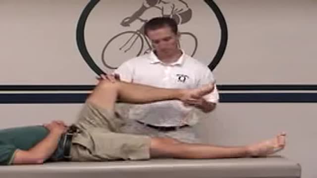
McMurray's maneuver is used to detect the presence of meniscal tears. To detect a medial meniscal injury, the patient is put in a supine position with the knee in maximum flexion. One hand of the examiner is placed on the posteromedial margin of the involved knee and the other hand supports the foot. The tibia is then externally rotated, and the knee is extended slowly. The test is positive if there is an audible or palpable click or popping sensation during extension of the involved knee.

One of the criteria to determine brain death is the irreversible absence of cerebral and brainstem reflexes including pupillary, oculocephalic, oculovestibular (caloric), corneal, gag, sucking, swallowing, and extensor posturing. Some of the other criteria for determination of brain death include: 1. Absence of respiratory drive (apnea) off the ventilator for a duration that is sufficient to produce hypercarbic drive (usually 10 to 20 minutes to achieve pC02 of 50 to 60 mmHg) ( 2. Body temperature below 34 C (93.2 F) 3. EEG isoelectric for 30 minutes at maximal gain 4. Absence of cerebral circulation by Doppler or magnetic resonance angiography 5. At least 24 hours of observation in adults with anoxic-ischemic brain damage with a negative drug screen
Questo Video 3D illustra la tecnica della Microlipocavitazione: sistema chirurgico ad ultrasuoni per ottenere l'emulsione del grasso in eccesso da eliminare. La Microlipocavitazione è una tecnica di chirurgia ambulatoriale, che richiede una modesta anestesia locale con un recupero delle proprie attività pressoché immediato.
Huge CYST in Abdomin
Chronic mesenteric ischemia (CMI) usually results from long-standing atherosclerotic disease of 2 or more mesenteric vessels. [1] Other nonatheromatous causes of CMI include the vasculitides, such as Takayasu arteritis. Symptoms are caused by the gradual reduction in blood flow to the intestine. [2] (See Presentation.) In 1958, Shaw and Maynard described the first thromboendarterectomy of the superior mesenteric artery (SMA) for the treatment of both acute mesenteric ischemia (AMI) and CMI. Several other surgical procedures have since been attempted, ranging from reimplantation of the visceral branch into the adjacent aorta to using an autogenous vein graft. In 1972, Stoney and Wylie introduced transaortic visceral thromboendarterectomy and aortovisceral bypass, which have proved to be highly effective techniques.

Watch that 200 lb Tumor in Man's Body Removal Surgery

Plastic Eyelid Beauty surgery

Bronchiolitis is a common lung infection in young children and infants. It causes inflammation and congestion in the small airways (bronchioles) of the lung. Bronchiolitis is almost always caused by a virus. Typically, the peak time for bronchiolitis is during the winter months. Bronchiolitis starts out with symptoms similar to those of a common cold but then progresses to coughing, wheezing and sometimes difficulty breathing. Symptoms of bronchiolitis can last for several days to weeks, even a month.
