- Physical Examination
- Surgical Examination
- Ophthalmology
- Clinical Skills
- Orthopedics
- Surgery Videos
- Laparoscopy
- Pediatrics
- Funny Videos
- Cardiothoracic Surgery
- Nursing Videos
- Plastic Surgery
- Otorhinolaryngology
- Histology and Histopathology
- Neurosurgery
- Dermatology
- Pediatric Surgery
- Urology
- Dentistry
- Oncology and Cancers
- Anatomy Videos
- Health and Fitness
- Radiology
- Anaesthesia
- Physical Therapy
- Pharmacology
- Interventional Radiology
- Cardiology
- Endocrinology
- Gynecology
- Emergency Medicine
- Psychiatry and Psychology
- Childbirth Videos
- General Medical Videos
- Nephrology
- Physiology
- Diet and Food Health
- Diabetes Mellitus
- Neurology
- Women Health
- Osteoporosis
- Gastroenterology
- Pulmonology
- Hematology
- Rheumatology
- Toxicology
- Nuclear Medicine
- Infectious Diseases
- Vascular Disease
- Reproductive Health
- Burns and Wound Healing
- Other
Top videos
Dr. Katherine Scovner from the Division of Nephrology at Massachusetts General Hospital discusses kidney dialysis.

Are you a first time would be mom? If yes, then you must be very excited to feel the first movement and kick from your baby. It is undoubtedly the most exciting experience for many expecting moms. It is an indication that there is a little angel growing inside you. There are interesting facts about baby kicks during pregnancy that you need to know.

World-renowned surgeons at Shriners Hospitals for Children – Northern California provide complex pediatric surgery for children one-year and older with congenital and acquired conditions. Children from throughout the Western United States with chest wall malformations, gastro-intestinal disease, ano-rectal disorders, urologic conditions and other complex surgical needs benefit from the expert care. The pediatric surgery team is devoted to the development of innovative and minimally invasive surgical techniques.
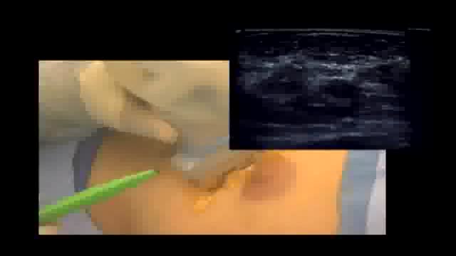
Excision of breast cancer that is visible only on mammogram. diagnosis is typically established on stereotactic biospy and excision is done with wire localization. This techniques involves localization by sonography of the hematoma that is left behind at the time of biopsy. It provides not only accu...rate location of the tumor but ensures adequate margins of excision.
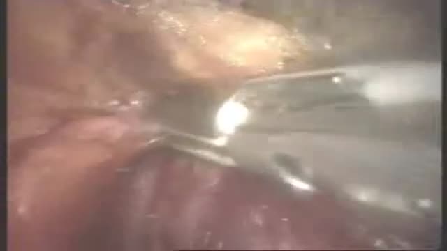
Splenectomy surgery video

This animation demonstrates how a unilateral complete cleft lip repair is performed. This video is meant for educational purposes for patients and families. There are many ways to fix a complete cleft lip, but the technique shown here is the most common known as the Millard Rotation Advancement Repair.
Ten percent of all pregnancies are complicated by hypertension. Eclampsia and preeclampsia account for about half of these cases worldwide, and these conditions have been recognized and described for years despite the general lack of understanding of the disease. [1] In the fifth century, Hippocrates noted that headaches, convulsions, and drowsiness were ominous signs associated with pregnancy. In 1619, Varandaeus coined the term eclampsia in a treatise on gynecology. [2, 3]
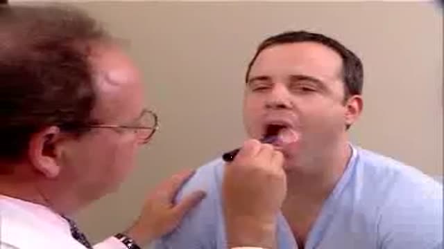
Clinical complete examination of the mouth and throat
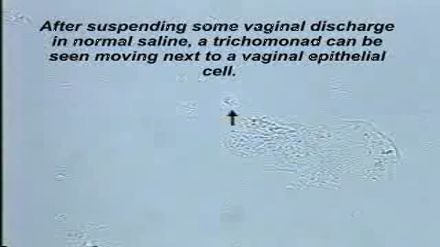
Trichomonas is best seen on the Normal Saline slide.These protozoans are about the same size as a white blood cell (a little smaller than a vaginal epithelial cell), but their violent motion is striking and unmistakable.

External ring Invagination
Internal ring occlusion test
History Inspection Palpation
taxis
Zieman
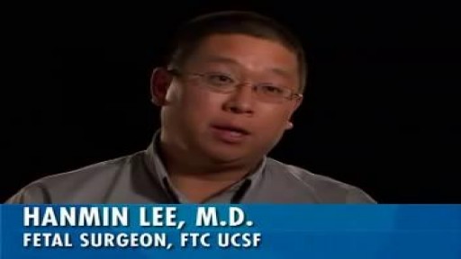
Sacrococcygeal teratoma (SCT) is an unusual tumor that, in the newborn, is located at the base of the tailbone (coccyx). This birth defect is more common in female than in male babies. Although the tumors can grow very large, they are usually not malignant (that is, cancerous).
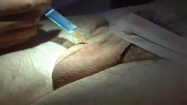
The operation of vasectomy
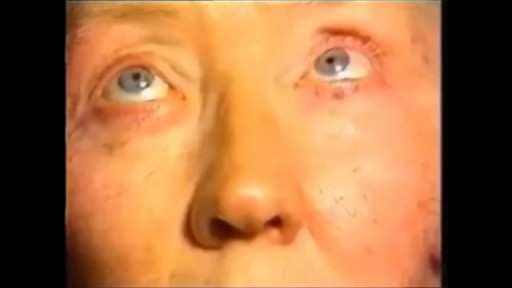
Nystagmus is a vision condition in which the eyes make repetitive, uncontrolled movements. These movements often result in reduced vision and depth perception and can affect balance and coordination. These involuntary eye movements can occur from side to side, up and down, or in a circular pattern.
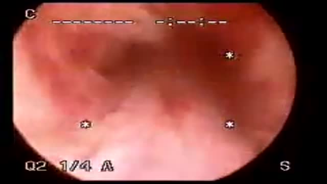
Cystoscopy
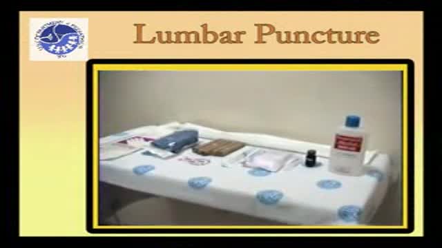
Pediatric Lumbar puncture
Watch as Dr. Benjamin Carson performs risky brain surgery on young Payton to remove a brain tumor. Dr. Carson, director of pediatric neurosurgery, is just one of the many reasons why Johns Hopkins Children's Center was recently ranked #1 in neurology and neurosurgery in America's Best Children's Hospitals 2008
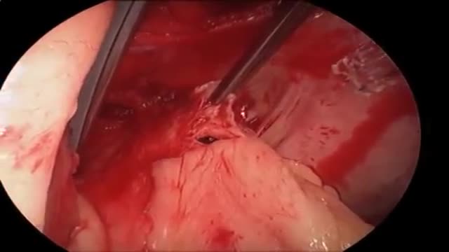
Endoscopic fenestration of suprasellar cyst in a 4 years old girl
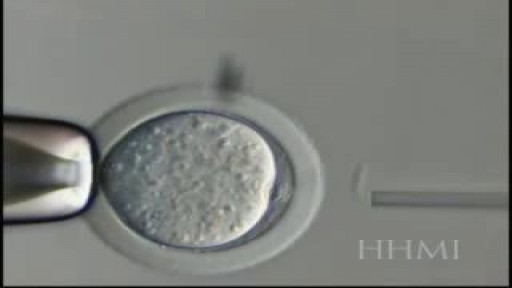
Nuclear Transfer is a form of cloning. The steps involve removing the DNA from an oocyte and while(unfertilized egg), and injecting the nucleus which contains the DNA to be cloned. In rare instances, the newly constructed cell will divide normally, replicating the new DNA while remaining
Pulmonary embolism symptoms can vary greatly, depending on how much of your lung is involved, the size of the clots, and whether you have underlying lung or heart disease. Common signs and symptoms include: Shortness of breath. This symptom typically appears suddenly and always gets worse with exertion. Chest pain. You may feel like you're having a heart attack. The pain may become worse when you breathe deeply (pleurisy), cough, eat, bend or stoop. The pain will get worse with exertion but won't go away when you rest. Cough. The cough may produce bloody or blood-streaked sputum. Other signs and symptoms that can occur with pulmonary embolism include: Leg pain or swelling, or both, usually in the calf Clammy or discolored skin (cyanosis) Fever Excessive sweating Rapid or irregular heartbeat Lightheadedness or dizziness

Mobile scanner detects disease from a drop of blood by nanotechnology
