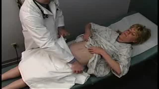
- Physical Examination
- Surgical Examination
- Ophthalmology
- Clinical Skills
- Orthopedics
- Surgery Videos
- Laparoscopy
- Pediatrics
- Funny Videos
- Cardiothoracic Surgery
- Nursing Videos
- Plastic Surgery
- Otorhinolaryngology
- Histology and Histopathology
- Neurosurgery
- Dermatology
- Pediatric Surgery
- Urology
- Dentistry
- Oncology and Cancers
- Anatomy Videos
- Health and Fitness
- Radiology
- Anaesthesia
- Physical Therapy
- Pharmacology
- Interventional Radiology
- Cardiology
- Endocrinology
- Gynecology
- Emergency Medicine
- Psychiatry and Psychology
- Childbirth Videos
- General Medical Videos
- Nephrology
- Physiology
- Diet and Food Health
- Diabetes Mellitus
- Neurology
- Women Health
- Osteoporosis
- Gastroenterology
- Pulmonology
- Hematology
- Rheumatology
- Toxicology
- Nuclear Medicine
- Infectious Diseases
- Vascular Disease
- Reproductive Health
- Burns and Wound Healing
- Other
Top videos
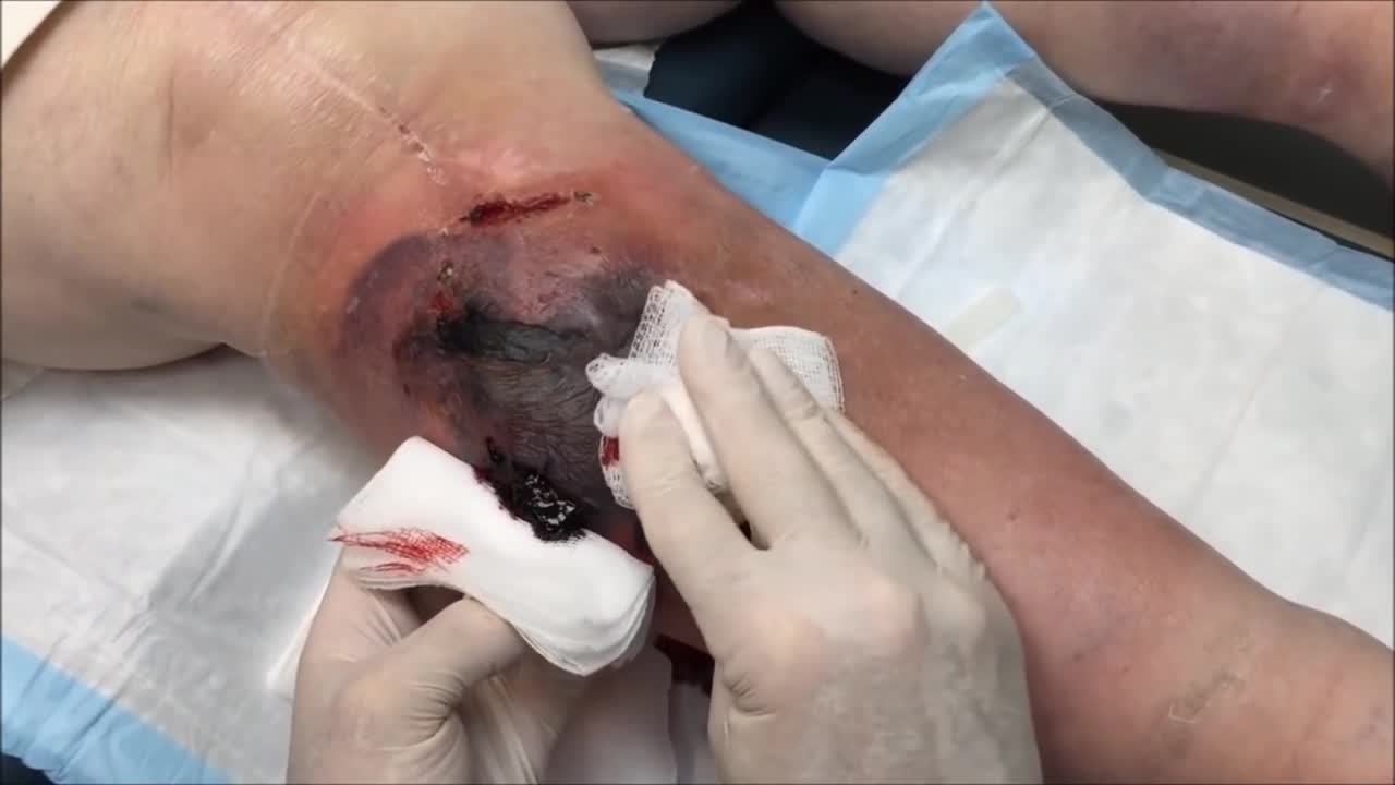
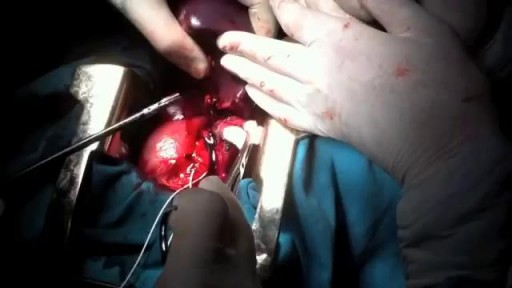
Sebaceous Cyst, Hematoma and Growth Removal
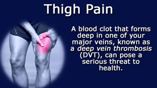
Thigh pain is most often caused by injuries to bones, joints, muscles, tendons, ligaments, and other soft tissues or blood vessels. These injuries are often caused during sports competition, or strain from overuse, obesity, or pregnancy.
The cardiac cycle is the sequence of events that occurs when the heart beats. As the heart beats, it circulates blood through pulmonary and systemic circuits of the body. There are two phases of the cardiac cycle. In the diastole phase, the heart ventricles are relaxed and the heart fills with blood

At URBN Dental, we offer the best dental services and highest quality care for your gum tissue health. Proper flossing techniques prevent your gum tissue from swelling, which often occurs from food and debris catching between your teeth. A routine dental cleaning every 6 months is recommended to maintain gum tissue health. Skipping bi annual checkups and improper flossing techniques often lead to periodontal disease which usually require a dental deep cleaning to undo tissue damage.
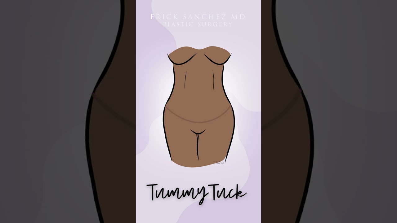
During a standard abdominoplasty, Dr. Sanchez removes the excess skin of the lower abdomen. He repairs separated muscles, and pulls the skin down nice and tight. Lastly, a new hole is cut into the skin for the belly button. Let us know your questions!
To request a consultation with Dr. Sanchez, visit sanchezplasticsurgery.com and click Request a Consultation. Fill out the form and someone will get in touch with you to answer all your questions.
Plasma cell dyscrasias are disorders of the plasma cells. Plasma cell dyscrasias are produced as a result of abnormal proliferation of a monoclonal population of plasma cells that may or may not secrete detectable levels of a monoclonal immunoglobulin or immunoglobulin fragment (paraprotein or M protein).
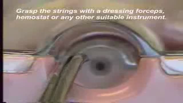
How to remove the Intra Uterine Device (IUD)
You may have heard that some positions, such as your partner on top (missionary position), are better than others for getting pregnant. In fact, there's no evidence to back these theories up. Experts just haven't done the research yet. What experts have done, though, is use scanning to show what's going on inside when you're doing the deed. The research looked at two positions: the missionary position and doggy style. (Doggy style being when you're on all fours, and your partner enters you from behind). Common sense tells us that these positions allow for deep penetration. This means that they're more likely to place sperm right next to your cervix (the opening of your uterus). The scans confirm that the tip of the penis reaches the areas between the cervix and vaginal walls in both of these positions. The missionary position allows the penis to reach the area at the front of the cervix. The rear entry position reaches the area at back of the cervix. It's amazing what some experts spend their time doing, isn't it! Other positions, such as standing up, or woman on top, may be just as good for getting sperm right next to the cervix. We just don't know yet. So, in the meantime, enjoy some variety in your sex life and keep it fun while you're trying for a baby. And talk to others who are hoping to get pregnant by joining our Actively trying group. Do I have to have an orgasm to conceive? Obviously, it's very important for your partner to reach orgasm if you are trying for a baby. There is no evidence, however, that you need to orgasm to conceive. The female orgasm is all about pleasure and satisfaction. It doesn't really help to get the sperm to the egg. Gentle contractions in your uterus can help the sperm along, but these happen without you having an orgasm. So, it's really not vital for you to reach orgasm after your partner, or even to reach orgasm at all, for you to conceive.
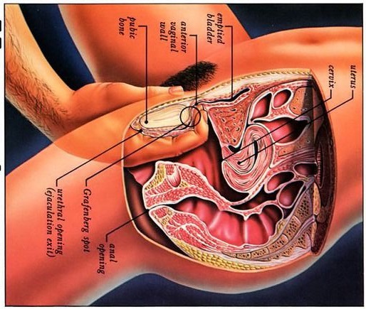
Watch that video to know what G spot is
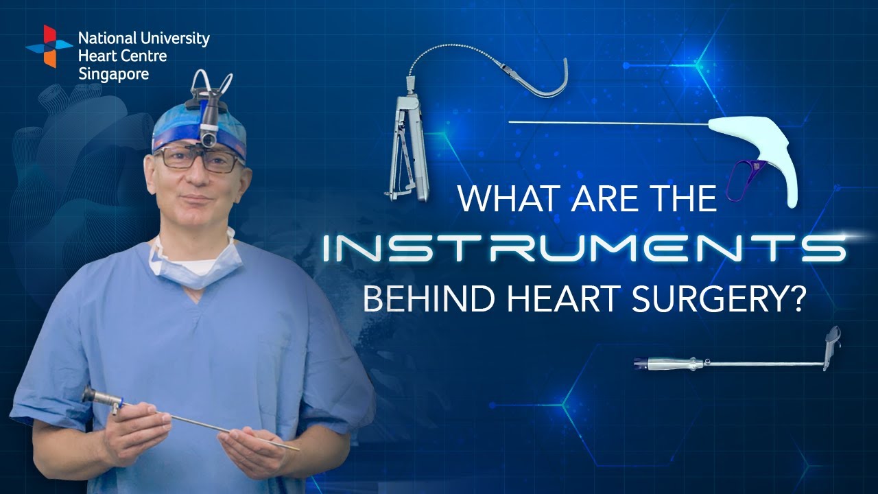
Instruments at work, innovation at play. 🔍
Watch on to discover the behind-the-scenes instruments utilised by our NUHCS cardiac surgery expert, A/Prof Theodoros Kofidis, Head of NUHCS' Department of Cardiac, Thoracic & Vascular Surgery (CTVS), for keyhole heart operations. 🔑
To find out more about Minimally Invasive Heart Surgery @ NUHCS, visit: https://[a]www.nuhcs.com.sg%2FOur-Services%2FSpecialties%2FPages%2FMinimally-Invasive-Cardiac-Surgery-Programme.aspx[/a]
Connect with us:
Instagram: @nuhcsofficial
Facebook: www.facebook.com/nuhcs
Website: www.nuhcs.com.sg
LinkedIn: www.linkedin.com/company/nuhcs
To make an appointment with the NUHCS Heart Clinic, email us at appointment@nuhs.edu.sg
#NUHCS #cardiacsurgery #heartsurgery #keyholesurgery #minimallyinvasive
You are most fertile at the time of ovulation, (when an egg is released from your ovaries) which usually occurs 12-14 days before your next period starts. This is the time of the month when you are most likely to get pregnant. It is unlikely that you will get pregnant just after your period, although it can happen.
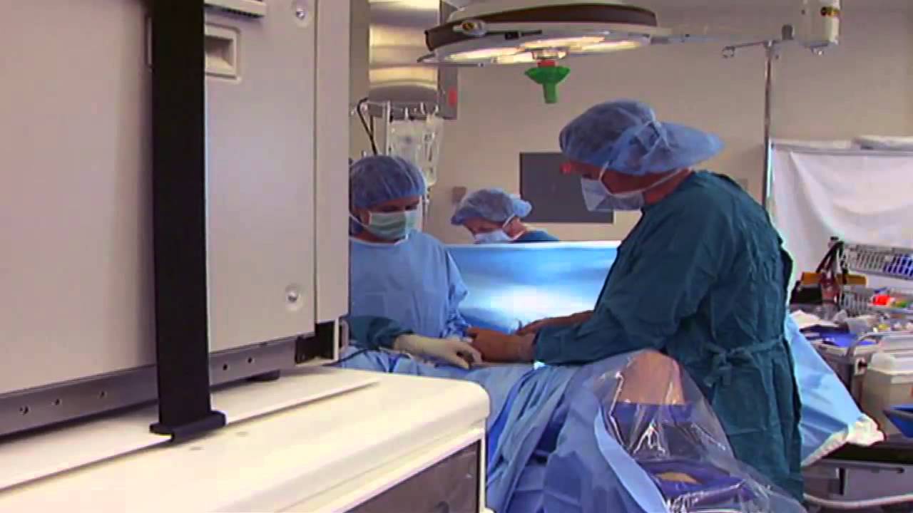
It used to be when a woman needed a hysterectomy she could expect full abdominal surgery with a long recovery time. Dr. Melissa Lee uses less invasive methods that can cut the patients downtime in half.
"We were trained in more laparoscopic and minimally invasive cases so of course that's what I'm more comfortable with doing right now."
She sees a new generation of patients opting for laparoscopic surgery.
"Laparoscopy is the use of small cameras with small incisions and instruments that are guided by the hand, and you're able to see directly into the abdomen without actually fully opening the abdomen," says Dr. Lee, an obstetrician-gynecologist with Lee Memorial Health System.
Nowadays, even a large mass or uterus can be removed using the slender tools.
"There are multiple different laparoscopic instruments that you can use. Whether they're blunt dissections or just dissectors that hold and retract back or actual scissors or cutting instruments, there are multiple different options," says Dr. Lee.
While a standard abdominal hysterectomy requires a four to eight inch incision, the laparoscope needs only a quarter to half inch. It's enough to make a big difference in terms of recovery.
"They're able to get up and move around faster. They're able to recover faster, their pain level and their need for pain medicine is much lower," says Dr. Lee.
The laparoscopic procedure also cuts down on scarring and more importantly, shortens the hospital stay. The trend now is home within 24 hours.
"Where the patient is done early in the morning, they're doing well they're tolerating oral intake they're able to getup and move around. And those patients a lot of times will feel comfortable to go home that same nigh after a major surgery," says Dr. Lee.
New studies show women who've had a laparoscopic hysterectomy viewed their quality of life as better than those who had an open abdominal procedure, making this a good option for the right patient.
View More Health Matters video segments at leememorial.org/healthmatters/
Lee Memorial Health System in Fort Myers, FL is the largest network of medical care facilities in Southwest Florida and is highly respected for its expertise, innovation and quality of care. For nearly a century, we've been providing our community with everything from primary care treatment to highly specialized care services and robotic assisted surgeries.
Visit leememorial.org

http://www.landging.com/accident-animation-sports-injury-soccer.html
This accident animation demonstrates sports injury in soccer game.
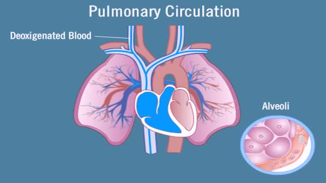
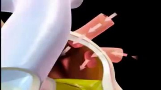
Pulmonary circulation is the portion of the cardiovascular system which carries deoxygenated blood away from the heart, to the lungs, and returns oxygenated (oxygen-rich) blood back to the heart. The function of pulmonary circulation is to exchange carbon dioxide for oxygen in the blood. It is the passage of blood from the heart to the capillaries of the lungs, where the gases are exchanged, and back to the heart to be pumped around the body.
Nasal polyps are soft, painless, noncancerous growths on the lining of your nasal passages or sinuses. They hang down like teardrops or grapes. They result from chronic inflammation due to asthma, recurring infection, allergies, drug sensitivity or certain immune disorders. Nasal polyps are polypoidal masses arising mainly from the mucous membranes of the nose and paranasal sinuses. They are overgrowths of the mucosa that frequently accompany allergic rhinitis, and are freely movable and nontender.
Pulmonary fibrosis is a condition in which the tissue deep in your lungs becomes scarred over time. This tissue gets thick and stiff. That makes it hard for you to catch your breath, and your blood may not get enough oxygen. Causes of pulmonary fibrosis include environmental pollutants, some medicines, some connective tissue diseases, and interstitial lung disease. Interstitial lung disease is the name for a large group of diseases that inflame or scar the lungs. In most cases, the cause cannot be found. This is called idiopathic pulmonary fibrosis
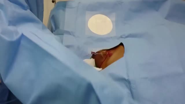
hemisplenectomy is removal of the half of the spleen.It was done firstly in Azerbaijan by prof. Dr Med Qurban Muslimov in 12 years old child with simple syst of the spleen.
inspection, auscultation and palpation
Insertion of a Palindrome TDC in the right internal jugular vein under ultrasound and fluoroscopic guidance at a restructured hospital in Singapore
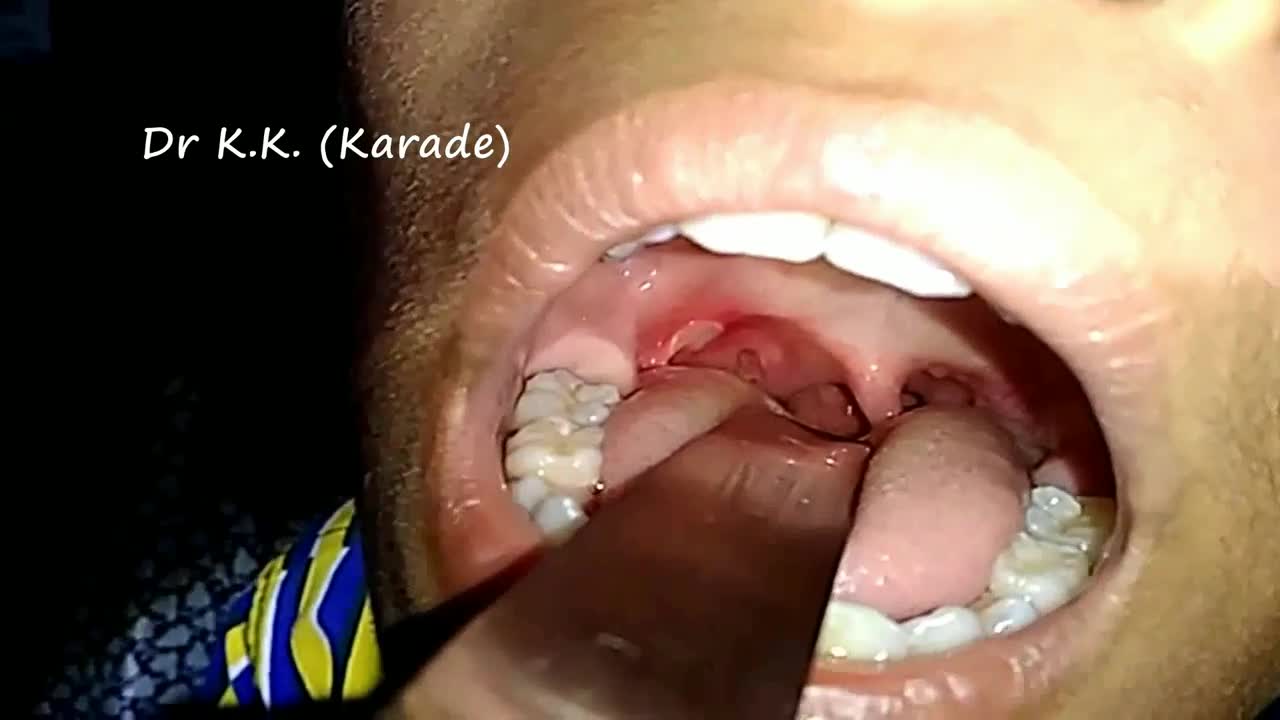
Canker sores (Aphthous ulcer) are small, painful ulcers on the inside of the mouth, tongue, lips, or throat.Canker sores are white or yellow and surrounded by a bright red area. They are not cancerous.
