- Physical Examination
- Surgical Examination
- Ophthalmology
- Clinical Skills
- Orthopedics
- Surgery Videos
- Laparoscopy
- Pediatrics
- Funny Videos
- Cardiothoracic Surgery
- Nursing Videos
- Plastic Surgery
- Otorhinolaryngology
- Histology and Histopathology
- Neurosurgery
- Dermatology
- Pediatric Surgery
- Urology
- Dentistry
- Oncology and Cancers
- Anatomy Videos
- Health and Fitness
- Radiology
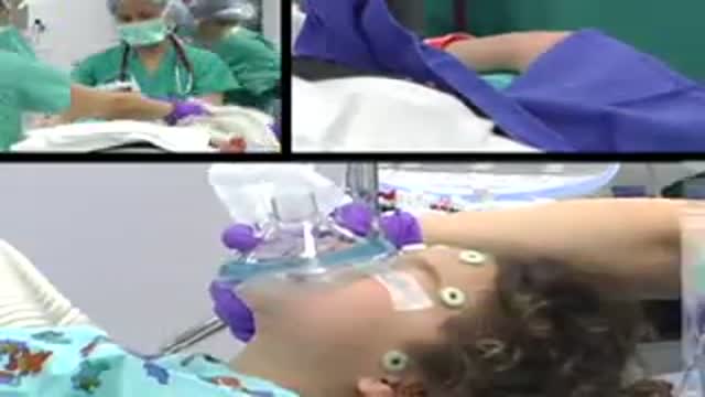
- Anaesthesia
- Physical Therapy
- Pharmacology
- Interventional Radiology
- Cardiology
- Endocrinology
- Gynecology
- Emergency Medicine
- Psychiatry and Psychology
- Childbirth Videos
- General Medical Videos
- Nephrology
- Physiology
- Diet and Food Health
- Diabetes Mellitus
- Neurology
- Women Health
- Osteoporosis
- Gastroenterology
- Pulmonology
- Hematology
- Rheumatology
- Toxicology
- Nuclear Medicine
- Infectious Diseases
- Vascular Disease
- Reproductive Health
- Burns and Wound Healing
- Other
Top videos
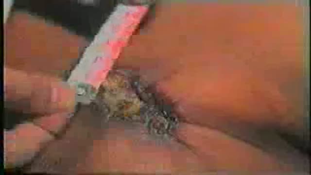
Surgical Examination of an ulcer in a proper way
Handal Plastic Surgery at the Sanctuary Surgery Center is the leading cosmetic surgery center of the Southeast Florida region, providing excellent consultation, surgery, and post operative services. Headed by Doctor Arthur G. Handal, top plastic & cosmetic surgeon in Boca Raton, the professional staff of the Sanctuary Surgery Center offers the best in patient care.
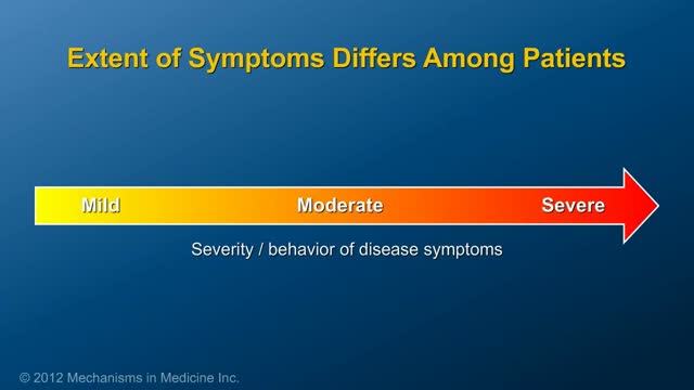
This animation describes the goals of inflammatory bowel disease (IBD) management and how patients can take an active role in managing their disease.
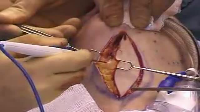
A direct neck lift is an uncommon procedure in plastic surgery for the sagging neck. It is reserved for older patients who do not want a facelift or who do not want or can not go through a bigger facelift operation.
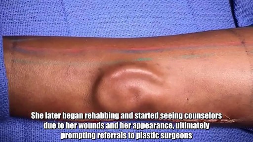
Shamika Burrage survived a near-fatal car accident two years ago, but not without losing something pretty important: her left ear. Now, thanks to a novel procedure performed at an Army medical center in Texas, Burrage is getting that ear back in a most unusual way. Plastic surgeons harvested cartilage from Burrage's ribs to create a new ear and then grew it under the skin of her forearm. Then the doctors at William Beaumont Army Medical Center in El Paso successfully transplanted the ear from her arm to her head. The technique -- a first time in the Army -- is called prelaminated forearm free flap, said Lt. Col. Owen Johnson III, chief of plastic and reconstructive surgery at William Beaumont Army Medical Center. Some of the big advantages of it is that it reduced the chance of more scarring around Burrage's ear. Also, growing the ear under the skin of her forearm allows new blood vessels to form. "(The ear) will have fresh arteries, fresh veins and even a fresh nerve so she'll be able to feel it," Johnson said on the US Army's website. Burrage, a 21-year-old private, still has to endure two more surgeries, but she's feeling more optimistic about the future than ever in the years since her accident. "It's been a long process for everything, but I'm back," said Burrage.
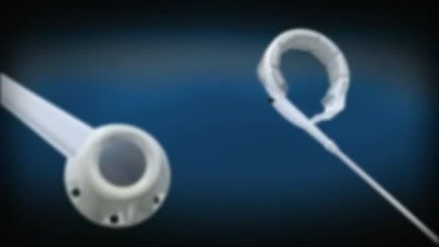
complications from using a urinary catheter include: allergic reaction to the material used in the catheter, such as latex. bladder stones. blood in the urine. injury to the urethra. kidney damage (with long-term indwelling catheters) septicemia, or infection of the urinary tract, kidneys, or blood.

The common obsession among men and women of having a perfect body has lead them to many serious neurotic disorders. They are constantly exposed to the ideas of having the perfect body. We are bombarded with the images on social media which create a hype among men and women, to achieve the exact same ratio of fats in them. Body image is merely an image of your thoughts and perceptions. The way you think how people notices you can greatly impact yourself and your way of thinking about yourself. It becomes quite a big deal when you start to feel low about yourself. It leads you towards having a low self esteem and it becomes hard for you to feel worthy and confident. On contrary when you have good self esteem you feel empowered and confident. It is not the consequence of just liking your own body but its about accepting who you are and making people accept you as you are.
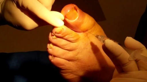
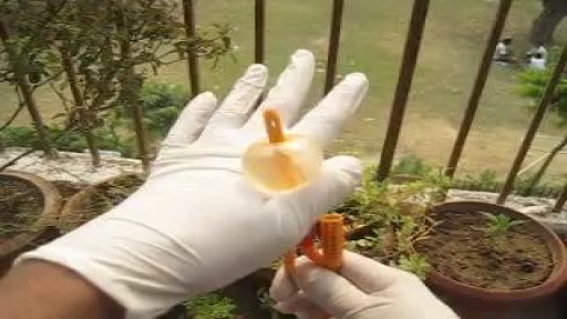
Ingrown toenails occur in both men and women. According to the National Health Services (NHS), ingrown toenails may be more common in people with sweaty feet, such as teenagers. Older people may also be at higher risk because toenails thicken with age. Many things can cause an ingrown toenail, including: cutting toenails incorrectly (Cut straight across, since angling the sides of the nail can encourage the nail to grow into the skin.) irregular, curved toenails footwear that places a lot of pressure on the big toes, such as socks and stockings that are too tight or shoes that are too tight, narrow, or flat for your feet toenail injury, including stubbing your toe, dropping something heavy on your foot, or kicking a ball repeatedly poor posture improper foot hygiene, such as not keeping your feet clean or dry genetic predisposition
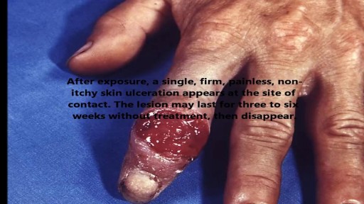
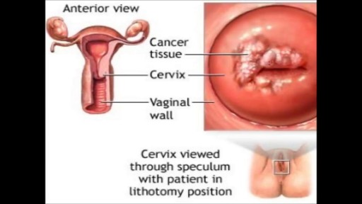
STDs are infections that are transmitted during vaginal, anal, and oral sex. They are very common and many people who have them don't show any symptoms.
Nosebleeds are common due to the location of the nose on the face, and the large amount of blood vessels in the nose. The most common causes of nosebleeds are drying of the nasal membranes and nose picking (digital trauma), which can be prevented with proper lubrication of the nasal passages and not picking the nose.
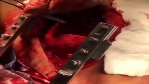
CARDIAC TAMPONADE!!! The surgeon in this video is performing a pericardiotomy in a 30-year-old man that presented to ER after a motor vehicle crash....
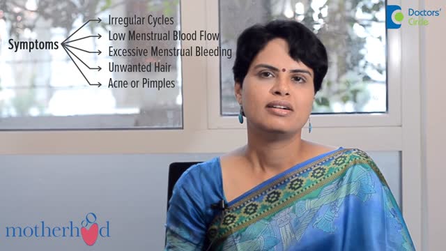
Polycystic ovary syndrome (PCOS) is a common endocrine system disorder among women of reproductive age. Women with PCOS may have enlarged ovaries that contain small collections of fluid — called follicles — located in each ovary as seen during an ultrasound exam. Infrequent or prolonged menstrual periods, excess hair growth, acne, and obesity can all occur in women with polycystic ovary syndrome. In adolescents, infrequent or absent menstruation may raise suspicion for the condition. The exact cause of polycystic ovary syndrome is unknown. Early diagnosis and treatment along with weight loss may reduce the risk of long-term complications, such as type 2 diabetes and heart disease.

The procedure was performed under wrist block regional anesthesia with tourniquet control. A single Chinese finger trap was used on the thumb with 5 to 8 lb of ongitudinal traction. The arm was held down with wide tape around the tourniquet securing it to the hand table to serve as countertraction. A shoulder holder, rather than a traction tower, was used to facilitate fluoroscopic intervention more easily. The Trapeziometacarpal joint was detected by palpation. Joint distension was achieved by injecting 1 to 3 mL of normal saline (Fig. 1). It is important to distally direct the needle approximately 20 degrees to clear the dorsal flare of the metacarpal base and enter the joint capsule. This course should be reproduced upon entering with arthroscopic sleeve/ trocar assembly to minimize iatrogenic cartilage injury. Fluid distention is important to facilitate this. The incision for the 1-R (radial) portal, used for proper assessment of the dorsoradial ligament, posterior oblique ligament, and ulnar collateral ligament, was placed just volar to the abductor pollicis longus tendon. The incision for the 1-U (ulnar) portal, for better evaluation of the anterior oblique ligament and ulnar collateral ligament, was made just ulnar to the extensor pollicis brevis tendon. A short-barrel, 1.9-mm, 30- degree inclination arthroscope was used for complete visualization of the CMC joint surfaces, capsule, and ligaments, and then appropriate management was done, as dictated by the stage of the arthritis detected (Fig. 2A). A full-radius mechanical shaver with suction was used in all the cases, particularly for initial debridement and visualization. Most of the cases were augmented with radiofrequency ablation to perform a thorough synovectomy and radiofrequency was also used to perform chondroplasty in the cases with focal articular cartilage wear or fibrillation. Chondroplasty refers to thedebridement of the fibrillated cartilage to improve vascularity of the cartilage and enhance the growth of fibrocartilage. Ligamentous laxity and capsular attenu- ation were treated with thermal capsulorraphy using a radiofrequency shrinkage probe. We were careful to avoid thermal necrosis; hence, a striping technique was used to tighten the capsule of the lax joints. The striping technique refers to thermal shrinkage performed in longitudinal stripes on the lax capsule, so as to leave vascular zones between the stripes; hence, thermal necrosis is prevented. Arthroscopic stage I disease was characterized by synovitis without any cartilage wear, wherein a synovectomy coupled with thermal capsulor- raphy as described was performed.

MUSC Children’s Health offers South Carolina’s only Level 1 Children’s Surgery Center, representing excellence in inpatient surgery at MUSC Shawn Jenkins Children’s Hospital, as well as outpatient surgery at R. Keith Summey Medical Pavilion. These two state-of-the-art facilities are equipped with a team of pediatric board-certified providers utilizing pediatric-specific devices and the most technologically advanced tools.
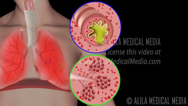
Chronic obstructive pulmonary disease Email this page to a friend Print Facebook Twitter Google+ Chronic obstructive pulmonary disease (COPD) is a common lung disease. Having COPD makes it hard to breathe. There are two main forms of COPD: Chronic bronchitis, which involves a long-term cough with mucus Emphysema, which involves damage to the lungs over time Most people with COPD have a combination of both conditions. Causes Smoking is the main cause of COPD. The more a person smokes, the more likely that person will develop COPD. But some people smoke for years and never get COPD. In rare cases, nonsmokers who lack a protein called alpha-1 antitrypsin can develop emphysema. Emphysema Other risk factors for COPD are: Exposure to certain gases or fumes in the workplace Exposure to heavy amounts of secondhand smoke and pollution Frequent use of a cooking fire without proper ventilation Symptoms Symptoms may include any of the following: Cough, with or without mucous Fatigue Many respiratory infections Shortness of breath (dyspnea) that gets worse with mild activity Trouble catching one's breath Wheezing Because the symptoms develop slowly, some people may not know that they have COPD.
Watch as Dr. Benjamin Carson performs risky brain surgery on young Payton to remove a brain tumor. Dr. Carson, director of pediatric neurosurgery, is just one of the many reasons why Johns Hopkins Children's Center was recently ranked #1 in neurology and neurosurgery in America's Best Children's Hospitals 2008

A video discussing Causes of Itching in the Vulva
Medical examination of the abdomen from Loyola University, Chicago
Menorrhagia is the medical term for menstrual periods with abnormally heavy or prolonged bleeding. Although heavy menstrual bleeding is a common concern, most women don't experience blood loss severe enough to be defined as menorrhagia. With menorrhagia, you can't maintain your usual activities when you have your period because you have so much blood loss and cramping. If you dread your period because you have such heavy menstrual bleeding, talk with your doctor. There are many effective treatments for menorrhagia.
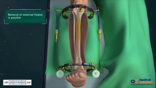
Massive bone defects (>8 cm) will not unite without an additional intervention. They require a predictable, durable, and efficient method to regrow bone. The Ilizarov method of tension stress, or distraction osteogenesis, first involves a low-energy osteotomy1 - 5. The bone segments are then pulled apart, most often using an external device at a specific rate and rhythm (distraction phase), after which the newly formed bone (the regenerate) requires time for consolidation. The consolidation phase is variable and usually requires a substantially greater amount of time before the external device can be removed. Our technique of tibial bone transport over an intramedullary nail using cable and pulleys combines internal and external fixation, allowing the external fixator to be removed at the end of the distraction phase. This increases the efficiency of limb reconstruction and decreases the external-fixator-associated complications.
