- Physical Examination
- Surgical Examination
- Ophthalmology
- Clinical Skills
- Orthopedics
- Surgery Videos
- Laparoscopy
- Pediatrics
- Funny Videos
- Cardiothoracic Surgery
- Nursing Videos
- Plastic Surgery
- Otorhinolaryngology
- Histology and Histopathology
- Neurosurgery
- Dermatology
- Pediatric Surgery
- Urology
- Dentistry
- Oncology and Cancers
- Anatomy Videos
- Health and Fitness
- Radiology
- Anaesthesia
- Physical Therapy
- Pharmacology
- Interventional Radiology
- Cardiology
- Endocrinology
- Gynecology
- Emergency Medicine
- Psychiatry and Psychology
- Childbirth Videos
- General Medical Videos
- Nephrology
- Physiology
- Diet and Food Health
- Diabetes Mellitus
- Neurology
- Women Health
- Osteoporosis
- Gastroenterology
- Pulmonology
- Hematology
- Rheumatology
- Toxicology
- Nuclear Medicine
- Infectious Diseases
- Vascular Disease
- Reproductive Health
- Burns and Wound Healing
- Other
Top videos
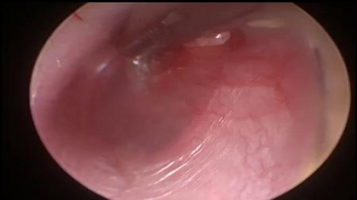
A myringotomy is a procedure in which your doctor creates a small hole in the eardrum so fluids such as water, blood, or pus can drain out. In many cases, your doctor will put in a tube so it won't get backed up again. The tube, which will usually fall out on its own in about six to 18 months, lets air flow through and keeps the middle ear dry. Tubes also: Reduce pain Improve hearing Cut down on the number of infections your child may have

How Liposuction Works in 15 seconds.
See how we illustrated this amazing technology by Alma Lasers.
Curious 🤔 about medical device 3D animation? ➜ http://www.arcreative-media.com

A flail chest occurs when a segment of the thoracic cage is separated from the rest of the chest wall. This is usually defined as at least two fractures per rib (producing a free segment), in at least two ribs. A segment of the chest wall that is flail is unable to contribute to lung expansion. Large flail segments will involve a much greater proportion of the chest wall and may extend bilaterally or involve the sternum. In these cases the disruption of normal pulmonary mechanics may be large enough to require mechanical ventilation.
Breast implants do not last forever, and during its lifetime, it may rupture. Dr. Linder, Beverly Hills breast surgeon specialist, breaks down how removing breast implants works. To learn more about Dr. Stuart Linder and his expertise, Visit: www.drlinder.com
Microcalcifications in the breast can be the first sign of cancer. They are, as the name says, very small and clustered. A precise biopsy without pain under stereotactic guidance is the standard procedure. What makes this Spirotome different from the vacuum assisted biopsies is that only a few biopsies are needed and that the approach of the needle towards the microcalcifications is direct and frontal. There is no damage to the surrounding tissues making this procedure rather painfree and with minimal bleeding.


What are the disadvantages of male condoms? a moderately high failure rate when used improperly or inconsistently. the potential for diminished sensation. skin irritation, such as contact dermatitis, due to latex sensitivity or allergy. allergic reactions to spermicides, lubes, scents, and other chemicals in the condoms.

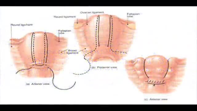
B-Lynch suture for uterine atony technique described

childbirth normal labor delivery 3d medical animation company healthcare 3d visualization san antoni

Lack of sunshine causes skin cancer, according to Andreas Moritz. In this video from 2009, he explains why being in the sun is actually good for you and your skin. Find out why your sunscreen is doing more harm than good. Also, you need vitamin D to prevent cancer, and sunscreen may interfere with your exposure to vitamin D from the sun.

Watch that video of Sperm Formation and Pathway Ejaculation

Inflammation of the uvula is known as uvulitis. Your uvula will appear red, puffy, and larger than normal. Other symptoms of uvulitis may include: itching burning a sore throat spots on your throat snoring difficulty swallowing trouble breathing If you have a swollen uvula along with a fever or abdominal pain, consult with your doctor right away. In rare cases, the uvula can swell enough to block your airway. Swelling of the throat is a life-threatening event. If this happens, seek immediate medical attention. What causes a swollen uvula? Causes Inflammation is your body’s response when it’s under attack. Triggers for inflammation include: environmental and lifestyle factors an infection trauma genetics Environmental and Lifestyle Factors The most common food allergies are peanuts tree nuts milk eggs wheat soy fish, including shellfish You could be having an allergic reaction to something you touched, swallowed, or breathed in. Some common allergens include: food irritants , such as dust, animal dander, or pollen medication exposure to chemicals or other toxic substances, including tobacco Infection You can get viral infections or bacterial infections. Examples of viral infections include: the common cold the flu mononucleosis chickenpox measles croup The most common bacterial infection is strep throat, which occurs due to Streptococcus pyogenes, which is a type of group A Streptococcus. If you have infected tonsils, or tonsillitis, severe inflammation can cause them to push against and irritate your uvula. Trauma Trauma to the uvula can happen if you need an intubation, such as during surgery. Your uvula can also be injured during a tonsillectomy. This is a procedure to remove your tonsils, which are located on both sides of your uvula. Your throat and uvula can also become irritated if you have acid reflux disease or if you vomit frequently. Genetics A condition called hereditary angioedema (HAE) can cause swelling of the uvula and throat, as well as swelling of the face, hands, and feet. Other symptoms include nausea, vomiting, and abdominal pain. It’s an uncommon genetic mutation that occurs in 1 in 10,000 to 1 in 50,000 people. It’s rare, but there are case reports of individuals who have an elongated uvula, which can also interfere with breathing. What are the risk factors for a swollen uvula? Risk Factors Anyone can get uvulitis, but adults get it less often than children do. You’re at increased risk if you: have allergies use tobacco products are exposed to chemicals and other irritants in the environment have a weakened immune system, making you more susceptible to infections How is a swollen uvula diagnosed? Diagnosis If you have fever or swelling of your throat, see your doctor. Be prepared to give a complete medical history. Tell your doctor: about all the over-the-counter and prescription medications you take if you’re a smoker or you chew tobacco if you’ve recently tried new foods if you’ve been exposed to chemicals or unusual substances about your other symptoms, such as abdominal pain, fever, or dehydration Your doctor may be able to make a diagnosis through a physical exam. It’s likely you’ll also need a throat swab to evaluate for strep or to obtain secretions for culture to determine if you have another bacterial or fungal infection. This test is known as the rapid strep test. You may also need a nasal swab to test for influenza. Blood testing can help identify or rule out some other infectious agents. If those tests are inconclusive, you may need to see an allergist. Blood and skin tests can help identify foods or other substances that cause a reaction. Learn more: Allergy testing » If necessary, imaging tests can provide a more detailed view of your throat and the surrounding area. What’s the treatment for a swollen uvula? Treatment When you have something like the common cold, swelling usually clears up on its own without treatment. Otherwise, treatment will depend on how severe your symptoms are, as well as what’s causing the inflammation. Infection Viral infections tend to clear up without treatment. The only upper respiratory infection for which an antiviral medication is available is influenza. Antibiotics can treat bacterial infections. Even after symptoms clear up, take all the medication as prescribed. If your condition may be contagious, stay home until your doctor tells you that you’re no longer at risk of spreading it to others. Allergy If you test positive for an allergy, try to avoid the allergen in the future. Doctors usually treat allergies with antihistamines or steroids. Anaphylaxis is a severe allergic reaction. Doctors use epinephrine to treat this reaction. Hereditary angioedema Your doctor may treat HAE with any of the following: anabolic steroids, or androgens antifibrinolytics C1 inhibitors, such as C1 esterase inhibitor (Berinert) or C1 esterase inhibitor (recombinant) (Ruconest) a plasma kallikrein inhibitor, such as ecallantide (Kalbitor) bradykinin receptor antagonist, such as icatibant injection (Firazyr) Tell your doctor if you have new or worsening symptoms, and follow up as necessary. Tips for relief home treatment If you have a swollen uvula or sore throat, it’s your body’s way of telling you that something is wrong. A few home remedies can help keep you strong and soothe your irritated throat. Make sure you’re getting enough fluids. If your throat hurts when you drink, try drinking small amounts throughout the day. Your urine should be light in color. If it’s dark yellow or brown, you’re not drinking enough and may be dehydrated. Additional tips include the following: Cool your throat by sucking on ice chips. Frozen juice bars or ice cream may also do the trick. Gargle with warm salt water to ease your dry, scratchy throat. Aim for a full night’s sleep, and nap during the day if you can. What’s the outlook? Outlook A swollen uvula isn’t a common occurrence. Most of the time it clears up without treatment. If you have an infection, prompt treatment should take care of the problem within a week or two. If you have allergies that lead to swelling of the uvula or throat, do your best to avoid that allergen. You should also be prepared to deal with an attack if you come into contact with the substance again. If you’ve ever had anaphylaxis, ask your doctor if you should carry injectable epinephrine (EpiPen) in case of emergency. People with HAE must learn to recognize triggers and early warning signs of an attack. Talk to your doctor about how to manage HAE. Article Resources Was this article helpful?Yes No Share Tweet Email Print Read This Next 9-Month-Old Baby: Developmental Milestones and Guidelines 9-Month-Old Baby: Developmental Milestones and Guidelines Read More » All of the ‘Firsts’ That Come with Breast-Feeding All of the ‘Firsts’ That Come with Breast-Feeding Read More » 5 Types of Health Professionals You Should Know About 5 Types of Health Professionals You Should Know About Read More » What’s the Difference Between a Fracture and a Break? What’s the Difference Between a Fracture and a Break? Read More » Is Corn a Vegetable? Is Corn a Vegetable? Read More » Advertisement Advertisement Advertisement

Watch that Functional Neck Dissection Surgery
The majority of fetuses are in a breech presentation early in pregnancy. By week 38th week of gestation, however, the fetus normally turns to a cephalic presentation. Although the fetal head is the widest single diameter, the fetus’s buttocks [ breech], plus the lower extremities, actually takes up more space. The fundus, being the largest part of the uterus, probably accounts for the fact that in approximately 97% of all pregnancies, the fetus turns so that the buttocks and lower extremities are in the fundus. Vaginal delivery of a breech presentation requires great skill if the fetus is not to be damaged. With the low rate of vaginal breech deliveries in the developed world, experience is being lost. 6% of women with breech presentation still have a vaginal breech delivery as they present too late - so units need to retain a high level of preparedness. Types of breech presentation: I. Complete breech [ flexed breech]: The fetal attitude is one of complete flexion, with hips and knees both flexed and the feet tucked in beside the buttocks. The presenting part consists of two buttocks, external genitalia and two feet. It is commonly present in multiparae. II. Incomplete breech: This is due to varying degrees of extension of thighs or legs at podalic pole. Three varieties are possible; - Breech with extended legs [ frank breech ]: The breech presents with the hips flexed and legs extended on the abdomen. 70% of breech presentations are of this type and it is particularly common in primigravidae whose good uterine muscle tone inhibits flexion of the legs and free turning of the fetus. - Footling breech: This is rare. One or both feet present because neither hips nor knees are fully flexed. The feet are lower than the buttocks, which distinguishes it from the complete breech. - Knee presentation: This is very rare. Thighs are extended but the knees are flexed, bringing the knees down to present at the brim.

A video showing the surgery of vaginal hysterectomy Operation

nkylosing spondylitis (pronounced ank-kih-low-sing spon-dill-eye-tiss), or AS, is a form of arthritis that primarily affects the spine, although other joints can become involved. It causes inflammation of the spinal joints (vertebrae) that can lead to severe, chronic pain and discomfort

Watch that video of Huge 132 lbs Testicles Tumor Removal Surgery
• Define and use related medical terminology.
• Describe and demonstrate techniques for imaging the thyroid gland.
• Discuss functional abnormalities of the thyroid gland.
• Correlate laboratory data relevant to the thyroid and parathyroid glands.
• Describe, and recognize on images, pathologies of the thyroid gland.
• Identify the anatomy of the parathyroid glands on diagrams and sonograms.
• Describe and demonstrate techniques for imaging the parathyroid glands.
• Describe, and recognize on images, pathologies of the parathyroid glands.
• List and describe other neck masses.
• Follow relevant protocols when scanning.
• Differentiate the sonographic appearances of the female reproductive organs in relation to the menstrual cycle, the use of contraceptives and hormone replacement, and following chemotherapy.
• Explain the Patient Privacy Rule (HIPAA) and Patient Safety Act (see reference).

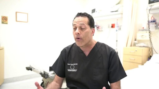
Curious about LASIK eye surgery? NVISION's Dr. Richard Mauer talks risks, life-changing benefits, and outcomes (plus why he loves what he does!).
Want to start your journey to better vision? Schedule your complimentary consult today! https://bit.ly/3H2i0FU
NVISION: The Eye Doctors' #1 Choice in LASIK and Laser Cataract Surgery
