- Physical Examination
- Surgical Examination
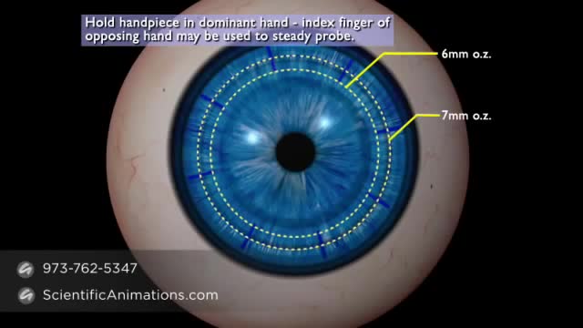
- Ophthalmology
- Clinical Skills
- Orthopedics
- Surgery Videos
- Laparoscopy
- Pediatrics
- Funny Videos
- Cardiothoracic Surgery
- Nursing Videos
- Plastic Surgery
- Otorhinolaryngology
- Histology and Histopathology
- Neurosurgery
- Dermatology
- Pediatric Surgery
- Urology
- Dentistry
- Oncology and Cancers
- Anatomy Videos
- Health and Fitness
- Radiology
- Anaesthesia
- Physical Therapy
- Pharmacology
- Interventional Radiology
- Cardiology
- Endocrinology
- Gynecology
- Emergency Medicine
- Psychiatry and Psychology
- Childbirth Videos
- General Medical Videos
- Nephrology
- Physiology
- Diet and Food Health
- Diabetes Mellitus
- Neurology
- Women Health
- Osteoporosis
- Gastroenterology
- Pulmonology
- Hematology
- Rheumatology
- Toxicology
- Nuclear Medicine
- Infectious Diseases
- Vascular Disease
- Reproductive Health
- Burns and Wound Healing
- Other
Top videos

very day, specialists deliver high-quality care in 68 disciplines in health centres across Canada. Yet many Canadians know very little about what many specialists actually do, and the important role these disciplines play in Canada’s health care system. This video provides a brief high-level overview of what Internal Medicine Specialists actually do, their training, and their role in Canadian health care.
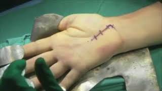
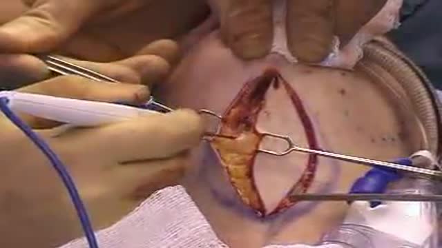
Carpal tunnel release (part 1). Skin incision and retraction. Procedure performed by Deepak Kapila, MD, Broward Health, Fort Lauderdale, FL. Courtesy of BroadcastMed (http://ortho.broadcastmed.com/....4229/videos/carpal-t
There are hundreds more procedural videos as well as news, features, resources and references on Medscape.com. Join today for free.
Cytoplasmic organelles are "little organs" that are suspended in the cytoplasm of the cell. Each type of organelle has a definite structure and a specific role in the function of the cell. Examples of cytoplasmic organelles are mitochondrion, ribosomes, endoplasmic reticulum, golgi apparatus, and lysosomes.
Multiple sclerosis (MS) is a disease of the central nervous system estimated to affect 2.3 million people worldwide. It is a chronic disease in which the immune system abnormally attacks the insulation and support around the nerve cells (myelin sheath) in the brain, spinal cord and optic nerves, causing inflammation and consequent damage. MS is a leading cause of non-traumatic disability in young people, usually striking between 20 and 40 years of age. There is no cure for MS, but research continues to better understand and treat the disease.
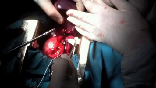
hemisplenectomy is removal of the half of the spleen.It was done firstly in Azerbaijan by prof. Dr Med Qurban Muslimov in 12 years old child with simple syst of the spleen.
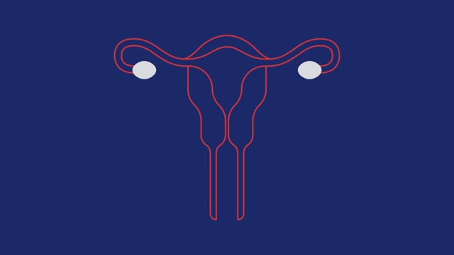
Uterine fibroids are noncancerous growths of the uterus that often appear during childbearing years. Also called leiomyomas (lie-o-my-O-muhs) or myomas, uterine fibroids aren't associated with an increased risk of uterine cancer and almost never develop into cancer. Fibroids range in size from seedlings, undetectable by the human eye, to bulky masses that can distort and enlarge the uterus. You can have a single fibroid or multiple ones. In extreme cases, multiple fibroids can expand the uterus so much that it reaches the rib cage. Many women have uterine fibroids sometime during their lives. But most women don't know they have uterine fibroids because they often cause no symptoms. Your doctor may discover fibroids incidentally during a pelvic exam or prenatal ultrasound.
What is a female condom? How do female condoms work to prevent pregnancy and STDs? Learn all about female condoms — also called internal condoms — in this video.
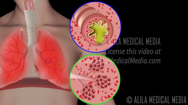
Chronic obstructive pulmonary disease Email this page to a friend Print Facebook Twitter Google+ Chronic obstructive pulmonary disease (COPD) is a common lung disease. Having COPD makes it hard to breathe. There are two main forms of COPD: Chronic bronchitis, which involves a long-term cough with mucus Emphysema, which involves damage to the lungs over time Most people with COPD have a combination of both conditions. Causes Smoking is the main cause of COPD. The more a person smokes, the more likely that person will develop COPD. But some people smoke for years and never get COPD. In rare cases, nonsmokers who lack a protein called alpha-1 antitrypsin can develop emphysema. Emphysema Other risk factors for COPD are: Exposure to certain gases or fumes in the workplace Exposure to heavy amounts of secondhand smoke and pollution Frequent use of a cooking fire without proper ventilation Symptoms Symptoms may include any of the following: Cough, with or without mucous Fatigue Many respiratory infections Shortness of breath (dyspnea) that gets worse with mild activity Trouble catching one's breath Wheezing Because the symptoms develop slowly, some people may not know that they have COPD.

Is that knee pain just a sprain or a more serious ACL injury? Orthopedic surgeon Paul Fadale, M.D., offers tips on how to tell the difference. http://www.orthopedicsri.org/
Handal Plastic Surgery at the Sanctuary Surgery Center is the leading cosmetic surgery center of the Southeast Florida region, providing excellent consultation, surgery, and post operative services. Headed by Doctor Arthur G. Handal, top plastic & cosmetic surgeon in Boca Raton, the professional staff of the Sanctuary Surgery Center offers the best in patient care.

Robotics is the engineering science and technology of robots, and their design, manufacture, application, and structural disposition.
A cornea transplant, also called keratoplasty, is a surgical procedure to replace part of your cornea with corneal tissue from a donor. Your cornea is the transparent, dome-shaped surface of your eye that accounts for a large part of your eye's focusing power. A cornea transplant can restore vision, reduce pain and improve the appearance of a damaged or diseased cornea. Most cornea transplant procedures are successful. But cornea transplant carries a small risk of complications, such as rejection of the donor cornea.
A direct neck lift is an uncommon procedure in plastic surgery for the sagging neck. It is reserved for older patients who do not want a facelift or who do not want or can not go through a bigger facelift operation.
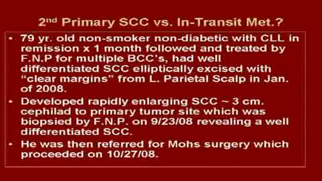
Graphic content of Mohs surgical removal of a large Squamous Cell Carcinoma on scalp followed by reconstruction with 10 week follow up. Visit us @ skincancercentre.com.
Electronystagmography (ENG) is a diagnostic test to record involuntary movements of the eye caused by a condition known as nystagmus. It can also be used to diagnose the cause of vertigo, dizziness or balance dysfunction by testing the vestibular system.
Cytomegalovirus is a genus of viruses in the order Herpesvirales, in the family Herpesviridae, in the subfamily Betaherpesvirinae. Humans and monkeys serve as natural hosts.
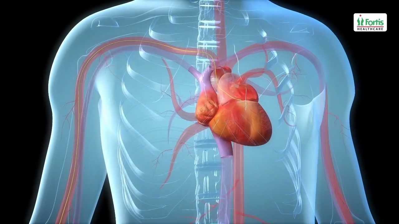
Angioplasty Procedure Animation Video
Emergency angioplasty is an operation that is performed directly after a heart attack, on admission to the hospital. It involves the insertion of a catheter into the blocked blood vessel that caused the heart attack. This opens it up and allows blood to flow again, thus minimizing damage to the heart.
If one or more arteries become clogged, it may result in a heart attack. This normally presents with chest pain, sweating and a feeling of anxiety, among other symptoms. Urgent medical assistance should be sought. A heart attack is a medical emergency requiring intervention as soon as possible.
Know more: http://www.emergencyangioplasty.com/
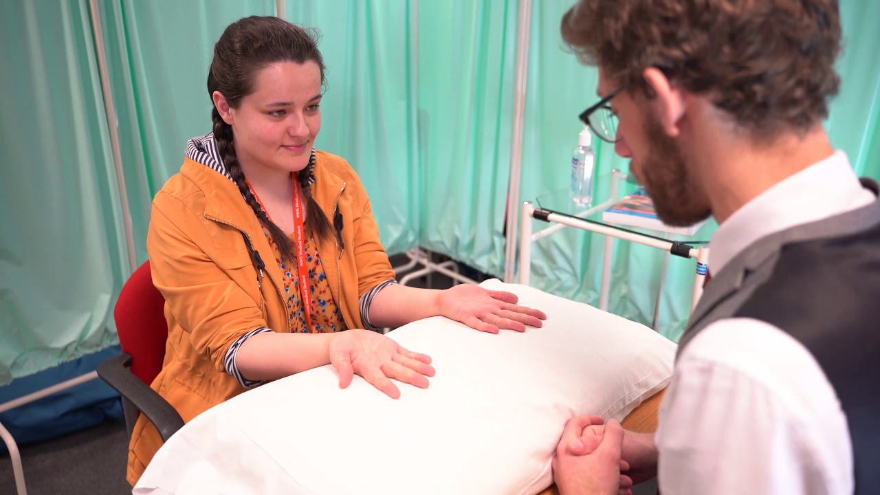
A clinical examination of the hands using the standard Look, Feel, Move approach. Specific examination structure derived from MacLeod's Clinical Examination 14th edition. Performed by Dr James Gill

General Examination - Clinical Skills OSCE - Dr Gill
The general examination is one of those early exams, which is essentially used to start medical students off with their clinical skills studies.
In the real world, it's mainly used with regard to gaining an overview of a patient, such as for a medical check up, or a baseline examination, for example, a health report.
They have been a couple of comments about the pulse monitor used in the video. For those who are interested. I’ve reached out to the manufacturer, and they’ve requested that the following code is provided to viewers, in order to get 20% off, if they decide on themselves.
Product model number: Vibeat SP20
Official Website: https://vibeatstore.com/produc....ts/sp20-handheld-pul
Special 20% OFF code: JAMES
--------------
Different medical schools, nursing colleges and other health professional courses will have their own preferred approach to a clinical assessment - you should concentrate on THEIR marks schemes for your assessments.
Some people watching this video may experience an ASMR effect
#DrGill #Asmr #Clinicalskills
#drgill #clinicalskills #asmr
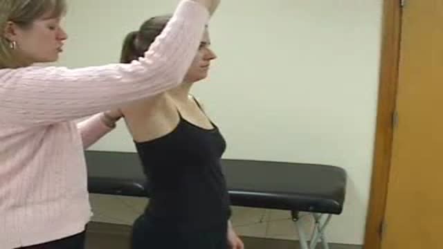
Neer's Sign
