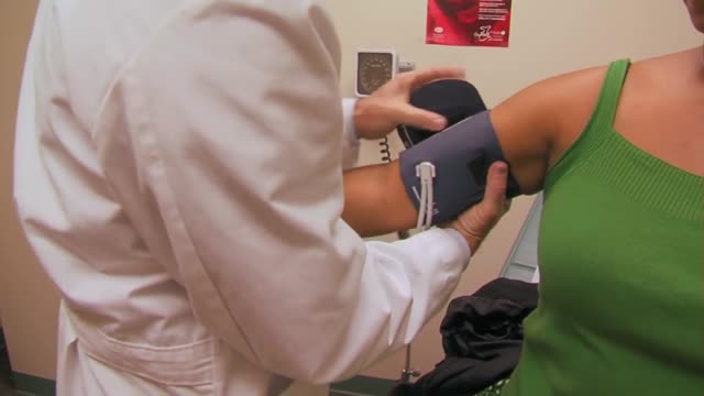
- Physical Examination
- Surgical Examination
- Ophthalmology
- Clinical Skills
- Orthopedics
- Surgery Videos
- Laparoscopy
- Pediatrics
- Funny Videos
- Cardiothoracic Surgery
- Nursing Videos
- Plastic Surgery
- Otorhinolaryngology
- Histology and Histopathology
- Neurosurgery
- Dermatology
- Pediatric Surgery
- Urology
- Dentistry
- Oncology and Cancers
- Anatomy Videos
- Health and Fitness
- Radiology
- Anaesthesia
- Physical Therapy
- Pharmacology
- Interventional Radiology
- Cardiology
- Endocrinology
- Gynecology
- Emergency Medicine
- Psychiatry and Psychology
- Childbirth Videos
- General Medical Videos
- Nephrology
- Physiology
- Diet and Food Health
- Diabetes Mellitus
- Neurology
- Women Health
- Osteoporosis
- Gastroenterology
- Pulmonology
- Hematology
- Rheumatology
- Toxicology
- Nuclear Medicine
- Infectious Diseases
- Vascular Disease
- Reproductive Health
- Burns and Wound Healing
- Other
Top videos

The pain is your feet trying to tell you something!

Alcoholic hepatitis can occur in people who drink heavily for many years. Symptoms include yellow skin and eyes along with increasing belly size due to fluid accumulation. Treatment involves hydration, nutritional care, and stopping alcohol use. Steroid drugs can help reduce liver inflammation.

Do you suffer with depression? Maybe you’ve recently been diagnosed or are a caregiver to someone with depression. Learn more about this common mood disorder, including depression causes, risk factors, and prevention. We’ll help you take control of your depression and live an active, healthy life.

M. Patrick Lowe, MD, renowned robotic surgeon and gynecologic oncologist at Northwestern Memorial Hospital, will demonstrate the use of robotic surgery to treat endometrial cancer.
Dr. Lowe, director of the robotics and minimally invasive surgical program for the Division of Gynecologic Oncology at Northwestern University's Feinberg School of Medicine, was among the early adopters of robotics to treat gynecologic malignancies, citing precision, improved dexterity and superior patient outcomes among the benefits.
"Women diagnosed with a gynecologic malignancy want the shortest route leading back to a degree of normalcy post treatment," says Lowe. "Robotic surgery offers the path of least resistance, combining shorter recovery times with superior outcomes."
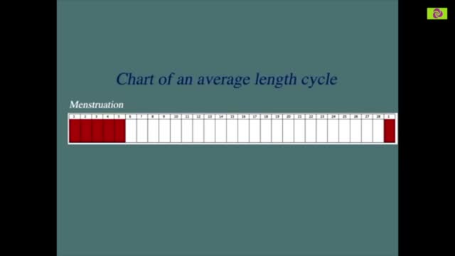
Our calculator can help you discover the most fertile days of your menstrual cycle or your “Estimated Fertility Window” based on information you provide.

Tudo Sobre Diabetes, Diabetes Tem Cura, O Que é Diabetes Tipo 2, Plantas Que Curam Diabetes
http://tudo-sobre-diabetes.good-info.co
Cura Naturalmente a Diabetes Tipo 2
A diabetes tipo II se tornou uma das doenças mais comuns nos tempos modernos. A boa notícia é que em pouco menos de um mês, seguindo um plano de alimentação e vida saudável, é possível equilibrar seu nível de açúcar no sangue e prevenir as terríveis consequências que esta doença tem.
A seguir, você encontrará este plano para nivelar o açúcar no sangue e dizer adeus para a diabetes.
Restrinja o consumo de todo o tipo de bebidas.
Realize atividade física de baixo impacto todo o dia, por um mínimo de meia hora.
Elimine por completo de suas refeições, todos os alimentos que contenham farinha branca.
Inclua em sua alimentação habitual, ácidos gordos essenciais (especialmente ácidos ômega 3), inclua também o consumo de frutas secas.
único Sistema Eficiente, Fácil E Natural Para Eliminar Para Sempre O Diabetes. Um Sistema Cientificamente Comprovado
Clique No Link Abaixo Para Verificá-la
http://tudo-sobre-diabetes.good-info.co
Assine O Nosso Canal
https://www.youtube.com/user/dicasdesaude11
https://www.youtube.com/watch?v=61MN7xSR9yA
Tudo Sobre Diabetes, Diabetes Tem Cura, O Que é Diabetes Tipo 2, Plantas Que Curam Diabetes,
diabetes gestacional,
diabetes mellitus tipo 2,
diabetes dieta,
sintomas de diabete,
diabetes tipo 1 e 2,
medicamentos para diabetes,
diabete sintomas,
causas da diabetes,
como evitar diabetes,
sintomas da diabetes tipo 2,
tratamento da diabetes,
o que diabetes,
os sintomas da diabete

A video from Physical Exam Series of Loyola University Health System, Chicago showing the medical examination of the abdomen
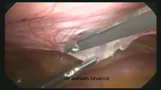
this video is showing the laparoscopic transabdominal preperitoneal herina repair for direct inguinal herina

For more than 25 years, The Children's Hospital of Philadelphia — the first Level 1 Pediatric Trauma Center in Pennsylvania — has provided unparalleled medical and surgical care for all injured children, including those with the most severe injuries.
Learn what makes the Trauma Center at CHOP a Level 1 Pediatric Trauma Center, and how our work toward trauma prevention, research advances and overall trauma awareness provides hope for reduced injuries in the future.
Learn more about the Trauma Center at CHOP: http://www.chop.edu/trauma.

Adrenoleukodystrophy, or ALD, is a deadly genetic disease that affects 1 in 18 000 people. It most severely affects boys and men. This brain disorder destroys myelin, the protective sheath that surrounds the brain's neurons -- the nerve cells that allow us to think and to control our muscles.
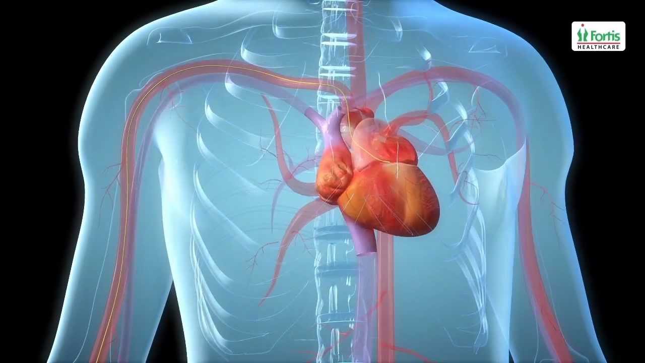
Angioplasty Procedure Animation Video
Emergency angioplasty is an operation that is performed directly after a heart attack, on admission to the hospital. It involves the insertion of a catheter into the blocked blood vessel that caused the heart attack. This opens it up and allows blood to flow again, thus minimizing damage to the heart.
If one or more arteries become clogged, it may result in a heart attack. This normally presents with chest pain, sweating and a feeling of anxiety, among other symptoms. Urgent medical assistance should be sought. A heart attack is a medical emergency requiring intervention as soon as possible.
Know more: http://www.emergencyangioplasty.com/
each type of heart problem requires different treatment but may share similar warning signs. It is important to see your doctor so that you can receive a correct diagnosis and prompt treatment. Learn to recognize the symptoms that may signal heart disease. Call your doctor if you begin to have new symptoms or if they become more frequent or severe. Symptoms of Coronary Artery Disease The most common symptom of coronary artery disease is angina, or chest pain. Angina can be described as a discomfort, heaviness, pressure, aching, burning, fullness, squeezing, or painful feeling in your chest. It can be mistaken for indigestion or heartburn. Angina may also be felt in the shoulders, arms, neck, throat, jaw, or back. Other symptoms of coronary artery disease include: Shortness of breath Palpitations (irregular heart beats, or a "flip-flop" feeling in your chest) A faster heartbeat Weakness or dizziness Nausea Sweating

Bunions can be very painful. ... Bunion removal is a surgical procedure that corrects a deformed area of the foot near the big toe. Bunion removal is sometimes called a bunionectomy, bunion surgery, or hallux valgus correction. Hallux valgus is a Latin phrase that means “foot deformity
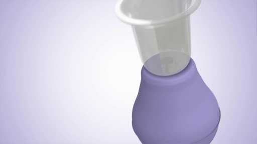
The Lansinoh Latch Assist has been designed to extend inverted nipples - watch this video to see how.?
Retinitis pigmentosa is a rare, inherited degenerative eye disease that causes severe vision impairment. Symptoms often begin in childhood. They include decreased vision at night or in low light and loss of side vision (tunnel vision).
Pompe disease is a rare multisystem genetic disorder that is characterized by absence or deficiency of the lysosomal enzyme alpha-glucosidase (GAA). This enzyme is required to breakdown (metabolize) the complex carbohydrate glycogen and convert it into the simple sugar glucose.
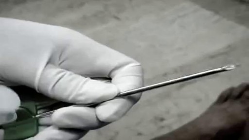
When placement of a urethral catheter is contraindicated or unsuccessful, percutaneous suprapubic urinary bladder catheterization is a commonly performed procedure to relieve urinary retention. [1, 2] This topic describes the Catheter over needle technique. The Seldinger technique is described in the Clinical Procedures topic Suprapubic Aspiration.
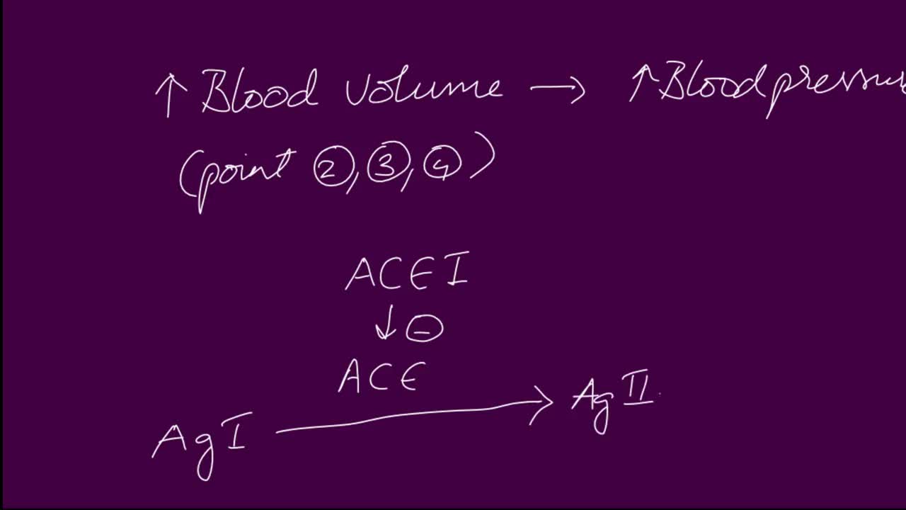
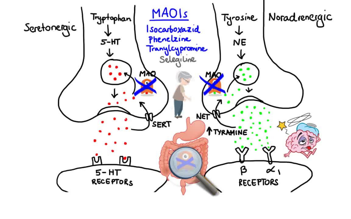
ACE Inhibitor Mechanisms. Angiotensin converting enzyme (ACE) inhibitors are agents used to relax blood vessels and lower blood pressure. They prevent an enzyme from producing angiotensin II, which narrows blood vessels and raises blood pressure, meaning the heart has to work harder to pump blood around the body.
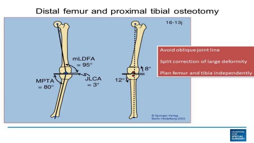
This video discusses knee arthritis, and when to do osteotomy, partial knee replacement, or total knee replacement.
