- Physical Examination
- Surgical Examination
- Ophthalmology
- Clinical Skills
- Orthopedics
- Surgery Videos
- Laparoscopy
- Pediatrics
- Funny Videos
- Cardiothoracic Surgery
- Nursing Videos
- Plastic Surgery
- Otorhinolaryngology
- Histology and Histopathology
- Neurosurgery
- Dermatology
- Pediatric Surgery
- Urology
- Dentistry
- Oncology and Cancers
- Anatomy Videos
- Health and Fitness
- Radiology
- Anaesthesia
- Physical Therapy
- Pharmacology
- Interventional Radiology
- Cardiology
- Endocrinology
- Gynecology
- Emergency Medicine
- Psychiatry and Psychology
- Childbirth Videos
- General Medical Videos
- Nephrology
- Physiology
- Diet and Food Health
- Diabetes Mellitus
- Neurology
- Women Health
- Osteoporosis
- Gastroenterology
- Pulmonology
- Hematology
- Rheumatology
- Toxicology
- Nuclear Medicine
- Infectious Diseases
- Vascular Disease
- Reproductive Health
- Burns and Wound Healing
- Other
Top videos
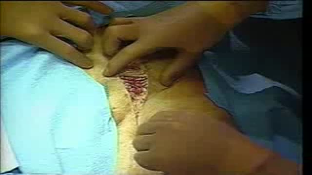
A surgical video showing Femoro-Popliteal Bypass with a Saphenous Vein Graft
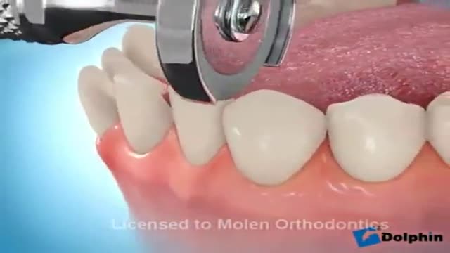
Dental Braces Animation

Mothers can do everything for her baby
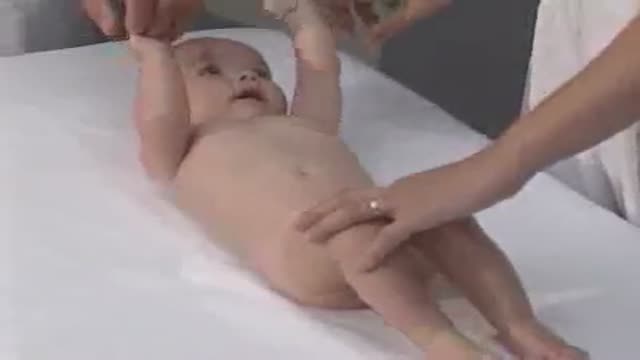
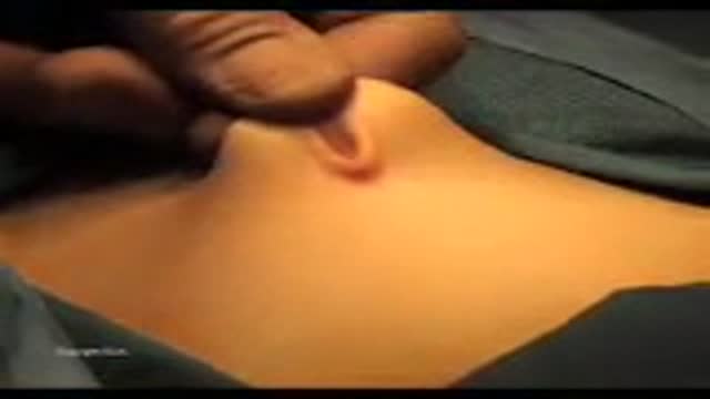
Pediatrics abdominal examination

Respiratory syncytial virus (RSV) is a virus that causes infections of the lungs and respiratory tract. It's so common that most children have been infected with the virus by age 2. Respiratory syncytial (sin-SISH-ul) virus can also infect adults. In adults and older, healthy children, the symptoms of respiratory syncytial virus are mild and typically mimic the common cold. Self-care measures are usually all that's needed to relieve any discomfort. Infection with respiratory syncytial virus can be severe in some cases, especially in premature babies and infants with underlying health conditions. RSV can also become serious in older adults, adults with heart and lung diseases, or anyone with a very weak immune system (immunocompromised).

Watch that video to know how to treat premature ejaculation naturally

Most condoms are made of latex rubber, but they can also be made from lamb cecum or polyurethane. In addition to their contraceptive value, condom use has been found effective in preventing the spread of sexually transmitted diseases.

WATCH MORE NURSING SKILLS HERE: https://nursing.com/course/nursing-skills/?utm_source=youtube&utm_medium=social
In our Nursing Skills course, we show you the most common and most important skills you will use as a nurse! We included everything from bed baths, to inserting a foley, to advanced skills like chest tube management.
Welcome to the NURSING Family, we call it the most supportive nursing cohort on the planet.
At NURSING.com, we want to help you remove the stress and overwhelm of nursing school so that you can focus on becoming an amazing nurse.
Check out our freebies and learn more at: (http://www.nursing.com)
Visit us at http://www.nursing.com/medical....-information-disclai for disclaimer information.
NCLEX®, NCLEX-RN® are registered trademarks of the National Council of State Boards of Nursing, INC. and hold no affiliation with NURSING.
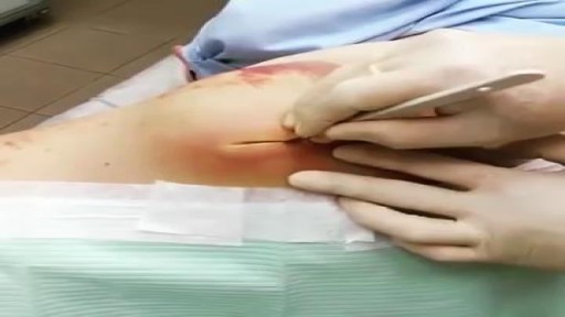
Watch that Large Jelly Like Hematoma Extraction
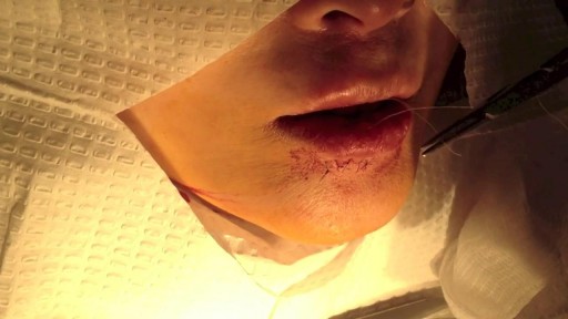
This video details the layered closure of a through-and-through facial laceration
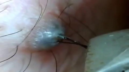
Ingrown Hair Removal Video
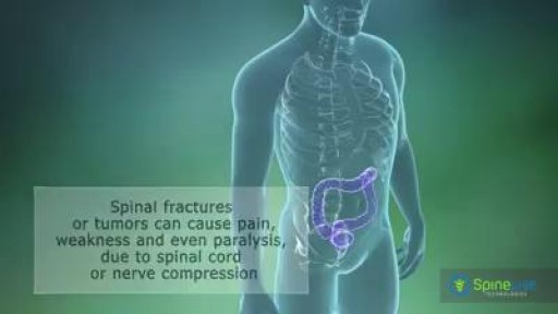
A spinal tumor is a growth that develops within your spinal canal or within the bones of your spine. It may be cancerous or noncancerous. Tumors that affect the bones of the spine (vertebrae) are known as vertebral tumors. Tumors that begin within the spinal cord itself are called spinal cord tumors. There are two main types of tumors that may affect the spinal cord: Intramedullary tumors begin in the cells within the spinal cord itself, such as astrocytomas or ependymomas. Extramedullary tumors develop within the supporting network of cells around the spinal cord. Although they don't begin within the spinal cord itself, these types of tumors may affect spinal cord function by causing spinal cord compression and other problems. Examples of extramedullary tumors that can affect the spinal cord include schwannomas, meningiomas and neurofibromas.
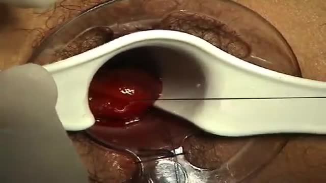
A surgeon begins the PPH stapled hemorrhoidectomy by inserting a circular anal dilator and obturator into the anal canal and then securing the dilator in place with four sutures. The surgeon then inserts a PPH anoscope into the obturator. Next, he places a circumferential purse-string suture of 2-0 Monocryl on a UR-6 needle 4 cm proximal to the dentate line. The surgeon opens a PPH stapler and places its anvil across the purse string. The stapler is then closed and fired; it is held closed for two minutes to improve hemostasis. Prior to firing the stapler in a female patient, the surgeon places a gloved finger in the vagina to ensure the vaginal mucosa and rectal-vaginal septum are not trapped within the jaws of the closed stapler. The surgeon then opens and removes the stapler.
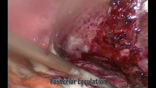
A liver abscess is a pus-filled mass inside the liver. Common causes are abdominal infections such as appendicitis or diverticulitis due to haematogenous spread through the portal vein. A pyogenic liver abscess (PLA) is a pocket of pus that forms in the liver in response to an infection or trauma. Pus is a fluid composed of white blood cells, dead cells, and bacteria that forms when your body fights off infection.Dec 11, 2015

Watch that video to know if it is safe to have sex during pregnancy
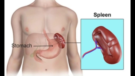
The spleen plays multiple supporting roles in the body. It acts as a filter for blood as part of the immune system. Old red blood cells are recycled in the spleen, and platelets and white blood cells are stored there. The spleen also helps fight certain kinds of bacteria that cause pneumonia and meningitis

G-Shot (G-Spot Amplification)
Sanjeev Dutta, MD, FACS discusses the fascinating new world of surgical technology. The pediatric general surgeon shares how medicine and technology have combined to achieve less invasive procedures and healthier outcomes for surgical patients.
Dr. Dutta is a pediatric general surgeon at Lucile Packard Children's Hospital. He is also an Associate Professor of Surgery at Stanford School of Medicine and Surgical Director of the Multidisciplinary Initiative for Surgical Technology Research.
Learn more about Stanford Children's Health. http://www.stanfordchildrens.org.

Am I missing something?
Subscribe to my fun weekly newsletter (for free!): http://eepurl.com/iaYycn
To check out a previous newsletter, click here: https://mailchi.mp/a9909f90cac....a/why-are-you-having
For more Doc Schmidt content, check out my website: https://www.docschmidt.org/
Check out my children's book here: https://www.amazon.com/Night-Before-Med-School-Medical/dp/B0B193KWXT/ref=sr_1_1?keywords=doc+schmidt&qid=1653339841&sprefix=doc+sc%2Caps%2C202&sr=8-1
Logo and graphics designed by iamlindaayoade.com and loigraphics.com (LOI Graphics Inc.)
Want me to make you a personalized video for you or your friend? Check me out on Cameo!
https://v.cameo.com/DFKBSe2HSib
Want to connect with me and watch more content?
Find me on TikTok!
https://vm.tiktok.com/ZMRFmqKts/
And Instagram!
https://instagram.com/docschmidtig?r=nametag
All content is intended as medical education or entertainment and is NOT intended to be medical advice. If you have any symptoms concerning you, please schedule an appointment with your doctor.
Join my channel to get access to perks! Click link below:
https://www.youtube.com/channe....l/UCLbidg2ZT49dWrxDk
Open Appendectomy Surgery Video
