- Physical Examination
- Surgical Examination
- Ophthalmology
- Clinical Skills
- Orthopedics
- Surgery Videos
- Laparoscopy
- Pediatrics
- Funny Videos
- Cardiothoracic Surgery
- Nursing Videos
- Plastic Surgery
- Otorhinolaryngology
- Histology and Histopathology
- Neurosurgery
- Dermatology
- Pediatric Surgery
- Urology
- Dentistry
- Oncology and Cancers
- Anatomy Videos
- Health and Fitness
- Radiology
- Anaesthesia
- Physical Therapy
- Pharmacology
- Interventional Radiology
- Cardiology
- Endocrinology
- Gynecology
- Emergency Medicine
- Psychiatry and Psychology
- Childbirth Videos
- General Medical Videos
- Nephrology
- Physiology
- Diet and Food Health
- Diabetes Mellitus
- Neurology
- Women Health
- Osteoporosis
- Gastroenterology
- Pulmonology
- Hematology
- Rheumatology
- Toxicology
- Nuclear Medicine
- Infectious Diseases
- Vascular Disease
- Reproductive Health
- Burns and Wound Healing
- Other
Top videos

Watch that video to know if oral sex causes cancer
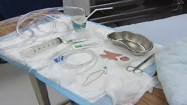
video shows how to insert a nasogatric tube.
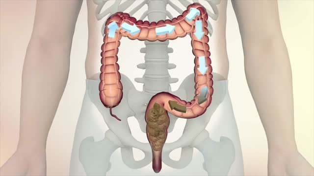
This information is collected from Oncolex. For more on colon and rectum (
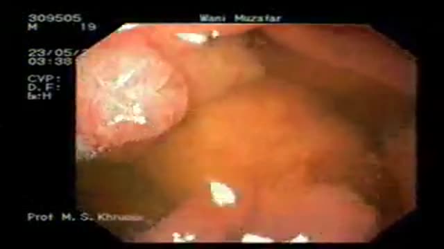
ENDOSCOPIC (NON-SURGICAL) REMOVAL OF MULTIPLE LARGE TUMORS FROM STOMACH IN A PATIENT WITH PEUTZ-JEGHERS SYNDROME
PEUTZ-JEGHERS SYNDROME: Peutz-Jeghers syndrome (PJS) is a familial syndrome consisting of mucocutaneous pigmentation, gastrointestinal polyposis and cancers of gut & other sites like breast, ovary, and testes. PJS has an autosomal dominant inheritance with variable and incomplete penetrance. Germline mutations of STK11/LKB1 gene on 19p cause this syndrome. Mucocutaneous pigmentation may be noted in early infancy. These deposits of melanin are most commonly found around the mouth, nose, lips, buccal mucosa, hands, and feet, and may also be present in perianal and genital areas. PJS polyps may be found in stomach, small intestine, or colon, but they tend to be prominent in the small intestine. These polyps may increase in size and cause small intestinal obstruction or intussusceptions that may occur in early infancy. Acute upper gastrointestinal bleeding and chronic faecal blood may complicate the disease.
PATIENT: The patient was a 25 yr male who had mucocutaneous pigmentation and multiple polyps in the stomach and duodenum. He presented with bleeding from gastric polyps. As the polyps in stomach were numerous, (more than 20 in number) and were large in size (some equal to small egg size), he had been advised to undergo surgery. Surgery planned was total gastrectomy.
PROCEDURE: The patient underwent video-endoscopy of the esophagus, stomach and duodenum. All polyps were examined for size and presence or absence of stalk. A plan to remove all the gastric polyps at endoscopy was made in the same sitting. He received light conscious sedation. Flat polyps were raised form the gastric wall by injection of saline in to polyp base to let these lesions have a stalk. This was done by needle injector. Each polyp was engaged in a snare and the polyp stalk was cut by coagulation cutting current. The cuts were clean without any bleeding. All polyps were recovered for histology. The histology revealed all polyps to be hamartomous lesions. None of the polyps were cancerous. Patient has been followed up for over one year and is doing fine without any further bleeding or pain.
Video shows the procedure of videoendoscpy and endoscopic removal of polyps.
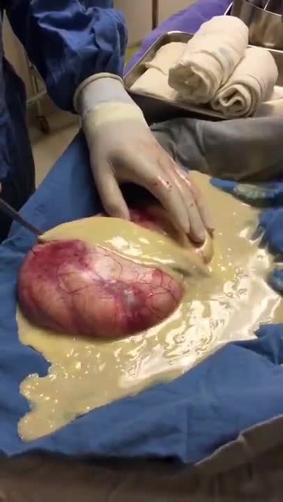
Ovarian teratoma is a type of germ cell tumour. Germ cell tumours are cancers that begin in egg cells in women or sperm cells in men. There are 2 main types of ovarian teratoma. Mature teratoma, which is benign. Immature teratoma, which is cancerous.
At Hutzel Women's Hospital, Dr. Giancarlo Mari performs breakthrough in-utero surgery to save the lives of high-risk twins developing with a rare "shared" circulatory problem. ~ Detroit Medical Center
watch that video of Horrifying Creatures Found Living Inside a Human Body
A video showing bone marrow biopsy
When foreign organisms such as bacteria enter the body, the immune system sends white blood cells to fight the infection. This causes swelling (inflammation) at the site of infection and the death of nearby tissue, creating a hole called a cavity, which fills with pus to form an abscess.
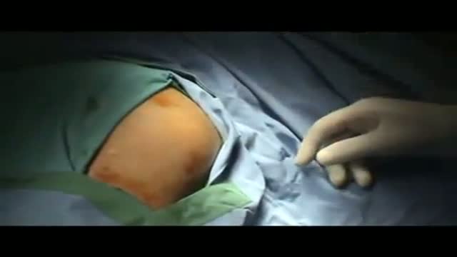
With the patient in the supine position; apply the antiseptic agent (betadine). Video is uploaded on www.MedicalVideos.us In this video the subclavian vein will be placed on the left side.
Watch that video of Doctors Removed 30 Pounds Of Poop From Man’s Colon
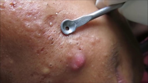
Comedone Extraction Video
Macrobiopsy of breast lesions is a complicated procedure when performed with vacuum assisted biopsy tools. The Spirotome is a hand-held needle set that doesn’t need capital investment, is ready to use and provides tissue samples of high quality in substantial amounts. In this way quantitative molecular biology is possible with one tissue sample. The Coramate is an automated version of this direct and frontal technology

Tonsillectomy 3D Animation
Watch that Big Size Fibrodenoma Removal Under Local Anesthesia
Surgery to treat men with prostate cancer is often followed by months of difficulty controlling urine flow, a condition known as urinary incontinence. But new research suggests that this problem may go away more quickly if the men perform certain exercises to strengthen their pelvic floor muscles.
Researchers from the Kaiser Permanente Medical Center in Los Angeles, California, found that men who were taught how to perform pelvic floor exercises before and after surgery were more likely to have regained continence three months later.
Men Doing Pelvic Exercises Recover Earlier
In the current study, the researchers randomly assigned 38 men scheduled for radical prostatectomy to either a treatment group or a control group. The men in the treatment group were referred to a physical therapist. They were instructed how to do Pelvic Floor Exercises both before and after surgery, using biofeedback to ensure they were using the proper muscles. The control group did not receive any formal instruction. All of the men completed questionnaires regarding bladder function at regular intervals over the next year.
Overall, 82% of the patients had regained continence (defined as not needing to use any absorbent pads) by the end of the year, including about equal numbers in both groups. But on average the men who had been educated about Pelvic exercises regained continence about one month earlier than those in the control group (at 12 weeks vs. 16 weeks).
Most of the men who did not regain continence within a year were still using at least three absorbent pads a day, indicating continued severe incontinence. The study authors explained that these men probably had extensive damage to the bladder sphincter or severe dysfunction of the bladder after surgery, and the exercises alone were unable to compensate for this.
But the exercises seemed to be effective. Pelvic floor exercise and education initiated prior to surgery is an effective noninvasive intervention useful for improving early return of urinary continence, the authors concluded. It would certainly have a positive impact on our patients undergoing radical prostatectomy in an effort to improve quality of life after major urological surgery.
The results of the study were published in the Journal of Urology (Vol. 170, No. 1: 130-133)
The Knee Exam
Observation:
1. Make sure that both knees are fully exposed. The patient should be in either a gown or shorts. Rolled up pant legs do not provide good exposure!
2. Watch the patient walk. Do they limp or appear to be in pain? When standing, is there evidence of bowing (varus) or knock-kneed (valgus) deformity? There is a predilection for degenerative joint disease to affect the medical aspect of the knee, a common cause of bowing. Varus Knee Deformity, more marked on the left leg. 3. Make note of any scars or asymmetry. Chronic/progressive damage, as in degenerative joint disease, may lead to abnormal contours and appearance. Is there obvious swelling as would occur in an effusion? Redness suggesting inflammation? 4. Is there evidence of atrophy of the quadriceps, hamstring, or calf muscle groups? Knee problems/pain can limit the use of the affected leg, leading to wasting of the muscles.
While both legs have well developed musculature,
the left calf and hamstring are bulkier than the right. 5. Look at the external anatomy, noting structures above and below the knee itself: 1. Patella 2. Patellar tendon 3. Quadriceps/Hamstring/Calf muscles 4. Medial and lateral joint lines. 5. Femur and Tibia 6. Tibial tuberosity
Ballotment (helpful if the effusion is large) 1. Slightly flex the knee which is to be examined.
2. Place one hand on the supra-pateallar pouch, which is above the patella and communicates with the joint space. Gently push down and towards the patella, forcing any fluid to accumulate in the central part of the joint.
3. Gently push down on the patella with your thumb.
4. If there is a sizable effusion, the patella will feel as if it's floating and "bounce" back up when pushed down.
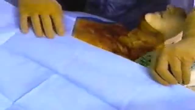
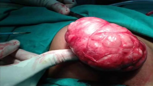
A video showing the procedure of lipoma excision
A Video showing a fine needle biopsy guided with ultrasound of a thyroid nodule
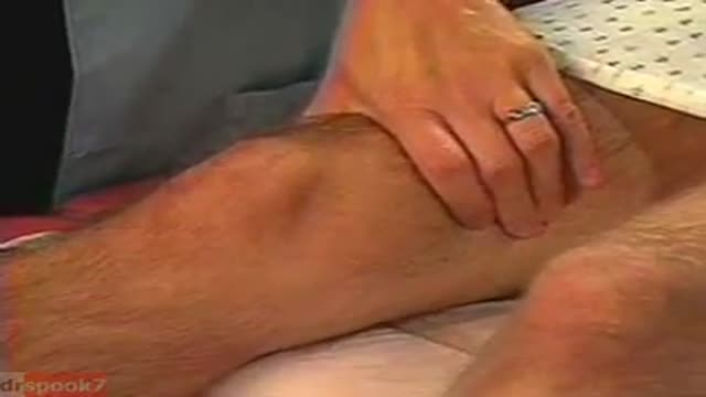
Examination of peripheral pulses of the lower limb
