- Physical Examination
- Surgical Examination
- Ophthalmology
- Clinical Skills
- Orthopedics
- Surgery Videos
- Laparoscopy
- Pediatrics
- Funny Videos
- Cardiothoracic Surgery
- Nursing Videos
- Plastic Surgery
- Otorhinolaryngology
- Histology and Histopathology
- Neurosurgery
- Dermatology
- Pediatric Surgery
- Urology
- Dentistry
- Oncology and Cancers
- Anatomy Videos
- Health and Fitness
- Radiology
- Anaesthesia
- Physical Therapy
- Pharmacology
- Interventional Radiology
- Cardiology
- Endocrinology
- Gynecology
- Emergency Medicine
- Psychiatry and Psychology
- Childbirth Videos
- General Medical Videos
- Nephrology
- Physiology
- Diet and Food Health
- Diabetes Mellitus
- Neurology
- Women Health
- Osteoporosis
- Gastroenterology
- Pulmonology
- Hematology
- Rheumatology
- Toxicology
- Nuclear Medicine
- Infectious Diseases
- Vascular Disease
- Reproductive Health
- Burns and Wound Healing
- Other
Top videos
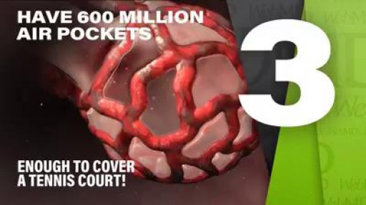
Your lungs are have 600 million air pockets -- enough to cover a tennis court.
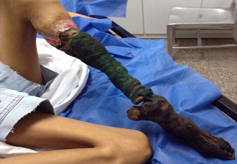
Watch that video of a Snake bite causes girl’s leg to rot away
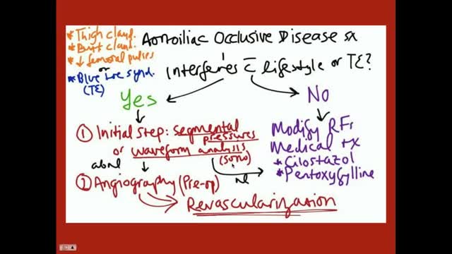
Aortoiliac occlusive disease (AIOD) occurs commonly in patients with PAD. Significant lesions in the aortoiliac arterial segment are exposed easily by palpation of the femoral pulses. Any diminution of the palpable femoral pulse indicates that a more proximal obstruction exists. Obstructive lesions may be present in the infrarenal aorta, common iliac, internal iliac (hypogastric), external iliac, or combinations of any or all of these vessels. Occasionally, degenerated nonstenotic atheromatous disease exists in these vessels and may manifest by atheroembolism to the foot, the "blue toe" or "trash foot" syndrome. Generally, patients with aortoiliac PAD have a poorer general prognosis than those with more distal PAD.
Robot-Assisted Laparoscopic Rectal resection for Endometriosis.Operation performed by D.Vitobello, director of divisione of Gynaecology, and G.Baldazzi,director of Surgical department. Abano Terme Hospital Padova (Italy)
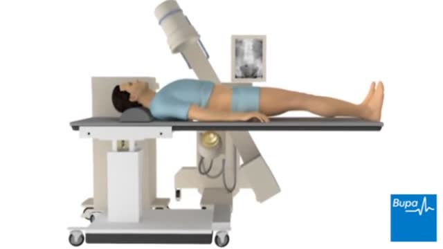
Extracorporeal shock wave lithotripsy (ESWL) uses shock waves to break a kidney stone into small pieces that can more easily travel through the urinary tract camera.gif and pass from the body. See a picture of ESWL camera.gif. You lie on a water-filled cushion, and the surgeon uses X-rays or ultrasound tests to precisely locate the stone. High-energy sound waves pass through your body without injuring it and break the stone into small pieces. These small pieces move through the urinary tract and out of the body more easily than a large stone. The process takes about an hour. You may receive sedatives or local anesthesia. Your surgeon may use a stent if you have a large stone. A stent is a small, short tube of flexible plastic mesh that holds the ureter open. This helps the small stone pieces to pass without blocking the ureter.

A drill. A mallet. A robot. Go inside the operating room to see how Northwestern Medicine Orthopaedic Surgeon Linda Idris Suleiman, MD, uses these tools for a total knee replacement.
#insidetheor
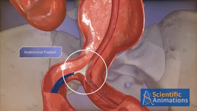
Abdominal aortic aneurysms can weaken the aorta, your body’s largest blood vessel. This can develop into a potentially serious heath problem that can be fatal if the aneurysm bursts, causing massive internal bleeding. Endovascular stent grafting, or endovascular aneurysm repair (EVAR), is a newer form of treatment for abdominal aortic aneurysm that is less invasive than open surgery. Endovascular stent grafting uses an endovascular stent graft to reinforce the wall of the aorta and to help keep the damaged area from rupturing.
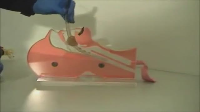
nee joint aspiration and injection are performed to aid in diagnosis and treatment of knee joint diseases. The knee joint is the most common and the easiest joint for the physician to aspirate. One approach involves insertion of a needle 1 cm above and 1 cm lateral to the superior lateral aspect of the patella at a 45-degree angle. Once the needle has been inserted 1 to 1½ inches, aspiration aided by local compression is performed. Local corticosteroid injections can provide significant relief and often ameliorate acute exacerbations of knee osteoarthritis associated with significant effusions. Among the indications for arthrocentesis are crystal-induced arthropathy, hemarthrosis, unexplained joint effusion, and symptomatic relief of a large effusion. Contraindications include bacteremia, inaccessible joints, joint prosthesis, and overlying infection in the soft tissue. Large effusions can recur and may require repeat aspiration. Anti-inflammatory medi

Testis operation

The MINI tummy-tuck is a lesser variant of the classic tummy tuck. The MINI tummy-tuck always involved skin excision (often a scar revision and skin excision of the flabby skin over a C-section scar or hysterectomy or laparotomy scar) but may also involve liposuction, umbilical floating, etc. Commonly it will not include any muscle repair otherwise it it now a classic tummy tuck (aka abdominoplasty). Cost varies depending on the components involved. Here, Toronto Aesthetic Plastic Surgeon Dr Marc DuPéré describes a MINI tummy-tuck done on a patient who had a Brazilian Butt Lift before (and skin harvesting from abdomen) and a recent 20 lbs weight loss, a patient who wants more liposuction to abdomen and flanks and whose skin has now lost elasticity, hence the requirement for this small skin excision. Dr DuPéré also explains what UMBILICAL floating means. Dr DuPéré performs more than 5 different techniques of tummy-tucks in Toronto and the technique chosen reflects the patient’s expectations and anatomy. Call us if interested in learning about YOUR options for a flatter tummy! 📱 416-929-9800
Patient consent obtained. Thank you to my patient.
Visage Clinic Toronto
https://www.visageclinic.com/
(416) 929-9800
101-133 Hazelton Avenue, Toronto, ON M5R 0A6
https://www.facebook.com/VisageClinic/
https://www.instagram.com/VisageClinicDrDuPere/
The Combitube is a twin lumen device designed for use in emergency situations and difficult airways. It can be inserted without the need for visualization into the oropharynx, and usually enters the esophagus. It has a low volume inflatable distal cuff and a much larger proximal cuff designed to occlude the oro- and nasopharynx.
If the tube has entered the trachea, ventilation is achieved through the distal lumen as with a standard ETT. More commonly the device enters the esophagus and ventilation is achieved through multiple proximal apertures situated above the distal cuff. In the latter case the proximal and distal cuffs have to be inflated to prevent air from escaping through the esophagus or back out of the oro- and nasopharynx.
Symptoms Burning stomach pain Feeling of fullness, bloating or belching Fatty food intolerance Heartburn Nausea The most common peptic ulcer symptom is burning stomach pain. Stomach acid makes the pain worse, as does having an empty stomach. The pain can often be relieved by eating certain foods that buffer stomach acid or by taking an acid-reducing medication, but then it may come back. The pain may be worse between meals and at night. Nearly three-quarters of people with peptic ulcers don't have symptoms. Less often, ulcers may cause severe signs or symptoms such as: Vomiting or vomiting blood — which may appear red or black Dark blood in stools, or stools that are black or tarry Trouble breathing Feeling faint Nausea or vomiting Unexplained weight loss Appetite changes

The dentin is a hard tissue that forms the bulk of the tooth. It is similar to bone but is slightly harder, although softer than enamel. The dentin has numerous dentinal tubules that run across its length. Each dentinal tubule houses the cytoplasmic process of an odontoblast (odontoblastic process).
📄Notes for the video: https://www.hackdentistry.com/....bundles/revision-nin
💻Website: https://www.hackdentistry.com/
📰Blog: https://hackdentistry.substack.com/
Study resources on our website-
📖Oral pathology Revision Ninja (Notes, Videos & MCQs): https://www.hackdentistry.com/bundles/oral-pathology-revision-ninja
📖Oral Histology Revision Ninja (Notes, Videos & MCQs): https://www.hackdentistry.com/....bundles/revision-nin
📖Periodontics Revision Ninja (Notes & MCQs): https://www.hackdentistry.com/bundles/perio-rn
📖Question Bank: https://www.hackdentistry.com/bundles/question-bank
📖Access all content: https://www.hackdentistry.com/bundles/all-access-premium
References and further reading:
💡Berkovitz BKB, Hollan GR, Moxham BJ. Oral Anatomy, Histology and Embryology. 4th ed. Mosby Elsevier; 2009.
💡Nanci A. Tencate’s Oral Histology. Development, Structure and Function. 8th ed. Elsevier; 2013.
💡Kumar GS. Orban’s Oral Histology and Embryology.13th ed. Elsevier; 2011.
💡Avery JK. Oral development and Histology. 3rd ed. Thieme Medical Publishers; 2002.
Log in to https://www.hackdentistry.com and get access to:
I) Numerous Notes/Cheatsheets and Videos II) Thousands of quiz questions from our vast Question Bank!
HackDentistry is an edtech company that aims make learning dentistry fun,engaging and light hearted.
1) It focuses on helping students understand and retain core concepts in dentistry through highly visual sketch/whiteboard style video animations.
2) The platform helps improve exam performance by providing Revision Bundles and allowing students to test themselves using thousands of Practice Questions from a vast Question Bank. (multiple choice format).
3) It also provides for a community platform where students can come together, and engage with fellow dental students and dentists across the globe!
Facebook:
https://www.facebook.com/hackdentistry
Instagram:
https://www.instagram.com/hack.dentistry/
Twitter:
https://twitter.com/hckdentistry

Mini-Laparoscopic Cholecystectomy with Intraoperative Cholangiogram for Symptomatic Cholelithiasis (Gallstones) - Extended
Authors: Brunt LM1, Singh R1, Yee A2
Published: September 26, 2017
AUTHOR INFORMATION
1 Department of Surgery, Washington University, St. Louis, Missouri
2 Division of Plastic and Reconstructive Surgery, Washington University, St. Louis, Missouri
DISCLOSURE
No authors have a financial interest in any of the products, devices, or drugs mentioned in this production or publication.
ABSTRACT
Minimal invasive laparoscopic cholecystectomy is the typical surgical treatment for cholelithiasis (gallstones), where patients present with a history of upper abdominal pain and episodes of biliary colic. The classic technique for minimal invasive laparoscopic cholecystectomy involves four ports: one umbilicus port, two subcostal ports, and a single epigastric port. The Society of American Gastrointestinal and Endoscopic Surgeons (SAGES) has instituted a six-step strategy to foster a universal culture of safety for cholecystectomy and minimize risk of bile duct injury. The technical steps are documented within the context of the surgical video for (1) achieving a critical view of safety for identification of the cystic duct and artery, (2) intraoperative time-out prior to management of the ductal structures, (3) recognizing the zone of significant risk of injury, and (4) routine intraoperative cholangiography for imaging of the biliary tree. In this case, the patient presented with symptomatic biliary colic due to a gallstone seen on the ultrasound in the gallbladder. The patient was managed a mini-laparoscopic cholecystectomy using 3mm ports for the epigastric and subcostal port sites with intraoperative fluoroscopic cholangiogram. Specifically, the senior author encountered a tight cystic duct preventing the insertion of the cholangiocatheter and the surgical video describes how the author managed the cystic duct for achieving a cholangiogram, in addition to the entire technical details of laparoscopic cholecystectomy.
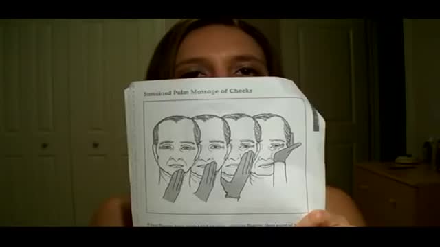
Bell's palsy is a form of facial paralysis resulting from damage or trauma to the facial nerves. The facial nerve-also called the 7th cranial nerve-travels through a narrow, bony canal (called the Fallopian canal) in the skull, beneath the ear, to the muscles on each side of the face. For most of its journey, the nerve is encased in this bony shell. Each facial nerve directs the muscles on one side of the face, including those that control eye blinking and closing, and facial expressions such as smiling and frowning. Additionally, the facial nerve carries nerve impulses to the lacrimal or tear glands, the saliva glands, and the muscles of a small bone in the middle of the ear called the stapes. The facial nerve also transmits taste sensations from the tongue. When Bell's palsy occurs, the function of the facial nerve is disrupted, causing an interruption in the messages the brain sends to the facial muscles. This interruption results in facial weakness or paralysis. Bell's palsy is named for Sir Charles Bell, a 19th century Scottish surgeon who described the facial nerve and its connection to the condition. The disorder, which is not related to stroke, is the most common cause of facial paralysis. Generally, Bell's palsy affects only one of the paired facial nerves and one side of the face, however, in rare cases, it can affect both sides.

http://www.landging.com/plane-animation.html
420 seconds 3d plane animation, designed for Expo 2010 Shanghai Aviation Pavilion.

You can now test your knowledge with a free lesson quiz on NURSING.com!
Click here for your free quiz: https://bit.ly/3HwJr8t
Stoma Care- Changing a Colostomy Bag (Nursing Skills)
FREE Nursing School Cheat Sheets at: http://www.NURSING.com
Get the full lesson on Stoma Care here:
05.01 Stoma Care (Colostomy bag) | NURSING.com
Check out our new Nurse Care Plan Lessons here:
https://bit.ly/3BPRfPL
Watch the Nursing Skills Course Introduction here:
https://nursing.com/lesson/ski....lls-00-01-course-int
Get Access to Thousands of Lessons here:
https://nursing.com/courses/
Welcome to the NURSING Family, we call it the most supportive nursing cohort on the planet.
At NURSING.com, we want to help you remove the stress and overwhelm of nursing school so that you can focus on becoming an amazing nurse.
Check out our freebies and learn more at: (http://www.nursing.com)
Stoma Care- Changing a Colostomy Bag (Nursing Skills)
In this video, we’re going to talk about stoma care. Now, the wafer and bag for an ostomy only NEEDS to be changed every 3 days, or if it’s leaking. But, you still need to be able to assess the stoma itself. In this case we’re going to show you how to replace the bag and clean and assess the stoma. Start by putting a towel under the patient on the side of the stoma. We love you guys! Go out and be your best selves today! And, as always, happy nursing!
Bookmarks:
0.05 Introduction to Stoma Care
0:20 Assessing the stoma
0:47 Cleaning the stoma
1:12 Inspecting the stoma
1:25 Measuring and cutting the stoma
2:00 Applying and sealing the bag
2:35 Documentation
2:41 Outro
Visit us at https://nursing.com/medical-disclaimer/ for disclaimer information.
NCLEX®, NCLEX-RN® are registered trademarks of the National Council of State Boards of Nursing, INC. and hold no affiliation with NURSING.com.
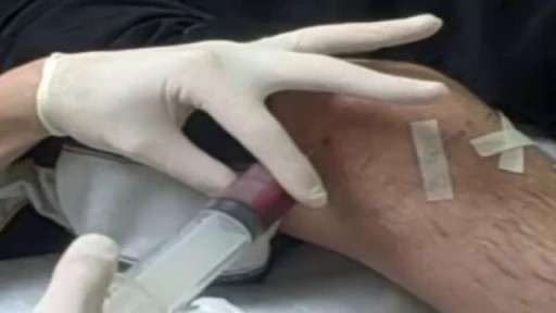
Subcutaneous Injection

https://bit.ly/3HIStRc #shorts
Tracheotomy and tracheostomy are surgical procedures that create an opening in the trachea (windpipe) to help patients breathe when they have difficulty doing so through the nose or mouth. Though they are similar in purpose, there are some key differences between them.
Tracheotomy is a temporary procedure that involves creating a small incision in the trachea to insert a breathing tube. The tube is typically removed once the patient no longer requires it, and the incision heals on its own. Tracheostomy, on the other hand, is a more permanent solution that involves creating a hole in the trachea and inserting a tracheostomy tube, which remains in place for an extended period.
Indications for these procedures include:
Airway obstruction due to trauma, tumors, or infection
Severe respiratory distress or failure
Prolonged mechanical ventilation
Inability to protect the airway due to neurological disorders or impaired consciousness
Steps for performing a tracheotomy and tracheostomy:
Preparation: The patient is positioned, and the neck area is cleaned and draped. Local anesthesia is often administered, although general anesthesia may be used in some cases.
Incision: A small incision is made in the neck, and the muscles and tissues are carefully separated to expose the trachea.
Tracheal opening: A small opening is made in the trachea, typically between the second and third tracheal rings.
Tube insertion: A tracheotomy tube is inserted through the incision and into the trachea for a tracheotomy, while a tracheostomy tube is inserted for a tracheostomy. Both tubes are secured in place.
Confirmation: Proper placement of the tube is confirmed by listening for breath sounds and checking for adequate ventilation.
Pre-operative care typically involves a thorough assessment of the patient's medical history, as well as any necessary imaging studies or lab tests to ensure the procedure is appropriate and safe. Informed consent should be obtained from the patient or their legal representative.
Post-operative care includes monitoring the patient's vital signs, ensuring the tube remains secure and patent, and managing any pain or discomfort. For tracheostomy patients, regular cleaning and maintenance of the stoma (the opening in the trachea) and the tracheostomy tube are essential to prevent infection and other complications. Long-term care may involve speech therapy, respiratory therapy, and support from a multidisciplinary team to address any ongoing needs.
It's crucial to remember that these procedures should only be performed by trained medical professionals in a clinical setting.
for additional information about this procedure check our article @ www.medicalartsshop.com
For more free resources, find us on Pinterest & Facebook pages:
https://www.pinterest.ca/medicalartsofficial/
https://www.facebook.com/Medicalartsofficial
https://www.youtube.com/@medic....alarts?sub_confirmat
https://www.instagram.com/medicalartsofficial/
https://www.tiktok.com/@medicalarts
This video and associated content are for entertainment and educational purposes only!!

Scientists don't know what causes canker sores. Most believe that there is a problem with the body's immune system. Emotional stress, menstruation or injury to the mouth are common triggers for simple canker sores. Certain foods such as citrus or acidic foods may trigger a canker sore or make one more uncomfortable.
