- Physical Examination
- Surgical Examination
- Ophthalmology
- Clinical Skills
- Orthopedics
- Surgery Videos
- Laparoscopy
- Pediatrics
- Funny Videos
- Cardiothoracic Surgery
- Nursing Videos
- Plastic Surgery
- Otorhinolaryngology
- Histology and Histopathology
- Neurosurgery
- Dermatology
- Pediatric Surgery
- Urology
- Dentistry
- Oncology and Cancers
- Anatomy Videos
- Health and Fitness
- Radiology
- Anaesthesia
- Physical Therapy
- Pharmacology
- Interventional Radiology
- Cardiology
- Endocrinology
- Gynecology
- Emergency Medicine
- Psychiatry and Psychology
- Childbirth Videos
- General Medical Videos
- Nephrology
- Physiology
- Diet and Food Health
- Diabetes Mellitus
- Neurology
- Women Health
- Osteoporosis
- Gastroenterology
- Pulmonology
- Hematology
- Rheumatology
- Toxicology
- Nuclear Medicine
- Infectious Diseases
- Vascular Disease
- Reproductive Health
- Burns and Wound Healing
- Other
Top videos
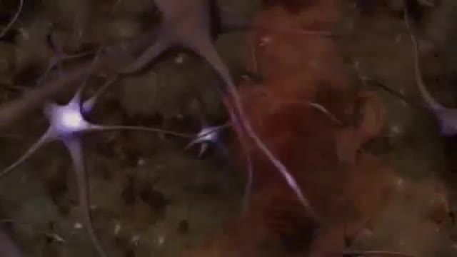
Genes are the building blocks of heredity. They are passed from parent to child. They hold DNA, the instructions for making proteins. Proteins do most of the work in cells. They move molecules from one place to another, build structures, break down toxins, and do many other maintenance jobs. Sometimes there is a mutation, a change in a gene or genes. The mutation changes the gene's instructions for making a protein, so the protein does not work properly or is missing entirely. This can cause a medical condition called a genetic disorder. You can inherit a gene mutation from one or both parents. A mutation can also happen during your lifetime.
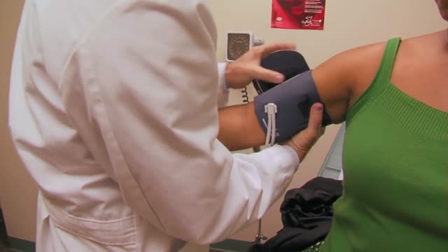
each type of heart problem requires different treatment but may share similar warning signs. It is important to see your doctor so that you can receive a correct diagnosis and prompt treatment. Learn to recognize the symptoms that may signal heart disease. Call your doctor if you begin to have new symptoms or if they become more frequent or severe. Symptoms of Coronary Artery Disease The most common symptom of coronary artery disease is angina, or chest pain. Angina can be described as a discomfort, heaviness, pressure, aching, burning, fullness, squeezing, or painful feeling in your chest. It can be mistaken for indigestion or heartburn. Angina may also be felt in the shoulders, arms, neck, throat, jaw, or back. Other symptoms of coronary artery disease include: Shortness of breath Palpitations (irregular heart beats, or a "flip-flop" feeling in your chest) A faster heartbeat Weakness or dizziness Nausea Sweating

Eczema, or atopic dermatitis, is a rash that primarily occurs in people with asthma or allergies. The rash is often reddish and itchy with a scaly texture. Psoriasis is a common skin condition that can cause a scaly, itchy, red rash to form along the scalp, elbows, and joints.Apr 13, 2016
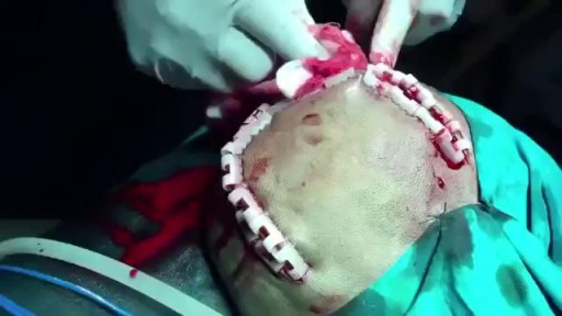
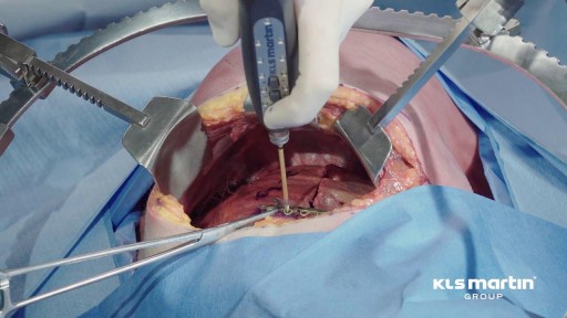
Epidural hematoma (EDH) is a traumatic accumulation of blood between the inner table of the skull and the stripped-off dural membrane. EDH results from traumatic head injury, usually with an associated skull fracture and arterial laceration.The inciting event often is a focused blow to the head, such as that produced by a hammer or baseball bat. In 85-95% of patients, this type of trauma results in an overlying fracture of the skull. Blood vessels in close proximity to the fracture are the sources of the hemorrhage in the formation of an epidural hematoma. Because the underlying brain has usually been minimally injured, prognosis is excellent if treated aggressively. Outcome from surgical decompression and repair is related directly to patient's preoperative neurologic condition. [1]

Bunions can be very painful. ... Bunion removal is a surgical procedure that corrects a deformed area of the foot near the big toe. Bunion removal is sometimes called a bunionectomy, bunion surgery, or hallux valgus correction. Hallux valgus is a Latin phrase that means “foot deformity
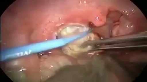
A tonsillolith lodged in the tonsillar crypt. Specialty. Otorhinolaryngology. Tonsilloliths, also known as tonsil stones, are clusters of calcified material that form in the tonsillar crypts, the crevices of the tonsils. While they occur most commonly in the palatine tonsils, they may also occur in the lingual tonsils.
A fractured rib is usually a result of a fall or accident. Prolonged coughing and sports with repetitive movement, such as golf, also can cause a rib fracture. Symptoms include pain when taking a deep breath, pressing on the injured area, or bending or twisting the body. In most cases, fractured ribs usually heal on their own in one or two months. Pain relievers can make it easier to breathe deeply.
Learn about the exciting results Baptist Healthcare System is experiencing with McKesson enterprise intelligence solutions in this video testimonial.
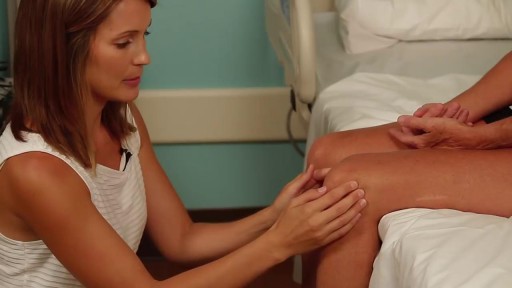
Detailed examination of the joints is usually not included in the routine medical examination. However, joint related complaints are rather common, and understanding anatomy and physiology of both normal function and pathologic conditions is critically important when evaluating the symptomatic patient. By gaining an appreciation for the basic structures and functioning of the joint, you'll be able to "logic" your way thru the exam, even if you can't remember the eponym attached to each specific test!
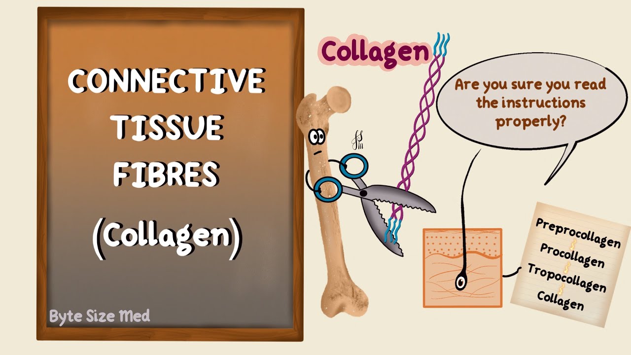
✨This video is on the protein fibres of connective tissue, the types, structure and synthesis of collagen and elastin. I hope it helps! ☀️
🌟What's in this video?
0:00 - Intro
0:07 - Connective Tissue Recap
0:39 - Connective Tissue Fibres
1:22 - Collagen
1:46 - Types of Collagen
3:40 - Structure of Collagen
4:40 - Collagen Synthesis
8:50 - Elastin
✨ Other videos you may need:
🔅 Connective Tissue : https://youtu.be/xw_ALdt5n-A
🔅 Cartilage : https://youtu.be/4inWF4H6pKE
🔅Epithelial Tissue: https://youtu.be/Gw5fC0zXaeU
🔅Structure of Blood Vessels: https://youtu.be/BAo2UqqyL3g
🔅Histology: https://www.youtube.com/playli....st?list=PL1rG930trF2
💫 For more videos like this, subscribe to my channel!
Byte Size Med: https://youtube.com/channel/UC....ZghvlgylH3r_CWfA18eF
📚Factual References & for Further Reading:
- DiFiore's Atlas of Histology
- Junqueira's Basic Histology
- Harper's Biochemistry
- Gartner's Concise Histology
- Openstax Anatomy and Physiology
https://openstax.org/details/b....ooks/anatomy-and-phy
- Openstax Biology
https://openstax.org/details/books/biology-2e
(The last two are links to open-source references. They are NOT affiliate links)
🌤 Note:
These are just a collection of my notes. So use them the way you would use borrowed notes from a friend. 📝
The images in this video are hand-drawn for illustration and explanation only.✍️ Hence, they may not be anatomically accurate. I am just one person making these videos. If there are any errors, that is unintentional. I try super hard to avoid them. Please let me know if you find any, so it gets clarified for other viewers. Science constantly evolves and changes. New discoveries are made everyday. So some of the information in these videos may become outdated. If you notice that, please let me know so I can update them.
⚡️Disclaimer:
These videos are NOT a substitute for a medical textbook. Textbooks are written by experts (which I do not claim to be), edited, proofread and referenced. Please use them.
The information has been sourced from multiple references as mentioned above. I draw all the pictures myself. But if I have inadvertently infringed on any copyright, that is completely unintentional. I only make these videos to impart education. If I have accidentally violated copyright in any way, do let me know so I can make the necessary changes or give credit to anyone who is owed the same.
These videos are NOT intended for patient education. They are NOT a substitute for diagnosis and treatment by a licensed medical professional. Always seek the advice of a qualified health care provider for any questions you may have regarding any medical condition, so that they can address your individual needs.
🔅They are ONLY meant to help students of medicine and health sciences with studying, and should be used for just that purpose and absolutely nothing else.
Byte Size Med. All Rights Reserved.

Genital warts are soft growths that appear on the genitals. Genital warts are a sexually transmitted infection (STI) caused by certain strains of the human papillomavirus (HPV). These skin growths can cause pain, discomfort, and itching. They are especially dangerous for women because some types of HPV can also cause cancer of the cervix and vulva.

Acute kidney failure occurs when your kidneys suddenly become unable to filter waste products from your blood. When your kidneys lose their filtering ability, dangerous levels of wastes may accumulate, and your blood's chemical makeup may get out of balance. Acute kidney failure — also called acute renal failure or acute kidney injury — develops rapidly over a few hours or a few days. Acute kidney failure is most common in people who are already hospitalized, particularly in critically ill people who need intensive care. Acute kidney failure can be fatal and requires intensive treatment. However, acute kidney failure may be reversible. If you're otherwise in good health, you may recover normal or nearly normal kidney function.

Learn about the structural unit of compact bone (the osteon) and it's four basic parts: central canal, lamellae, lacunae, and canaliculi

Mohs Surgeon Dr. Leslie Christenson shows entire Mohs Surgery from start to finish. This is the full procedure and includes the entire surgery. Dr. Christenson talks about the procedure as she removes the skin cancer.
Learn more about Dr. Christenson: https://www.mcfarlandclinic.co....m/doctors/leslie-chr
Learn more about Mohs Surgery: https://www.mcfarlandclinic.co....m/doctors/specialtie
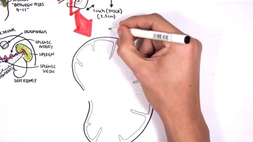
The spleen is an organ in the upper far left part of the abdomen, to the left of the stomach. The spleen varies in size and shape between people, but it’s commonly fist-shaped, purple, and about 4 inches long. Because the spleen is protected by the rib cage, you can’t easily feel it unless it’s abnormally enlarged. The spleen plays multiple supporting roles in the body. It acts as a filter for blood as part of the immune system. Old red blood cells are recycled in the spleen, and platelets and white blood cells are stored there. The spleen also helps fight certain kinds of bacteria that cause pneumonia and meningitis.

When United Airlines decides their employees flying to Kentucky is more important than a doctor or any passenger who paid for their ticket it is time to STOP FLYING UNITED!!! Here are United employees dragging the man off the plane like a criminal.
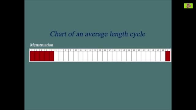
Our calculator can help you discover the most fertile days of your menstrual cycle or your “Estimated Fertility Window” based on information you provide.
A UK patient praises Dr.Venkatachalam for her Hip resurfacing surgery. She was able to get full movements after surgery which wouldn't have been possible with a hip replacement
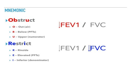
Obstructive lung diseases include conditions that make it hard to exhale all the air in the lungs. People with restrictive lung disease have difficulty fully expanding their lungs with air. Obstructive and restrictive lung disease share the same main symptom: shortness of breath with exertion.
Tudo Sobre Diabetes, Diabetes Tem Cura, O Que é Diabetes Tipo 2, Plantas Que Curam Diabetes
http://tudo-sobre-diabetes.good-info.co
Cura Naturalmente a Diabetes Tipo 2
A diabetes tipo II se tornou uma das doenças mais comuns nos tempos modernos. A boa notícia é que em pouco menos de um mês, seguindo um plano de alimentação e vida saudável, é possível equilibrar seu nível de açúcar no sangue e prevenir as terríveis consequências que esta doença tem.
A seguir, você encontrará este plano para nivelar o açúcar no sangue e dizer adeus para a diabetes.
Restrinja o consumo de todo o tipo de bebidas.
Realize atividade física de baixo impacto todo o dia, por um mínimo de meia hora.
Elimine por completo de suas refeições, todos os alimentos que contenham farinha branca.
Inclua em sua alimentação habitual, ácidos gordos essenciais (especialmente ácidos ômega 3), inclua também o consumo de frutas secas.
único Sistema Eficiente, Fácil E Natural Para Eliminar Para Sempre O Diabetes. Um Sistema Cientificamente Comprovado
Clique No Link Abaixo Para Verificá-la
http://tudo-sobre-diabetes.good-info.co
Assine O Nosso Canal
https://www.youtube.com/user/dicasdesaude11
https://www.youtube.com/watch?v=61MN7xSR9yA
Tudo Sobre Diabetes, Diabetes Tem Cura, O Que é Diabetes Tipo 2, Plantas Que Curam Diabetes,
diabetes gestacional,
diabetes mellitus tipo 2,
diabetes dieta,
sintomas de diabete,
diabetes tipo 1 e 2,
medicamentos para diabetes,
diabete sintomas,
causas da diabetes,
como evitar diabetes,
sintomas da diabetes tipo 2,
tratamento da diabetes,
o que diabetes,
os sintomas da diabete
