- Physical Examination
- Surgical Examination
- Ophthalmology
- Clinical Skills
- Orthopedics
- Surgery Videos
- Laparoscopy
- Pediatrics
- Funny Videos
- Cardiothoracic Surgery
- Nursing Videos
- Plastic Surgery
- Otorhinolaryngology
- Histology and Histopathology
- Neurosurgery
- Dermatology
- Pediatric Surgery
- Urology
- Dentistry
- Oncology and Cancers
- Anatomy Videos
- Health and Fitness
- Radiology
- Anaesthesia
- Physical Therapy
- Pharmacology
- Interventional Radiology
- Cardiology
- Endocrinology
- Gynecology
- Emergency Medicine
- Psychiatry and Psychology
- Childbirth Videos
- General Medical Videos
- Nephrology
- Physiology
- Diet and Food Health
- Diabetes Mellitus
- Neurology
- Women Health
- Osteoporosis
- Gastroenterology
- Pulmonology
- Hematology
- Rheumatology
- Toxicology
- Nuclear Medicine
- Infectious Diseases
- Vascular Disease
- Reproductive Health
- Burns and Wound Healing
- Other
Top videos

First Aid for a suspected Fracture
Multiple sclerosis (MS) involves an immune-mediated process in which an abnormal response of the body’s immune system is directed against the central nervous system (CNS). The CNS is made up of the brain, spinal cord and optic nerves. Within the CNS, the immune system causes inflammation that damages myelin — the fatty substance that surrounds and insulates the nerve fibers — as well as the nerve fibers themselves, and the specialized cells that make myelin. When myelin or nerve fibers are damaged or destroyed in MS, messages within the CNS are altered or stopped completely. Damage to areas of the CNS may produce a variety of neurological symptoms that will vary among people with MS in type and severity The damaged areas develop scar tissue which gives the disease its name – multiple areas of scarring or multiple sclerosis. The cause of MS is not known, but it is believed to involve genetic susceptibility, abnormalities in the immune system and environmental factors that combine to trigger the disease. People with MS typically experience one of four disease courses. There are over a dozen treatments to help modify the MS disease process.

This animated video explains what is meant by astigmatism, which is a very common problem with the eyes.
Massive PE causing hemodynamic instability (shock and/or low blood pressure, defined as a systolic blood pressure <90 mmHg or a pressure drop of 40 mmHg for >15 min if not caused by new-onset arrhythmia, hypovolemia or sepsis) is an indication for thrombolysis, the enzymatic destruction of the clot with medication.

The condition is caused by a blockage in the lymphatic system, part of the immune and circulatory systems. Lymphedema is most commonly caused by lymph node removal or damage due to cancer treatment. The main symptom is swelling in an arm or leg that may be accompanied by pain or discomfort. Exercise, wrapping, massage, and compression can help.
Pharyngitis is caused by swelling in the back of the throat (pharynx) between the tonsils and the voice box (larynx). Most sore throats are caused by colds, the flu, coxsackie virus or mono (mononucleosis). Bacteria that can cause pharyngitis in some cases: Strep throat is caused by group A streptococcus.
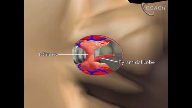
Minimally invasive open thyroidectomy (MIT) is similar to conventional thyroidectomy in its surgical approach. The major difference is the length of the neck incision. A smaller incision improves cosmesis and reduces discomfort. Typically, a skin incision less than 6 cm is considered minimally invasive. The remainder of the procedure is exactly the same as is used in conventional thyroidectomy. Adaptations to this technique include transection rather than lateral retraction of the strap muscles (the Sofferman technique). [1]

Reactive arthritis is a painful form of inflammatory arthritis (joint disease due to inflammation). It occurs in reaction to an infection by certain bacteria. Most often, these bacteria are in the genitals (Chlamydia trachomatis) or the bowel (Campylobacter, Salmonella, Shigella and Yersinia). Chlamydia most often transmits by sex. It often has no symptoms, but can cause a pus-like or watery discharge from the genitals. The bowel bacteria can cause diarrhea. If you develop arthritis within one month of diarrhea or a genital infection – especially with a discharge – see a health care provider. You may have reactive arthritis. - See more at: https://www.rheumatology.org/i-am-a/patient-caregiver/diseases-conditions/reactive-arthritis#sthash.PukaaQhj.dpuf

wide resection of giant cell tumor ,then strut grafting using free fibula graft,knowles pinning of the graft.

Glass ampules are often used to store medication, and as a nurse, you'll need to know how to use them.
In this video, I demonstrate how to clean an ampule using alcohol prep, how to open (or break) an ampule, as well as how to dispose of the ampule.
In addition, I show how to use an ample filter straw while drawing up (withdrawing) medication, how to use the syringe, and how to remove the air bubbles in the syringe.
This is another video in our series on clinical nursing skills.
Notes: https://www.registerednursern.....com/how-to-withdraw-
Website: https://www.registerednursern.com/
More Videos: https://www.youtube.com/watch?v=R2XMro13dD0&list=UUPyMN8DzkFl2__xnTEiGZ1w
Nursing Gear: https://teespring.com/stores/registerednursern
Instagram: https://www.instagram.com/registerednursern_com/
Facebook: https://www.facebook.com/RegisteredNurseRNs
Twitter: https://twitter.com/NursesRN
Popular Playlists:
NCLEX Reviews: https://www.youtube.com/playli....st?list=PLQrdx7rRsKf
Fluid & Electrolytes: https://www.youtube.com/playli....st?list=PLQrdx7rRsKf
Nursing Skills: https://www.youtube.com/playli....st?list=PLQrdx7rRsKf
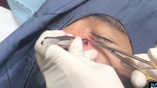
Nose Cyst Extraction
Female Catheter Insertion

Circumcision Video 3D
Testicular Self Exam

Medical students at Johns Hopkins University are getting a real-life birthing experience when a robot goes into labor. Kasey-Dee Gardner reports.
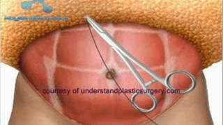
http://www.bodysculptor.com. Dr. Otto Placik, Board Certified Chicago based plastic surgeon demonstrates the results of a muscle separation(rectus diastasis) repair using 3 dimesional CAT scan and photographic images
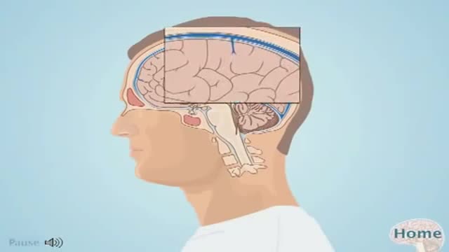
An intracranial hematoma occurs when a blood vessel ruptures within your brain or between your skull and your brain. The collection of blood (hematoma) compresses your brain tissue. An intracranial hematoma may occur because the fluid that surrounds your brain can't absorb the force of a sudden blow or a quick stop. Then your brain may slide forcefully against the inner wall of your skull and become bruised. Although some head injuries — such as one that causes only a brief lapse of consciousness (concussion) — can be minor, an intracranial hematoma is potentially life-threatening and often requires immediate treatment. An intracranial hematoma often, but not always, requires surgery to remove the blood.
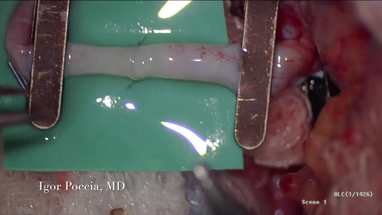
A circulatory anastomosis is a connection (an anastomosis) between two blood vessels, such as between arteries (arterio-arterial anastomosis), between veins (veno-venous anastomosis) or between an artery and a vein (arterio-venous anastomosis). An end artery (or terminal artery) is an artery that is the only supply of oxygenated blood to a portion of tissue. Examples of an end artery include the splenic artery that supplies the spleen and the renal artery that supplies the kidneys.

As one of the first pediatric centers in the United States to use a new state-of-the-art MRI machine designed especially for kids, Children's Hospital of Michigan continues to deliver world-class, patient-friendly health care. ~ Detroit Medical Center

Prompt treatment to break up the clot greatly reduces the risk of death. This can be done with blood thinners and drugs or procedures. Compression stockings and physical activity can help prevent clots from forming in the first place.
