- Physical Examination
- Surgical Examination
- Ophthalmology
- Clinical Skills
- Orthopedics
- Surgery Videos
- Laparoscopy
- Pediatrics
- Funny Videos
- Cardiothoracic Surgery
- Nursing Videos
- Plastic Surgery
- Otorhinolaryngology
- Histology and Histopathology
- Neurosurgery
- Dermatology
- Pediatric Surgery
- Urology
- Dentistry
- Oncology and Cancers
- Anatomy Videos
- Health and Fitness
- Radiology
- Anaesthesia
- Physical Therapy
- Pharmacology
- Interventional Radiology
- Cardiology
- Endocrinology
- Gynecology
- Emergency Medicine
- Psychiatry and Psychology
- Childbirth Videos
- General Medical Videos
- Nephrology
- Physiology
- Diet and Food Health
- Diabetes Mellitus
- Neurology
- Women Health
- Osteoporosis
- Gastroenterology
- Pulmonology
- Hematology
- Rheumatology
- Toxicology
- Nuclear Medicine
- Infectious Diseases
- Vascular Disease
- Reproductive Health
- Burns and Wound Healing
- Other
Top videos

Lasic in 10 years old girl for Myopia
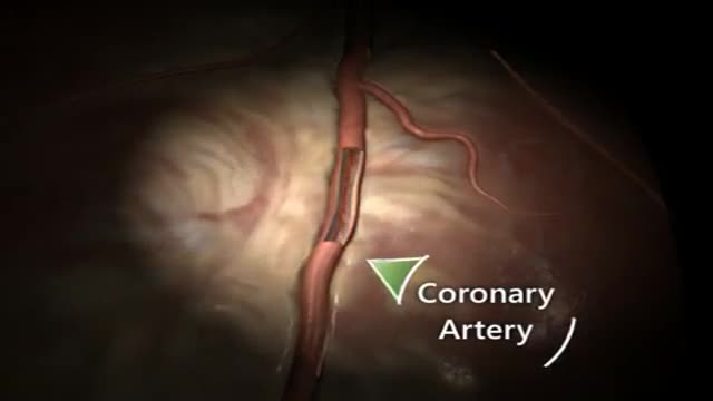
A heart attack occurs when blood flow to a part of your heart is blocked for a long enough time that part of the heart muscle is damaged or dies. The medical term for this is myocardial infarction.
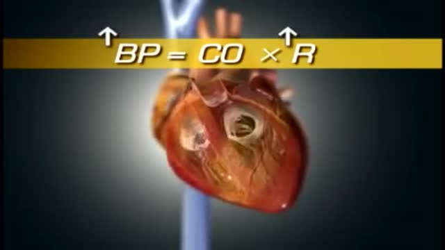
High blood pressure is a common condition in which the long-term force of the blood against your artery walls is high enough that it may eventually cause health problems, such as heart disease. Blood pressure is determined both by the amount of blood your heart pumps and the amount of resistance to blood flow in your arteries. The more blood your heart pumps and the narrower your arteries, the higher your blood pressure. You can have high blood pressure (hypertension) for years without any symptoms. Even without symptoms, damage to blood vessels and your heart continues and can be detected. Uncontrolled high blood pressure increases your risk of serious health problems, including heart attack and stroke. High blood pressure generally develops over many years, and it affects nearly everyone eventually. Fortunately, high blood pressure can be easily detected. And once you know you have high blood pressure, you can work with your doctor to control it.

Sialadenitis is an infection of the salivary glands. It is usually caused by a virus or bacteria . The parotid (in front of the ear) and submandibular (under the chin) glands are most commonly affected. Sialadenitis may be associated with pain, tenderness, redness, and gradual, localized swelling of the affected area.
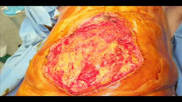
Laparostomy is a surgical condition in which the abdomen is left open to contrast a condition named Abdominal Compartment Syndrome
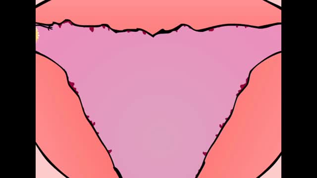
The menstrual cycle is the regular natural change that occurs in the female reproductive system like the uterus and ovaries that make pregnancy possible. The cycle is required for the production of ovocytes, and for the preparation of the uterus for pregnancy.
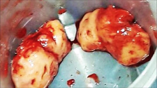
wide resection of giant cell tumor ,then strut grafting using free fibula graft,knowles pinning of the graft.
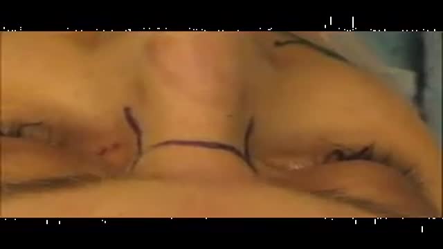
This procedure describes one of the most versatile approaches to the anterior skull base for large tumors of the sinonasal cavity. It may be used with or without a craniofacial resection. The benefits of this approach are: wide access around the tumor; good postoperative cosmesis; & decreased operative & postoperative morbidity. We have used this approach for many bilateral tumors of the nasal & sinus cavities that approach &/or invade the skull base & brain. This video show the resection of a large esthesioneuroblastoma.
Circumcision Video 3D

Open heart (coronary artery bypass, or CABG) surgery is performed in order to reroute, or "bypass," blood around blocked arteries, thereby improving the supply of oxygen-rich blood to the heart. Surgeons usually use an artery from the chest wall to construct the "detour" around the blocked part of the artery. Veins from the legs are also used.
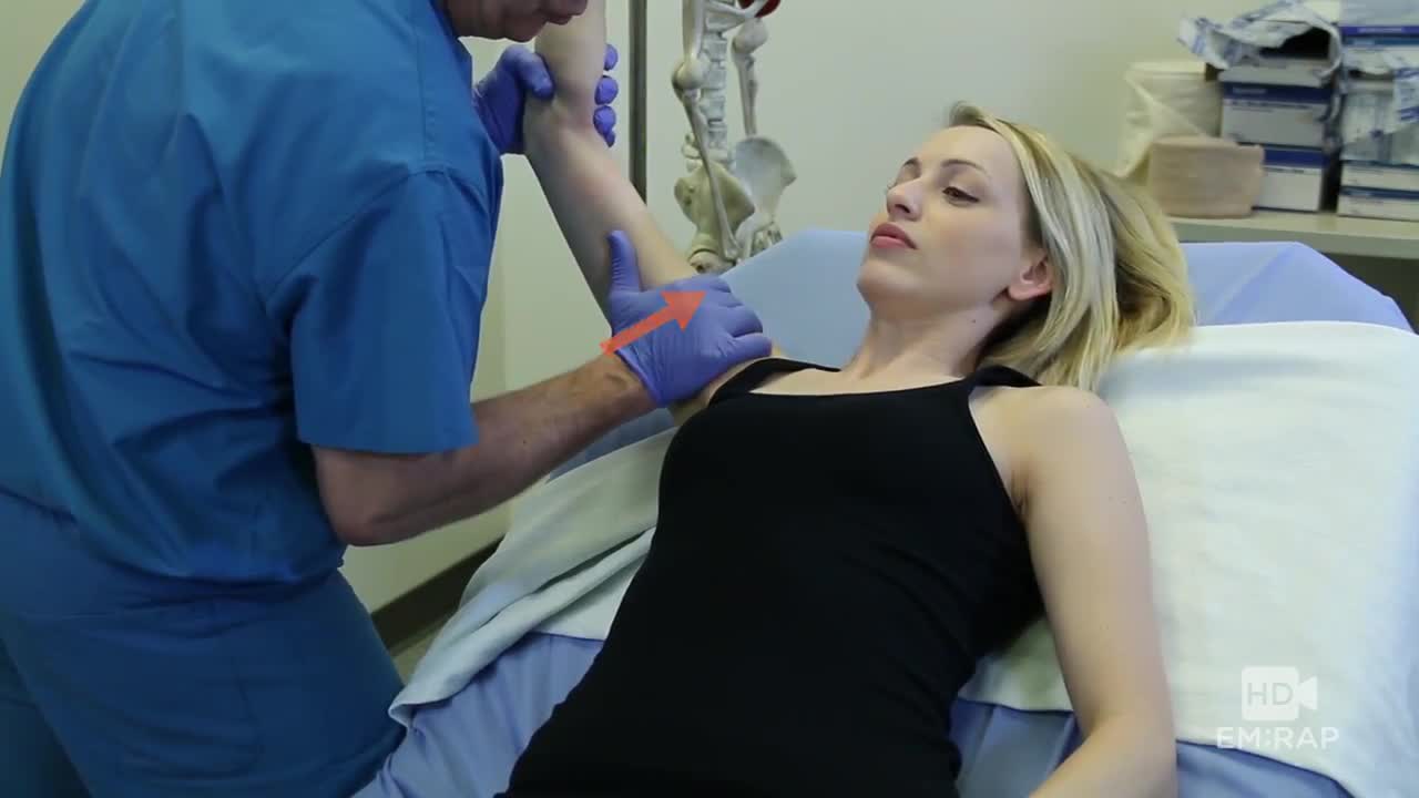
A technique for reducing an inferior shoulder dislocation. watch to learn more
Temporal Arteritis: what is it? how to treat it? follow up?
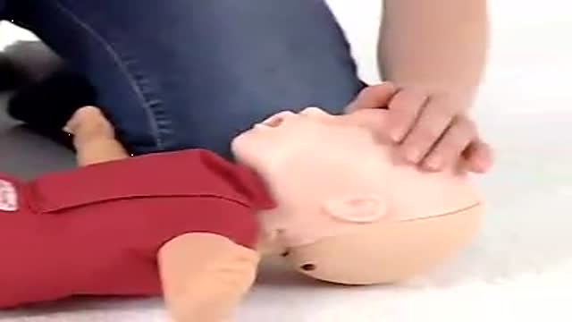
Baby CPR
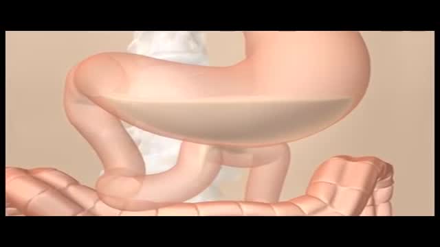
Constipation is a common problem. It means either going to the toilet less often than usual to empty the bowels, or passing hard or painful stools (faeces). Constipation may be caused by not eating enough fibre, or not drinking enough fluids. It can also be a side-effect of certain medicines, or related to an underlying medical condition. In many cases, the cause is not clear. Laxatives are a group of medicines that can treat constipation. Ideally, laxatives should only be used for short periods of time until symptoms ease. Note: there is a separate leaflet on constipation in children. What is constipation? Constipation is common. If you are constipated it causes one or more of the following: Stools (faeces) become hard and difficult or painful to pass. The time between toilet trips increases compared with your usual pattern. (Note: there is a large range of normal bowel habit. Some people normally go to the toilet to pass stools 2-3 times per day. For others, 2-3 times per week is normal. It is a change from your usual pattern that may mean that you are constipated.) Sometimes, crampy pains occur in the lower part of your tummy (abdomen) You may also feel bloated and feel sick if you have severe constipation. What are the causes of constipation? Known causes include the following: Not eating enough fibre (roughage) is a common cause. The average person in the UK eats about 12 g of fibre each day. But, 18 g per day is recommended by the British Nutrition Foundation. Fibre is the part of plant food that is not digested. It remains in your gut. It adds bulk to the stools (faeces) and helps your bowels to work well. Foods high in fibre include fruit, vegetables, cereals and wholemeal bread. Not drinking much may make constipation worse. Stools are usually soft and easily passed if you eat enough fibre and drink enough fluid. However, some people need more fibre and/or fluid than others in order to avoid constipation. Some special slimming diets are low in fibre and may cause constipation. Some medicines can cause constipation as a side-effect. Examples are painkillers (particularly those with codeine, such as co-codamol, or very strong painkillers, such as morphine), some antacids, some antidepressants (including amitriptyline) and iron tablets; however, there are many others. See the list of possible side-effects on the leaflet that comes with any medicine that you may be taking. Tell a doctor if you suspect a medicine is making you constipated. A change of medication may be possible. Various medical conditions can cause constipation. For example, an underactive thyroid gland, irritable bowel syndrome, some gut disorders and conditions that cause poor mobility, particularly in the elderly. Pregnancy. About 1 in 5 pregnant women will become constipated. It is due to the hormonal changes of pregnancy that slow down the gut movements. In later pregnancy, it can simply be due to the baby taking up a lot of room in the tummy and the bowels being pushed to one side.
Pneumonia is an infection that inflames the air sacs in one or both lungs. The air sacs may fill with fluid or pus (purulent material), causing cough with phlegm or pus, fever, chills, and difficulty breathing. A variety of organisms, including bacteria, viruses and fungi, can cause pneumonia.
Digoxin is used to treat heart failure, usually along with other medications. It is also used to treat a certain type of irregular heartbeat (chronic atrial fibrillation). Treating heart failure may help maintain your ability to walk and exercise and may improve the strength of your heart. Treating an irregular heartbeat can decrease the risk for blood clots, an effect that may reduce your risk for a heart attack or stroke.
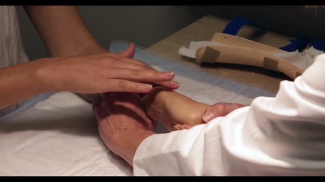
De Quervain's tenosynovitis (dih-kwer-VAINS ten-oh-sine-oh-VIE-tis) is a painful condition affecting the tendons on the thumb side of your wrist. If you have de Quervain's tenosynovitis, it will probably hurt when you turn your wrist, grasp anything or make a fist. Although the exact cause of de Quervain's tenosynovitis isn't known, any activity that relies on repetitive hand or wrist movement — such as working in the garden, playing golf or racket sports, or lifting your baby — can make it worse. Symptoms ShareTweet June 13, 2015 References Products and Services Mayo Clinic Sports Medicine Newsletter: Mayo Clinic Health Letter See also Prednisone risks, benefits Prednisone withdrawal: Why taper down slowly? Integrative approaches to treating pain Lifestyle strategies for pain management Nutrition and pain Pain rehabilitation Self-care approaches to treating pain Show more Advertisement Mayo Clinic does not endorse companies or products. Advertising revenue supports our not-for-profit mission. Advertising & Sponsorship PolicyOpportunitiesAd Choices Mayo Clinic Store Check out these best-sellers and special offers on books and newsletters from Mayo Clinic. NEW! – The Mayo Clinic Diet, Second Edition Healthy Heart for Life! Mayo Clinic on Better Hearing and Balance Treatment Strategies for Arthritis The Mayo Clinic Diet Online
Tonsil stones are hard yellow or white formations that are located on or within the tonsils. It’s common for people with tonsil stones to not even realize they have them. Tonsil stones aren’t always easily visible and they can range from rice- to pea-sized. Tonsil stones rarely cause larger health complications. However, sometimes they can grow into larger tonsilloliths which can cause your tonsils to swell
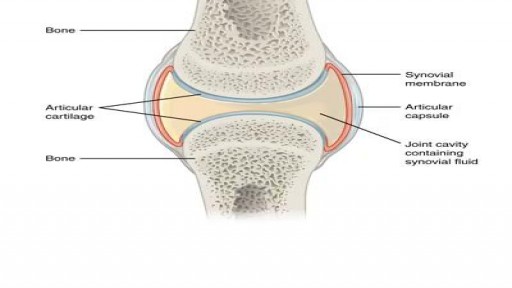
What happens when you crack your joints?

Restrictive cardiomyopathy (RCM) is a rare form of heart muscle disease that is characterized by restrictive filling of the ventricles. In this disease the contractile function (squeeze) of the heart and wall thicknesses are usually normal, but the relaxation or filling phase of the heart is very abnormal.
