- Physical Examination
- Surgical Examination
- Ophthalmology
- Clinical Skills
- Orthopedics
- Surgery Videos
- Laparoscopy
- Pediatrics
- Funny Videos
- Cardiothoracic Surgery
- Nursing Videos
- Plastic Surgery
- Otorhinolaryngology
- Histology and Histopathology
- Neurosurgery
- Dermatology
- Pediatric Surgery
- Urology
- Dentistry
- Oncology and Cancers
- Anatomy Videos
- Health and Fitness
- Radiology
- Anaesthesia
- Physical Therapy
- Pharmacology
- Interventional Radiology
- Cardiology
- Endocrinology
- Gynecology
- Emergency Medicine
- Psychiatry and Psychology
- Childbirth Videos
- General Medical Videos
- Nephrology
- Physiology
- Diet and Food Health
- Diabetes Mellitus
- Neurology
- Women Health
- Osteoporosis
- Gastroenterology
- Pulmonology
- Hematology
- Rheumatology
- Toxicology
- Nuclear Medicine
- Infectious Diseases
- Vascular Disease
- Reproductive Health
- Burns and Wound Healing
- Other
Top videos
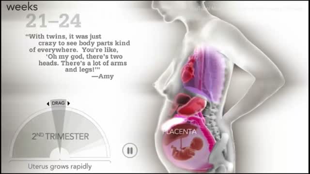
How a woman's body changes during Pregnancy
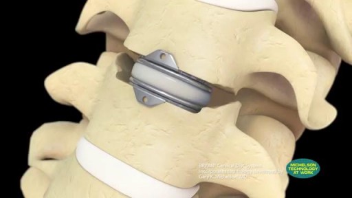
Cervical artificial disc replacement using the BRYAN Disc, a trusted product of Medtronic. Spine care offices are located in New Jersey, New York, and Florida. Are you experiencing neck pain? Do you think you may have a herniate disc? Learn more about our doctors' 97% success rate for performing BRYAN disc replacement:
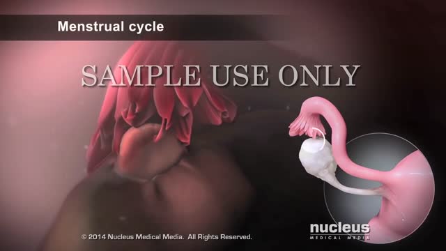
Intrauterine insemination (IUI) is a fertility treatment that involves placing sperm inside a woman's uterus to facilitate fertilization. The goal of IUI is to increase the number of sperm that reach the fallopian tubes and subsequently increase the chance of fertilization.
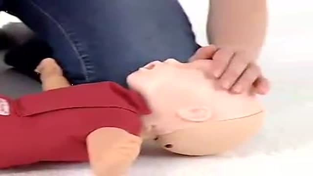
Baby CPR
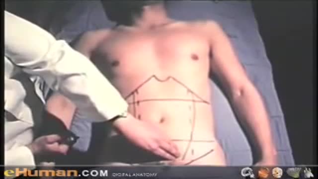
Clinical Anatomy Lecture Illustrate The Anatomy Of The Abdominal Wall
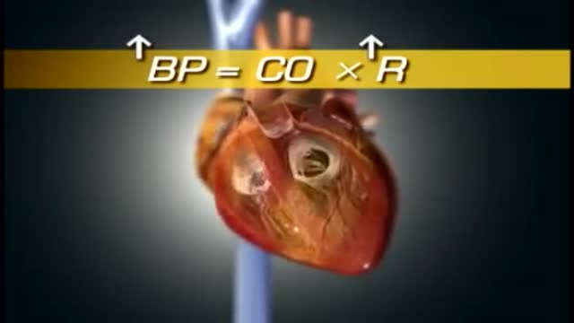
High blood pressure is a common condition in which the long-term force of the blood against your artery walls is high enough that it may eventually cause health problems, such as heart disease. Blood pressure is determined both by the amount of blood your heart pumps and the amount of resistance to blood flow in your arteries. The more blood your heart pumps and the narrower your arteries, the higher your blood pressure. You can have high blood pressure (hypertension) for years without any symptoms. Even without symptoms, damage to blood vessels and your heart continues and can be detected. Uncontrolled high blood pressure increases your risk of serious health problems, including heart attack and stroke. High blood pressure generally develops over many years, and it affects nearly everyone eventually. Fortunately, high blood pressure can be easily detected. And once you know you have high blood pressure, you can work with your doctor to control it.

Ellis demonstrates how to perform a central venous catheter (CVC) dressing change. Please note, you would want to perform hand hygiene after removing the clean gloves before donning the sterile gloves.
Our Critical Nursing Skills video tutorial series is taught by Ellis Parker MSN, RN-BC, CNE, CHS and intended to help RN and PN nursing students study for your nursing school exams, including the ATI, HESI and NCLEX.
#NCLEX #CVC #ClinicalSkills #HESI #Kaplan #ATI #NursingSchool #NursingStudent #Nurse #RN #PN #Education #LVN #LPN #nurseeducator
00:00 CVC Dressing Change
00:26 Preparing patient for CVC Change
00:54 Removing previous dressing
1:56 Removing gloves CVC Change
2:06 Opening CVC Change Kit
2:05 Sterile gloving CVC Change
3:22 Exploring CVC Change kit
3:36 Snap the scrubber CVC Change
3:49 Scrub site CVC Change
4:18 Applying antimicrobial patch CVC Change
4:41 Applying transparent dressing CVC Change
:rocket: Introducing FLASHABLES: Our signature flashcard content in an on-the-go DIGITAL format with guided, personalized learning & progress tracking. :books::syringe:
:mega: Limited-Time Offer Alert: :mega:
:fire: Bundle Up & SAVE! :fire: Purchase any of our amazing flashcard sets, SAVE BIG on FLASHABLES :heavy_dollar_sign::sparkles:& Level Up your study game!
:star2: There’s more…EITHER get 1 MONTH of Membership :free: with any flashcard purchase OR get a WHOLE YEAR of Membership :free: when you grab the Comprehensive Collection or the Ultimate Nursing School Survival Kit! :tada::gift:
Conquer nursing school like a pro with FLASHABLES! :thermometer::pill:
Don’t miss out on this incredible offer - click the link below and start making your nursing school journey your own! :star2::books::mortar_board: https://bit.ly/Flashables
🚪 Access our Cram Courses, Quizzes and Videos all in one ad free space with Level Up RN Membership https://bit.ly/LevelUpRNMembership
Want more ways to MASTER Clinical Skills? Check out our flashcards & videos!
👇👇👇👇👇👇👇👇👇👇
👉 https://bit.ly/clinicalnursingskills 👈
☝️👆☝️👆☝️👆☝️👆☝️👆
This is your one-stop-shop for materials to help you LEARN & REVIEW so you can PASS Nursing School.
🤔🤔🤔 DO YOU WANT TO PASS your classes, proctored exams and the NCLEX? 🤔🤔🤔 Our resources are the best you can buy. They are built with a single goal: help you pass with no fluff. Everything you need, and nothing you don’t. Don’t take our word for it, though! Check out our hundreds of ⭐️⭐️⭐️⭐️⭐️ reviews from nurses who passed their exams and the NCLEX with Level Up RN.
🗂️ Our Ultimate Nursing School Survival kit is your number 1 resource to get through nursing school and to pass the NCLEX. Whether you're just starting school or you’re already prepping for the NCLEX, this bundle of flashcards is the best you can buy. It covers all the information you need to know to pass all your exams and it has FREE shipping!
➡️ https://bit.ly/TUNSSK ⬅️
L👀king for EVEN MORE resources to survive Nursing School? Make your Nursing School experience your own! Life’s difficult enough—learning shouldn’t be.
🪅 Games https://nursesquad.com
💻 Digital resources https://bit.ly/NursingStudyCourses
📅 Organizational tools https://bit.ly/OrganizingSchool
✨Want perks? Join our channel!
https://youtube.com/leveluprn/join
🏷 Head to https://leveluprn.com/specials for all our latest deals!🥳️
📧 LOOKING FOR FREE RESOURCES TO HELP WITH YOUR EXAMS? Get exclusive tips, latest video releases and more delivered to your email!
➡️ https://leveluprn.com/signup ⬅️
⚕ 👩 LEVEL UP NURSE SQUAD 👩⚕️
All of the nurses at Level Up RN are here to help! Cathy Parkes started helping her fellow classmates back when she was in nursing school, tutoring so they could pass their exams and graduate. After she got her BSN and started working as an RN at Scripps Encinitas Hospital, she started this YouTube channel to help nursing students around the world. Since then she has built a team of top-notch dedicated nurses and nurse educators who are focused on improving nursing education and supporting career advancement for nurses everywhere. With flashcards, videos, courses, organizational tools and more, we are singularly focused on helping students and nurses Level Up on their exams and nursing careers.
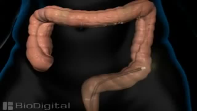
Colonoscopy is a test that allows your doctor to look at the inner lining of your large intestine (rectum and colon). He or she uses a thin, flexible tube called a colonoscope to look at the colon. A colonoscopy helps find ulcers, colon polyps, tumors, and areas of inflammation or bleeding.
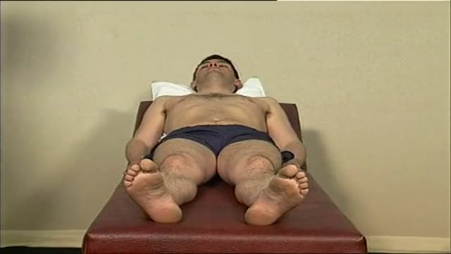
Hip Exam Video
One man is speaking out about the potential risks of laser eye surgery, after he says the procedure left his vision permanently impaired.
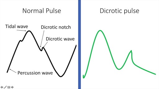
A detailed description of the Arterial Pulse including its waveform and pathological subtypes. Also discussed are the abnormal rates (tachycardia and bradycardia) and their causes, abnormal rhythm (including regularly regular and irregularly irregular pulses) and abnormal character (including pulses bisferiens, pulses parvus et tarsus, pulsus alternans, pulses paradoxus and others.) Description of pulse in various pathological states including Aortic stenosis and aortic regurgitation is also included. Finally there is also a description of the peripheral signs of aortic regurgitation.

Sialadenitis is an infection of the salivary glands. It is usually caused by a virus or bacteria . The parotid (in front of the ear) and submandibular (under the chin) glands are most commonly affected. Sialadenitis may be associated with pain, tenderness, redness, and gradual, localized swelling of the affected area.
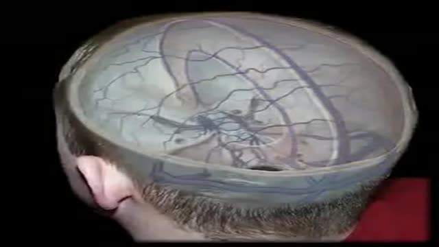
The dural venous sinuses are spaces between the endosteal and meningeal layers of the dura. They contain venous blood that originates for the most part from the brain or cranial cavity. The sinuses contain an endothelial lining that is continuous into the veins that are connected to them.
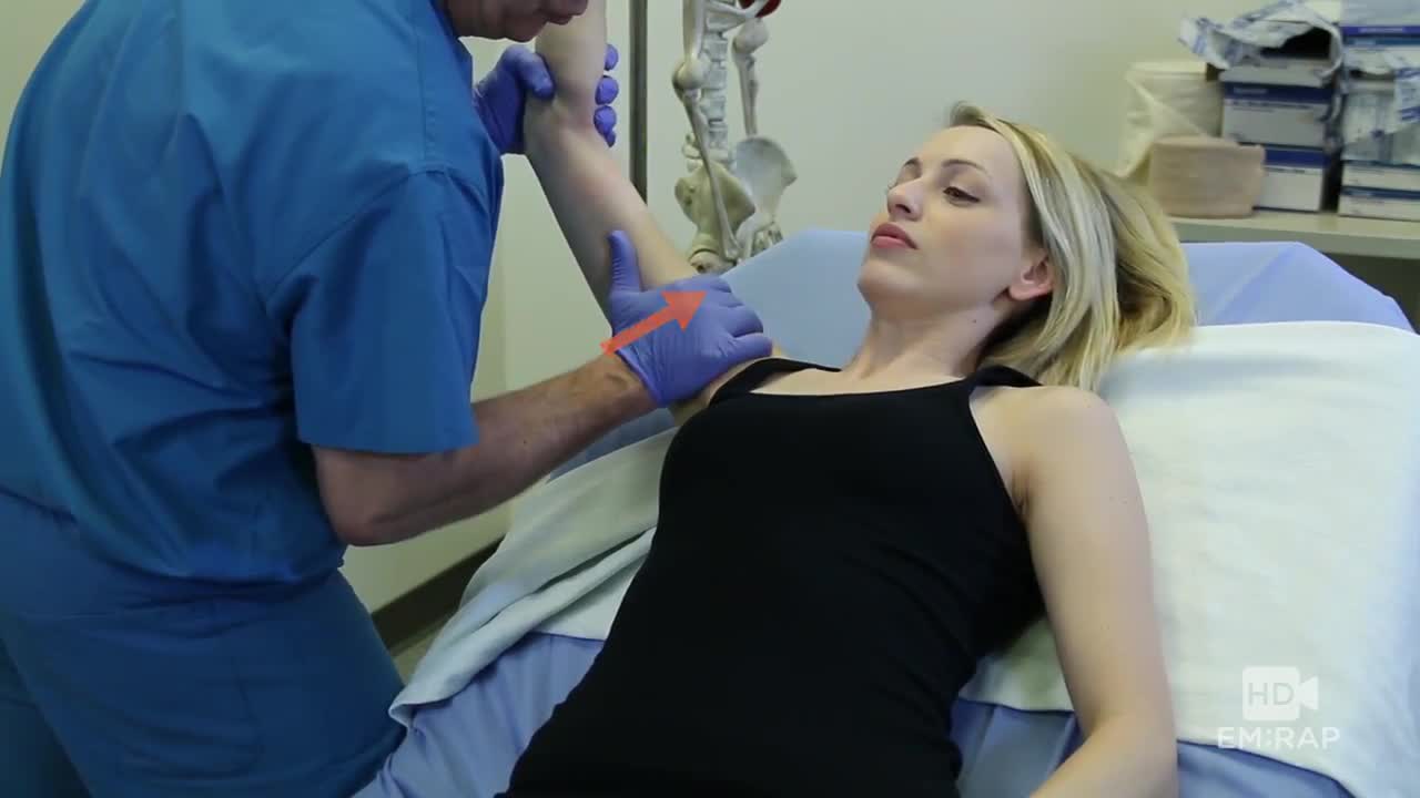
A technique for reducing an inferior shoulder dislocation. watch to learn more
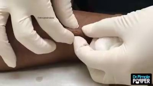
Epidermoid cysts, also called sebaceous, keratin, or epithelial cysts, are small, hard lumps that develop under the skin. These cysts are common. They grow slowly. They do not cause other symptoms and are nearly never cancerous. Epidermoid cysts are often found on the face, head, neck, back, or genitals
Clean hands can help prevent the spread of infectious diseases, such as flu. This podcast explains the proper way to wash your hands.
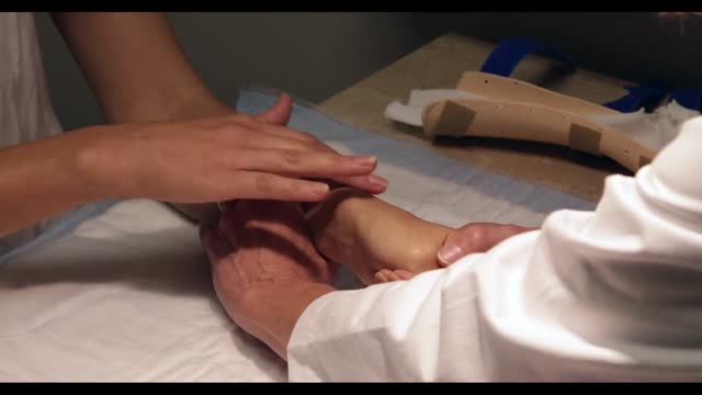
De Quervain's tenosynovitis (dih-kwer-VAINS ten-oh-sine-oh-VIE-tis) is a painful condition affecting the tendons on the thumb side of your wrist. If you have de Quervain's tenosynovitis, it will probably hurt when you turn your wrist, grasp anything or make a fist. Although the exact cause of de Quervain's tenosynovitis isn't known, any activity that relies on repetitive hand or wrist movement — such as working in the garden, playing golf or racket sports, or lifting your baby — can make it worse. Symptoms ShareTweet June 13, 2015 References Products and Services Mayo Clinic Sports Medicine Newsletter: Mayo Clinic Health Letter See also Prednisone risks, benefits Prednisone withdrawal: Why taper down slowly? Integrative approaches to treating pain Lifestyle strategies for pain management Nutrition and pain Pain rehabilitation Self-care approaches to treating pain Show more Advertisement Mayo Clinic does not endorse companies or products. Advertising revenue supports our not-for-profit mission. Advertising & Sponsorship PolicyOpportunitiesAd Choices Mayo Clinic Store Check out these best-sellers and special offers on books and newsletters from Mayo Clinic. NEW! – The Mayo Clinic Diet, Second Edition Healthy Heart for Life! Mayo Clinic on Better Hearing and Balance Treatment Strategies for Arthritis The Mayo Clinic Diet Online
Digoxin is used to treat heart failure, usually along with other medications. It is also used to treat a certain type of irregular heartbeat (chronic atrial fibrillation). Treating heart failure may help maintain your ability to walk and exercise and may improve the strength of your heart. Treating an irregular heartbeat can decrease the risk for blood clots, an effect that may reduce your risk for a heart attack or stroke.
A central venous catheter, also called a central line, is a long, thin, flexible tube used to give medicines, fluids, nutrients, or blood products over a long period of time, usually several weeks or more. A catheter is often inserted in the arm or chest through the skin into a large vein.
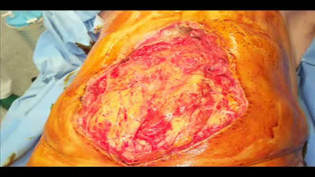
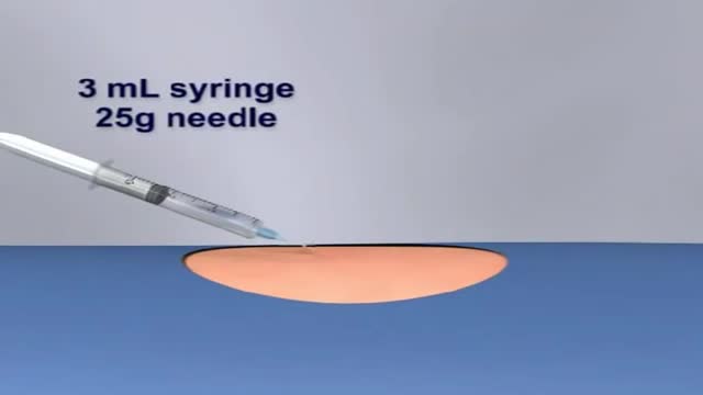
Laparostomy is a surgical condition in which the abdomen is left open to contrast a condition named Abdominal Compartment Syndrome
