- Physical Examination
- Surgical Examination
- Ophthalmology
- Clinical Skills
- Orthopedics
- Surgery Videos
- Laparoscopy
- Pediatrics
- Funny Videos
- Cardiothoracic Surgery
- Nursing Videos
- Plastic Surgery
- Otorhinolaryngology
- Histology and Histopathology
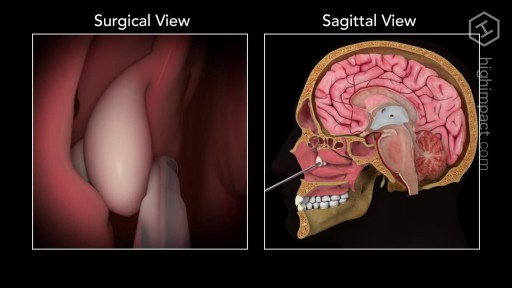
- Neurosurgery
- Dermatology
- Pediatric Surgery
- Urology
- Dentistry
- Oncology and Cancers
- Anatomy Videos
- Health and Fitness
- Radiology
- Anaesthesia
- Physical Therapy
- Pharmacology
- Interventional Radiology
- Cardiology
- Endocrinology
- Gynecology
- Emergency Medicine
- Psychiatry and Psychology
- Childbirth Videos
- General Medical Videos
- Nephrology
- Physiology
- Diet and Food Health
- Diabetes Mellitus
- Neurology
- Women Health
- Osteoporosis
- Gastroenterology
- Pulmonology
- Hematology
- Rheumatology
- Toxicology
- Nuclear Medicine
- Infectious Diseases
- Vascular Disease
- Reproductive Health
- Burns and Wound Healing
- Other
Top videos
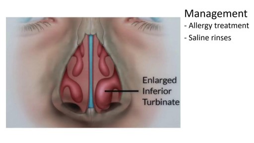
Nasal polyps are associated with inflammation of the lining of your nasal passages and sinuses that lasts more than 12 weeks (chronic rhinosinusitis, also known as chronic sinusitis). However, it's possible — and even somewhat more likely — to have chronic sinusitis without nasal polyps. Nasal polyps themselves are soft and lack sensation, so if they're small you may not be aware you have them. Multiple growths or a large polyp may block your nasal passages and sinuses.
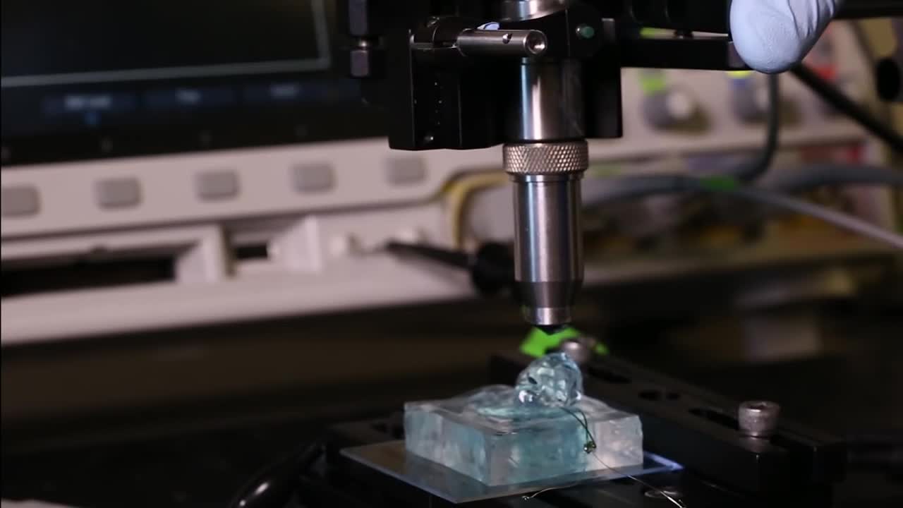
University of California, Berkeley engineers have built the first dust-sized, wireless sensors that can be implanted in the body, bringing closer the day when a Fitbit-like device could monitor internal nerves, muscles or organs in real time.
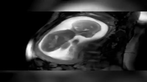
MRI scan of a 23-week-pregnancy

This video will cover, in detail, the motor, sensory, reflect components of a neurological examination.
This video is created for the UBC Medicine Neurology Clinical Skills curriculum as part of MEDD 419 FLEX projects.
Filmed, written, and directed by:
John Liu
Vincent Soh
Chris Calvin
Kashi (Siyoung) Lee
Kero (Yue) Yuen
Ge Shi
Doctor - Dr. Jason Valerio (Department of Neurology, UBC)
Supervised by:
Dr. Alex Henri-Bhargava (Department of Neurology, UBC)
Zac Rothman (UBC FOM Digital Solutions: Ed Tech)
Edited by:
Stephen Gillis
Produced by UBC FOM Digital Solutions EdTech team facilitates innovation by UBC Medicine learners and faculty.
Website: https://education.med.ubc.ca/
Subscribe: https://www.youtube.com/ubcmed....vid?sub_confirmation
UBCMLN Podcast Network: https://tinyurl.com/ubcmedicinelearningnetwork
----------------------------------------------------------------------------------------------------------------------------------------------------------
The Vancouver Fraser Medical Program and the Vancouver Academic Campus of the University of British Columbia are situated on the traditional territory of the Musqueam, Squamish and Tsleil-Waututh peoples.
The Southern Medical Program and the Okanagan Academic Campus of the University of British Columbia are situated on the territory of the Syilx Okanagan Nation.
The Northern Medical Program and the University of Northern BC are situated on the traditional territory of the Lheidli T’enneh, part of the Dakelh (Carrier) First Nations.
With respect the Lekwungen peoples on whose traditional territory the Island Medical Program and the University of Victoria stand and the Songhees, Esquimalt and WSÁNEĆ peoples whose historical relationships with the land continue to this day.
We acknowledge our traditional hosts and honour their welcome and graciousness to the students who seek knowledge here.
© UBC Faculty of Medicine
All rights reserved. Reproduction and distribution of this presentation without written permission from UBC Faculty of Medicine is strictly prohibited.
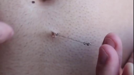
Longest Ingrown Hair Removal

Outpatient -- or same-day -- knee replacement surgery is more convenient than traditional knee replacement surgery and often can help you recover faster.
Outpatient -- or same-day -- knee replacement surgery is more convenient than traditional knee replacement surgery and often can help you recover faster. At Duke Ambulatory Surgery Center Arringdon, your knee replacement will be followed immediately by physical therapy to get you moving and start your recovery process right away. Our expert joint replacement team ensures your knee replacement surgery is safe and effective so you can return to the comfort of your home as soon as possible.

Histology lab video reviewing the structure and cells of thin skin, thick skin, and skin sensory structures on digital histology slides. This video is a part of our Histology Video Course (https://youtube.com/playlist?l....ist=PLnr1l7WuQdDynxT
All Histology Videos: https://youtube.com/playlist?l....ist=PLnr1l7WuQdDynxT
Thank you to our sponsor Doc2Doc Lending, the Personal Lending platform designed for Doctors, by Doctors. Check out https://doc2doclending.com/davinci to learn more today.
DaVinci Academy Merch - Coffee mugs, T-shirts, hoodies and more: https://my-store-d90f46.creator-spring.com
Additional YouTube Content
Biochemistry videos: https://youtube.com/playlist?l....ist=PLnr1l7WuQdDzCUC
Anatomy Videos: https://youtube.com/playlist?l....ist=PLnr1l7WuQdDz2dK
DaVinci Cases Videos: https://youtube.com/playlist?l....ist=PLnr1l7WuQdDyJUl
The DaVinci Hour Podcast: https://youtube.com/playlist?l....ist=PLnr1l7WuQdDwSm9
DaVinci Academy Website: https://www.dviacademy.com/

This video shows how to perform the McMurray test, one of the most commonly used clinical assessment tools to assess for meniscal injuries in the knee.
This video clip is part of the FIFA Diploma in Football Medicine and the FIFA Medical Network. To enrol or to find our more click on the following link http://www.fifamedicalnetwork.com
The Diploma is a free online course designed to help clinicians learn how to diagnose and manage common football-related injuries and illnesses. There are a total of 42 modules created by football medicine experts. Visit a single page, complete individual modules or finish the entire course.
The network provides the opportunity for clinicians around the world to meet and share ideas relating to football medicine. Ask about an interesting case, debate current practice and discuss treatment strategies. Create a profile and log on to interact with other health professionals from around the globe.
This is not medical advice. The content is intended as educational content for health care professionals and students. If you are a patient, seek care of a health care professional.
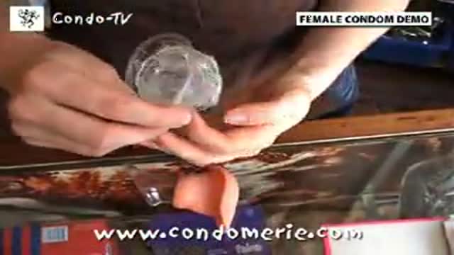
A usage instruction on how to use a female condom (also know as a Femidom). Female Condom Application and Removal.
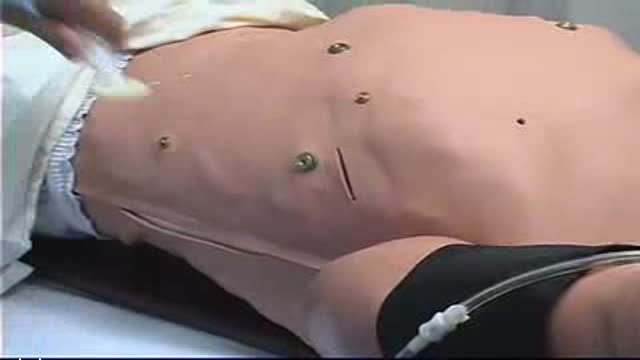
Chest Tube Placement

WORLD'S FIRST TRULY ANATOMIC MULTI-ROOTED ZIRCONIA DENTAL IMPLANT SOLUTION dentistry video

This video shows you how to examine the hand and wrist and how to identify common causes of pain.
This video clip is part of the FIFA Diploma in Football Medicine and the FIFA Medical Network. To enrol or to find our more click on the following link http://www.fifamedicalnetwork.com
The Diploma is a free online course designed to help clinicians learn how to diagnose and manage common football-related injuries and illnesses. There are a total of 42 modules created by football medicine experts. Visit a single page, complete individual modules or finish the entire course.
The network provides the opportunity for clinicians around the world to meet and share ideas relating to football medicine. Ask about an interesting case, debate current practice and discuss treatment strategies. Create a profile and log on to interact with other health professionals from around the globe.
This is not medical advice. The content is intended as educational content for health care professionals and students. If you are a patient, seek care of a health care professional.
houlder examination frequently appears in OSCEs. You’ll be expected to pick up the relevant clinical signs using your examination skills. This shoulder examination OSCE guide provides a clear step by step approach to examining the shoulder, with an included video demonstration.
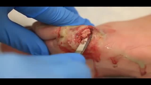
Treatment may not be needed for an eschar if it is part of the natural healing process. However, if an eschar looks like it may have a wound infection – symptoms can include oozing fluid such as pus or blood, your clinician will likely recommend topical treatment or debridement to help control and remove the infection.
Shingles is a viral infection that causes a painful rash. Although shingles can occur anywhere on your body, it most often appears as a single stripe of blisters that wraps around either the left or the right side of your torso. Shingles is caused by the varicella-zoster virus — the same virus that causes chickenpox. After you've had chickenpox, the virus lies inactive in nerve tissue near your spinal cord and brain. Years later, the virus may reactivate as shingles. While it isn't a life-threatening condition, shingles can be very painful. Vaccines can help reduce the risk of shingles, while early treatment can help shorten a shingles infection and lessen the chance of complications.

Vitiligine News, Vitiligine Foto, Vitiligine Come Si Manifesta, La Vitiligine, Rimedi Vitiligine --- http://vitiligine-cura.good-info.co --- Non Importa Quanto Sia Grave La Tua Vitiligine, Puoi Iniziare A Utilizzare Questo Sistema Potente PROPRIO ORA Per Ottenere La Libertà Dalla Vitiligine Che Hai Sempre Sognato! Funziona In Tutti I Casi Seguenti: Vitiligine Leggera, Moderata O Grave Vitiligine Focale Vitiligine Segmentale Vitiligine Mucoidale Vitiligine acrofacciale Vitiligine vulgaris Vitiligine universale I trattamenti anti-vitiligine che la maggior parte della gente usa NON FUNZIONANO! Il 95% di tutti quelli che trattano la vitiligine finisce peggio di quando ha iniziato! Una Presentazione Video Gratuita Spiega Un Singolare Consiglio Per Eliminare La Vitiligine Per Sempre http://vitiligine-cura.good-info.co
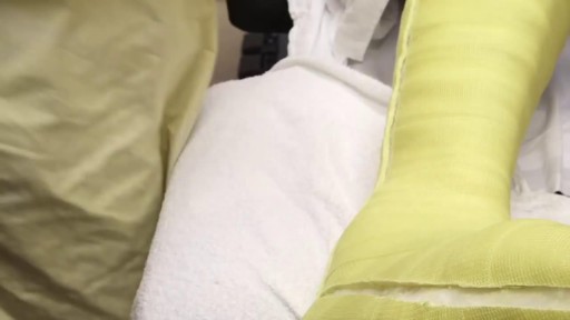
Total Contact Casting is the gold standard for treating diabetic foot ulcers; it's the most evidence-based treatment available. The Wound Care team at IU Health Methodist Hospital provides custom Total Contact Casting that completely offloads the wound, allowing it to heal in a matter of weeks.
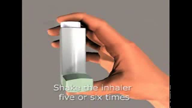
show your patients how to use an inhaler
