- Physical Examination
- Surgical Examination
- Ophthalmology
- Clinical Skills
- Orthopedics
- Surgery Videos
- Laparoscopy
- Pediatrics
- Funny Videos
- Cardiothoracic Surgery
- Nursing Videos
- Plastic Surgery
- Otorhinolaryngology
- Histology and Histopathology
- Neurosurgery
- Dermatology
- Pediatric Surgery
- Urology
- Dentistry
- Oncology and Cancers
- Anatomy Videos
- Health and Fitness
- Radiology
- Anaesthesia
- Physical Therapy
- Pharmacology
- Interventional Radiology
- Cardiology
- Endocrinology
- Gynecology
- Emergency Medicine
- Psychiatry and Psychology
- Childbirth Videos
- General Medical Videos
- Nephrology
- Physiology
- Diet and Food Health
- Diabetes Mellitus
- Neurology
- Women Health
- Osteoporosis
- Gastroenterology
- Pulmonology
- Hematology
- Rheumatology
- Toxicology
- Nuclear Medicine
- Infectious Diseases
- Vascular Disease
- Reproductive Health
- Burns and Wound Healing
- Other
Top videos

Watch that video of Popping Huge Epidermoid Cyst

A Cesarean section (C-section) is surgery to deliver a baby. The baby is taken out through the mother's abdomen. In the United States, almost one in three women has their babies this way. Some C-sections are planned, but many are done when unexpected problems happen during delivery. Reasons for a C-section may include Health problems in the mother The mother carrying more than one baby The size or position of the baby The baby's health is in danger Labor is not moving along as it should

Watch that video to know if oral sex can cause cancer

A report of Female Genital Mutilationn FGM (female circucision) in Menya In Egypt تقرير من مدينة المنيا في صعيد مصر عن ختان لاناث
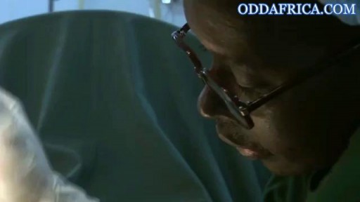
Correcting fgm https://oddafrica.com/videos/female-genital-mutilation-in-africa/
Full examination of the female from head to toe by Loyola Medical School, Chicago part 1

Watch that video to know if it is safe to have sex during pregnancy
Acute sinusitis can be triggered by a cold or allergies and may resolve on its own. Chronic sinusitis lasts up to eight weeks and may be caused by an infection or growths. Symptoms include headache, facial pain, runny nose, and nasal congestion. Acute sinusitis usually doesn't require any treatment beyond symptomatic relief with pain medications, nasal decongestants, and nasal saline rinses. Chronic sinusitis may require antibiotics.
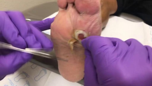
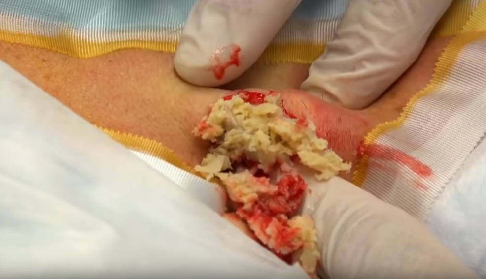
This is a diabetic foot ulcer. The patient reportedly went on vacation and noticed this ulcer upon their return. Debridement (removal of damaged tissue) to the level of healthy bleeding tissue is medically necessary as damaged tissue acts an impediment to wound healing. Due to their diabetic neuropathy, they did not feel any pain or indication that a wound was forming. This ulcer appeared to have penetrated to the level of subcutaneous tissue or even fascia, but turned out to be much deeper than that. These are serious wounds and are the beginnings of what lead to foot and leg amputations if they are not treated promptly by your healthcare provider, AKA Podiatrist.

Gynecological Examination

A spontaneous vaginal delivery (SVD) occurs when a pregnant woman goes into labor with or without use of drugs or techniques to induce labor, and delivers her baby in the normal manner, without forceps, vacuum extraction, or a cesarean section. Assisted vaginal delivery (AVD) occurs when a pregnant woman goes into labor with or without the use of drugs or techniques to induce labor, and requires the use of special instruments such as forceps or a vacuum extractor to deliver her baby vaginally.
Bartholin gland Marsupialization in Primary Bartholin Cyst

Vaginismus Pain Management
A video discussing Causes of Itching in the Vulva

a video of abdominal physical examination including all the required items:
-Inspection
-Palpation
-Percussion
-Auscultation
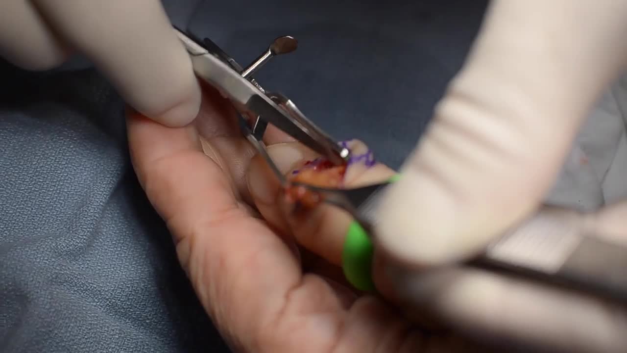
GIANT CELL TUMOR REMOVAL Plastic, Cosmetic and Reconstructive

This video - produced by students at Oxford University Medical School - demonstrates how to perform an examination of the respiratory system. It also indicates common pathologies encountered. It is part of a series of videos covering basic clinical examinations and is linked to Oxford Medical Education (www.oxfordmedicaleducation.com).

Watch that video of Butt Implants Gone Completely Wrong

Colposcopy (kol-POS-kuh-pee) is a procedure to closely examine your cervix, vagina and vulva for signs of disease. During colposcopy, your doctor uses a special instrument called a colposcope. Your doctor may recommend colposcopy if your Pap test has shown abnormal results.
