- Physical Examination
- Surgical Examination
- Ophthalmology
- Clinical Skills
- Orthopedics
- Surgery Videos
- Laparoscopy
- Pediatrics
- Funny Videos
- Cardiothoracic Surgery
- Nursing Videos
- Plastic Surgery
- Otorhinolaryngology
- Histology and Histopathology
- Neurosurgery
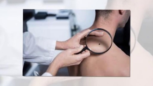
- Dermatology
- Pediatric Surgery
- Urology
- Dentistry
- Oncology and Cancers
- Anatomy Videos
- Health and Fitness
- Radiology
- Anaesthesia
- Physical Therapy
- Pharmacology
- Interventional Radiology
- Cardiology
- Endocrinology
- Gynecology
- Emergency Medicine
- Psychiatry and Psychology
- Childbirth Videos
- General Medical Videos
- Nephrology
- Physiology
- Diet and Food Health
- Diabetes Mellitus
- Neurology
- Women Health
- Osteoporosis
- Gastroenterology
- Pulmonology
- Hematology
- Rheumatology
- Toxicology
- Nuclear Medicine
- Infectious Diseases
- Vascular Disease
- Reproductive Health
- Burns and Wound Healing
- Other
Top videos
Female ejaculation is characterized as an expulsion of fluid from or near the vagina during or before an orgasm
A video showing the process of childbirth via vaginal delivery.
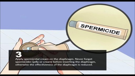
A diaphragm is a shallow, bendable cup that you put inside your vagina. It covers your cervix during sex to prevent pregnancy.
A video discussing Causes of Itching in the Vulva
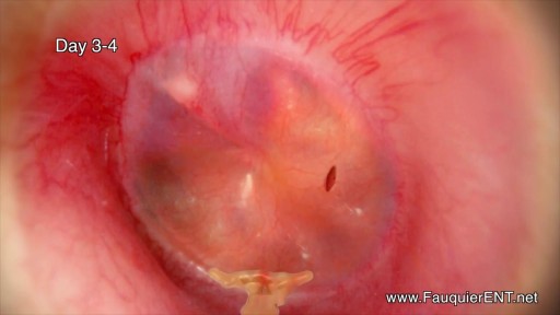
Ear Infection Drainage Time Lapse Video

http://www.vaginal-ultrasound.com A demonstration of a vaginal ultrasound.
http://www.proctoscopeexam.com This is a demonstration of a proctoscope examination of the rectum.

DermaClinix, The world's leading hair Transplantation and aesthetic dermatology clinic that offers the best hair transplant in Chennai. The clinic is fully equipped with infrastructure and the ultra-modern devices in the field of cosmetology and hair restoration. At DermaClinx Chennai offers a number of treatments including chemical peels for acne, pigmentation treatment, glow facials, hair growth therapies and many more. Hair transplant Doctors at DermaClinix Nungambakkam, Chennai have more than 12 years of experience in this field & holds the International accreditations and member of ISHRS (USA). Visit us today to know more about the hair growth treatments. https://www.dermatologistchennai.in/hair-transplant-surgeon-in-chennai.php Address: No:19 1st Floor TMA Tower Dr.Thirumurthy Nagar main road, Nungambakkam, Chennai, Tamil Nadu 600034 Call us at - +918939636222, +91 89398 81919 For more: Website - https://www.hairtransplantchennai.org/ Email - enquiry@hairtransplantchennai.org
Overview HIV is a virus that affects the immune system, specifically the CD4 cells. The CD4 cells help protect the body from illness. Unlike other viruses that the immune system can fight off, HIV can’t be eliminated by the immune system. The symptoms of HIV can vary greatly from person to person. No two people with HIV will likely experience the exact same symptoms. However, HIV will generally follow this pattern: acute illness asymptomatic period advanced infection Acute illness Approximately 80 percent of people who contract HIV experience flu-like symptoms within two to four weeks. This flu-like illness is known as acute HIV infection. Acute HIV infection is the primary stage of HIV and lasts until the body has created antibodies against the virus. The most common symptoms of this stage of HIV include: body rash fever sore throat severe headaches Less common symptoms may include: fatigue swollen lymph nodes ulcers in the mouth or on the genitals muscle aches joint pain nausea and vomiting night sweats Symptoms typically last one to two weeks. Anyone who has these symptoms and thinks they may have contracted HIV should consider scheduling an appointment with their healthcare provider to get tested. Symptoms specific to men Symptoms of HIV are generally the same in women and men. One HIV symptom that is unique to men is an ulcer on the penis. HIV may lead to hypogonadism, or poor production of sex hormones, in either sex. However, hypogonadism’s effects on men are easier to observe than its effects on women. Symptoms of low testosterone, one aspect of hypogonadism, can include erectile dysfunction (ED).

CORRECTION: After review of this video, it is clear that this video is of a baby who is near full term (40 weeks) based on the size. Late trimester "abortions" are defined only to viability of a baby (24 weeks) A 24 week baby is much smaller than this baby shown and by definition this is not a late "abortion" procedure. The proper labeling of this video should be management of a deceased breech baby with "head entrapment" as this was almost certainly a naturally occuring delivery and an OB nightmare (Reviewed by Dr. Frederick Bright)
No condom prevents pregnancy or sexually transmitted diseases (STDs) 100% of the time. But if you and your partner are having sex, nothing protects against STDs better than a properly used condom. For those having sex, condoms must always be used to protect against STDs even when using another method of birth control.
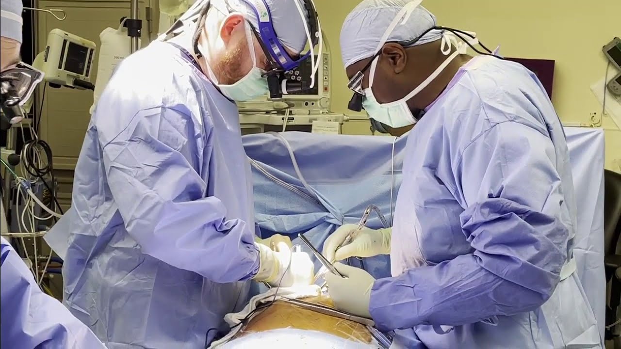
Dr. Erik Beyer, Florida Medical Center's chief of cardiac surgery, discusses performed a procedure called a micro-thoracotomy.

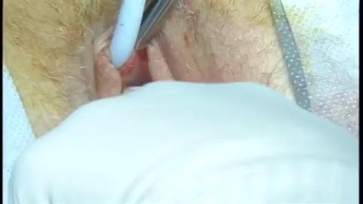
Surgical cutting and removal of a deep skin cyst Medical Videos

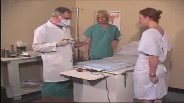
The products of a surgical abortion.
![Female Foley Insertion (Urinary Catheter) [How to Insert Nursing Skills]](https://i.ytimg.com/vi/Mq4Yh0-iozY/maxresdefault.jpg)
Pass your tests and improve your grades with the below FREE resources:
1) A FREE 140 Must Know Meds book
Click here to get your FREE copy of the 140 Must Know Meds Book: https://bit.ly/41rxSt0
2) A FREE test-taking tips webinar
Join us for our free test-taking tips webinar to boost your exam scores: https://bit.ly/nursingtesttaking
You can now test your knowledge with a free lesson quiz on NURSING.com!
Click here to take a free quiz: https://bit.ly/3HwJr8t
FREE Nursing School Cheat Sheets at: http://www.NURSING.com
Get the full lesson on Female Foley Insertion here:
https://nursing.com/lesson/ski....lls-03-01-inserting-
Get the Male Foley Insertion lesson here:
https://nursing.com/lesson/ski....lls-03-02-inserting-
Get the Sterile glove application lesson here:
https://nursing.com/lesson/ski....lls-01-04-sterile-gl
Check out our new Nurse Care Plan Lessons here:
https://bit.ly/3BPRfPL
Get Access to Thousands of Lessons here:
https://nursing.com/courses/
Welcome to the NURSING Family, we call it the most supportive nursing cohort on the planet.
At NURSING.com, we want to help you remove the stress and overwhelm of nursing school so that you can focus on becoming an amazing nurse.
Check out our freebies and learn more at: (http://www.nursing.com)
Female Foley Insertion (Urinary Catheter)- Nursing Skills
In this video, we’re going to look at inserting a Foley catheter in a female. Of course make sure you’ve verified your order and told the patient what’s happening. You’ll also typically want to perform perineal care before you start. Then, you’ll want to assist the patient into the appropriate position. For females, that’s supine with their knees bent and feet close to their hips – allowing their knees to fall to the side. You may need a helper to help hold the patient in this position. We love you guys! Go out and be your best selves today! And, as always, happy nursing!
Bookmarks:
0.05 Female Foley insertion introduction
0.15 Patient positioning
0.27 Opening the sterile kit
1.41 Setting up the sterile field
2.25 Prepping the remaining Foley kit items
2.34 Catheter lubrication
3.00 Saline syringe attachment
3.10 Iodine, swabs and cleansing the area
3.52 Catheter insertion (into urethra)
4.06 Balloon inflation
4.25 Final catheter setting
4.31 Securing the catheter and bag
4.48 Discarding your supplies
5.00 Documentation
5.08 Foley insertion outro
Visit us at https://nursing.com/medical-disclaimer/ for disclaimer information.
NCLEX®, NCLEX-RN® are registered trademarks of the National Council of State Boards of Nursing, INC. and hold no affiliation with NURSING.com.
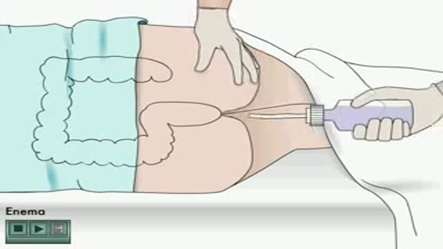
Enema how to apply Animation

Coin extraction from the upper esophagus in a child.
Dr. Mohamed Abeid
From the " Endoscopy Atlas " :
http://www.facebook.com/group.php?gid=16900943915&ref=ts

Patient Greg Grindley communicates with host Bryant Gumbel and his wife for the first time while undergoing deep brain stimulation surgery at University Hospital's Case Medical Center in Cleveland, Ohio.
➡ Subscribe: http://bit.ly/NatGeoSubscribe
About National Geographic:
National Geographic is the world's premium destination for science, exploration, and adventure. Through their world-class scientists, photographers, journalists, and filmmakers, Nat Geo gets you closer to the stories that matter and past the edge of what's possible.
Get More National Geographic:
Official Site: http://bit.ly/NatGeoOfficialSite
Facebook: http://bit.ly/FBNatGeo
Twitter: http://bit.ly/NatGeoTwitter
Instagram: http://bit.ly/NatGeoInsta
Greg's First In-Surgery Conversation | Brain Surgery Live
https://youtu.be/zvqV_2zncNU
National Geographic
https://www.youtube.com/natgeo
The use of breast MRI as part of the screening for breast cancer.
