- Physical Examination
- Surgical Examination
- Ophthalmology
- Clinical Skills
- Orthopedics
- Surgery Videos
- Laparoscopy
- Pediatrics
- Funny Videos
- Cardiothoracic Surgery
- Nursing Videos
- Plastic Surgery
- Otorhinolaryngology
- Histology and Histopathology
- Neurosurgery
- Dermatology
- Pediatric Surgery
- Urology
- Dentistry
- Oncology and Cancers
- Anatomy Videos
- Health and Fitness
- Radiology
- Anaesthesia
- Physical Therapy
- Pharmacology
- Interventional Radiology
- Cardiology
- Endocrinology
- Gynecology
- Emergency Medicine
- Psychiatry and Psychology
- Childbirth Videos
- General Medical Videos
- Nephrology
- Physiology
- Diet and Food Health
- Diabetes Mellitus
- Neurology
- Women Health
- Osteoporosis
- Gastroenterology
- Pulmonology
- Hematology
- Rheumatology
- Toxicology
- Nuclear Medicine
- Infectious Diseases
- Vascular Disease
- Reproductive Health
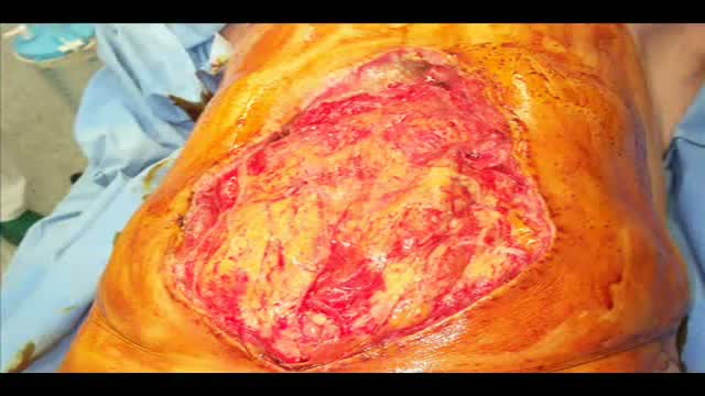
- Burns and Wound Healing
- Other
Top videos


Cholesterol is a fat-like, waxy substance that can be found in all parts of your body. It helps your body make cell membranes, many hormones, and vitamin D. The cholesterol in your blood comes from two sources: the foods you eat and your liver. But your liver makes all the cholesterol your body needs.

Surgical Scrub How To
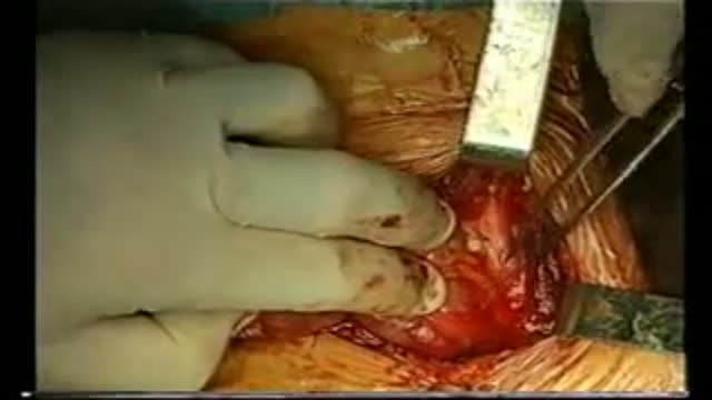
A video showing the total thyroidectomy operation
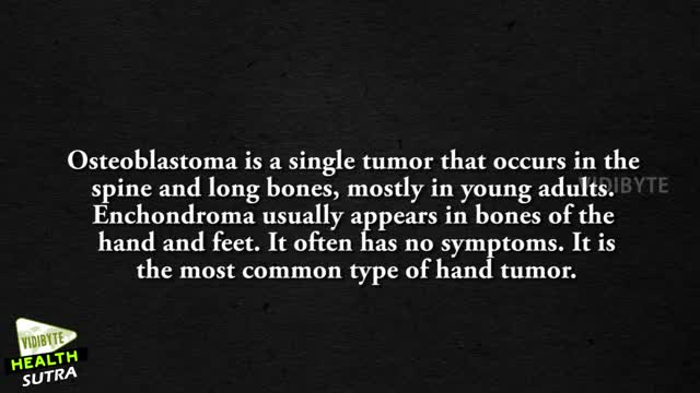
These are a few common types of benign bone tumors: Osteochondroma is the most common benign bone tumor. ... Giant cell tumor is a benign tumor, typically affecting the leg (malignant types of this tumor are uncommon). Osteoid osteoma is a bone tumor, often occurring in long bones, that occurs commonly in the early 20s.
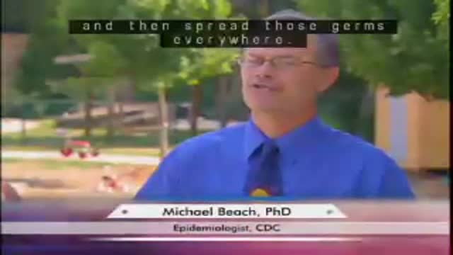
Clean hands can help prevent the spread of infectious diseases, such as flu. This podcast explains the proper way to wash your hands.
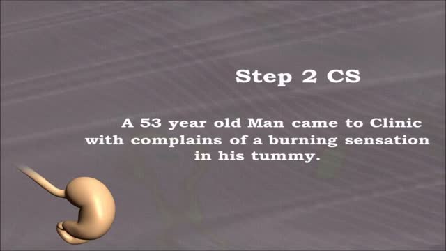
USMLE Step 2 CS - EPIGASTRIC This is just preview video. To get full access please visit our website : www.usmletutoring.com
Symptoms Burning stomach pain Feeling of fullness, bloating or belching Fatty food intolerance Heartburn Nausea The most common peptic ulcer symptom is burning stomach pain. Stomach acid makes the pain worse, as does having an empty stomach. The pain can often be relieved by eating certain foods that buffer stomach acid or by taking an acid-reducing medication, but then it may come back. The pain may be worse between meals and at night. Nearly three-quarters of people with peptic ulcers don't have symptoms. Less often, ulcers may cause severe signs or symptoms such as: Vomiting or vomiting blood — which may appear red or black Dark blood in stools, or stools that are black or tarry Trouble breathing Feeling faint Nausea or vomiting Unexplained weight loss Appetite changes

If you’ve suffered a sporting knee injury, how do you know when it’s serious? In this short video, Yorkshire Knee Clinic’s Dave Duffy reveals the two key tests that tell you whether your knee needs urgent, specialist attention.
𝗡𝗼𝘁𝗲𝘀 𝗳𝗼𝗿 𝘁𝗵𝗲 𝘀𝗾𝘂𝗲𝗮𝗺𝗶𝘀𝗵: This video features only features a model of the knee. There is no live footage from operations.
Discover more about sports knee injuries: https://yorkshirekneeclinic.com/sports-injuries/
Discover more about Dave Duffy: https://yorkshirekneeclinic.com/about/dave-duffy/
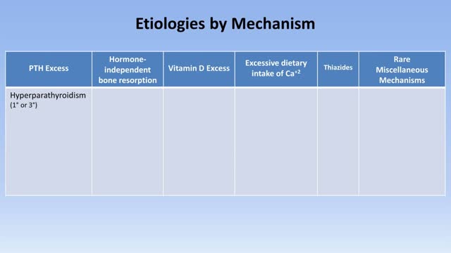
Hypercalcemia is a condition in which the calcium level in your blood is above normal. Too much calcium in your blood can weaken your bones, create kidney stones, and interfere with the way your heart and brain works. Hypercalcemia most commonly results from overactive parathyroid glands. These four tiny glands are each about the size of a grain of rice and are located on or near the thyroid gland. Other causes of hypercalcemia include cancer, certain other medical disorders, some medications, and excessive use of calcium and vitamin D supplements. Signs and symptoms of hypercalcemia may range from nonexistent to severe. Treatment depends on the underlying cause.
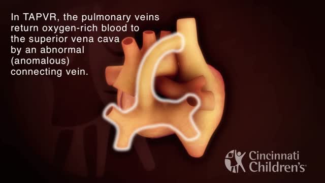
Total anomalous pulmonary venous return (TAPVR) is a rare congenital malformation in which pulmonary veins that return oxygen-rich blood from the lungs do not connect normally to the left atrium. Instead all four pulmonary veins drain abnormally to the right atrium. Heart models and animation were developed by the Cincinnati Children's Heart Institute in conjunction with Cincinnati Children's Critical Care Media Lab.

An excerpt from the award-winning documentary “Exposure: Environmental Links to Breast Cancer” about the effects of radiation. Featuring Olivia Newton-John, Dr. Rosalie Bertell and Dr. Susan Love.
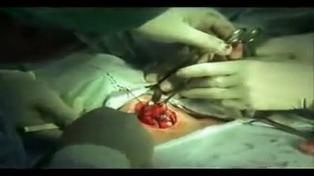
A video showing thyroidectomy surgery
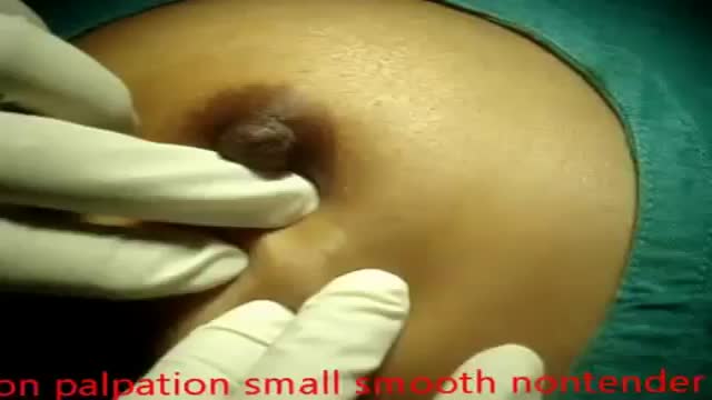
Fibroadenomas (fy-broe-ad-uh-NO-muhz) are solid, noncancerous breast tumors that occur most often in adolescent girls and women under the age of 30. You might describe a fibroadenoma as firm, smooth, rubbery or hard with a well-defined shape. Usually painless, a fibroadenoma might feel like a marble in your breast, moving easily under your skin when touched. Fibroadenomas vary in size, and they can get bigger or even shrink on their own. Fibroadenomas are among the most common breast lumps in young women. Treatment may include monitoring to detect changes in the size or feel of the fibroadenoma, a biopsy to evaluate the lump, or surgery to remove it.
Laparostomy is a surgical condition in which the abdomen is left open to contrast a condition named Abdominal Compartment Syndrome
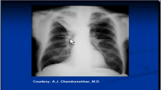
The video will describe features of right upper lobe collapse. Please see my website for disclaimer.
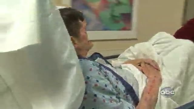
A man gets a face transplant from a Hollywood Exec and it's caught on tape by the people of Boston Med on ABC

Care for Your Knee After Knee Replacement Surgery
In this video, Dr. Mark Hammerberg, provides details on two important activities to help during recovery from knee replacement surgery.
Denver Health's Orthopedics department offers many different types of treatments to help you, including surgical and non-surgical options. To find out if surgery is right for you, visit DenverHealth.org/Orthopedics or call 303-602-1590 to make an appointment.
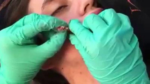
Always consult your doctor and seek help early enough to prevent complications
Megacolon, as well as megarectum, is a descriptive term. It denotes dilatation of the colon that is not caused by mechanical obstruction.[1, 2] Although the definition of megacolon has varied in the literature, most researchers use the measurement of greater than 12 cm for the cecum as the standard. Because the diameter of the large intestine varies, the following definitions would also be considered: greater than 6.5 cm in the rectosigmoid region and greater than 8 cm for the ascending colon. Megacolon can be divided into the following 3 categories: Acute megacolon ( pseudo-obstruction) Chronic megacolon, which includes congenital, acquired, and idiopathic causes Toxic megacolon
