- Physical Examination
- Surgical Examination
- Ophthalmology
- Clinical Skills
- Orthopedics
- Surgery Videos
- Laparoscopy
- Pediatrics
- Funny Videos
- Cardiothoracic Surgery
- Nursing Videos
- Plastic Surgery
- Otorhinolaryngology
- Histology and Histopathology
- Neurosurgery
- Dermatology
- Pediatric Surgery
- Urology
- Dentistry
- Oncology and Cancers
- Anatomy Videos
- Health and Fitness
- Radiology
- Anaesthesia
- Physical Therapy
- Pharmacology
- Interventional Radiology
- Cardiology
- Endocrinology
- Gynecology
- Emergency Medicine
- Psychiatry and Psychology
- Childbirth Videos
- General Medical Videos
- Nephrology
- Physiology
- Diet and Food Health
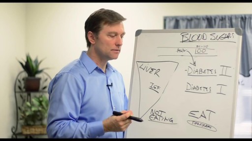
- Diabetes Mellitus
- Neurology
- Women Health
- Osteoporosis
- Gastroenterology
- Pulmonology
- Hematology
- Rheumatology
- Toxicology
- Nuclear Medicine
- Infectious Diseases
- Vascular Disease
- Reproductive Health
- Burns and Wound Healing
- Other
Top videos

Weight loss is the most effective nonpharmacologic measure to decrease blood pressure in overweight individuals. Weight loss with other nonpharmacologic measures can prevent or delay the onset of hypertension and reduce the overall risk of cardiovascular events in such patients. In some patients with established hypertension, lifestyle changes alone may control their blood pressure.

Capsaicin binds to pain receptors on our nerves called TRPV1. Normally, it reacts to heat by sending warning signals to the brain. Capsaicin causes TRPV1 to send those same signals. So, you react as if there's something hot in your mouth
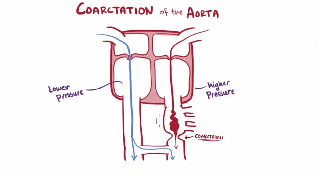
Coarctation of the aorta (CoA[1][2] or CoAo), also called aortic narrowing, is a congenital condition whereby the aorta is narrow, usually in the area where the ductus arteriosus (ligamentum arteriosum after regression) inserts. The word “coarctation” means narrowing. Coarctations are most common in the aortic arch. The arch may be small in babies with coarctations. Other heart defects may also occur when coarctation is present, typically occurring on the left side of the heart. When a patient has a coarctation, the left ventricle has to work harder. Since the aorta is narrowed, the left ventricle must generate a much higher pressure than normal in order to force enough blood through the aorta to deliver blood to the lower part of the body. If the narrowing is severe enough, the left ventricle may not be strong enough to push blood through the coarctation, thus resulting in lack of blood to the lower half of the body. Physiologically its complete form is manifested as interrupted aortic arch
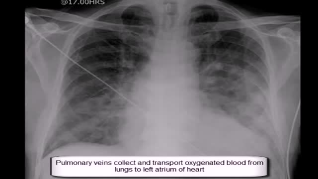
Expand Section. Pulmonary edema is often caused by congestive heart failure. When the heart is not able to pump efficiently, blood can back up into the veins that take blood through the lungs. As the pressure in these blood vessels increases, fluid is pushed into the air spaces (alveoli) in the lungs.

Classical PKU is an autosomal recessive disorder, caused by mutations in both alleles of the gene for phenylalanine hydroxylase (PAH), found on chromosome 12. In the body, phenylalanine hydroxylase converts the amino acid phenylalanine to tyrosine, another amino acid.

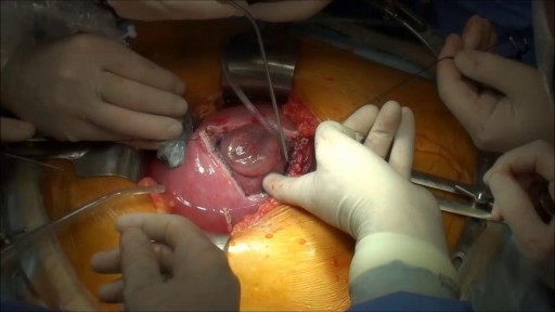
Note: This video contains graphic surgical footage so viewer discretion is advised.
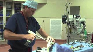
Director of the Penn Orthopaedics Robotics and Navigation Program, Dr. Christopher Travers, discusses robotic joint replacement surgery, which is one of the multiple options that Penn Orthopaedics offers for joint replacement surgery. He walks through a robotic knee replacement surgery, discussing what the procedure is, how it differs from traditional joint replacement surgery, and the benefits.
Refer a patient (physicians only):
https://www.pennmedicine.org/refer-your-patient
Learn more about the Penn Joint Replacement Program:
https://www.pennmedicine.org/f....or-patients-and-visi
Learn more about Dr. Travers:
https://www.pennmedicine.org/providers/profile/christopher-travers?fadf=pennmedicine&keyword=travers
#RoboticSurgery #JointReplacementSurgery #KneeReplacement #SurgicalFootage
Prenatal repair of myelomeningocele (MMC), the most common and severe form of spina bifida, is a delicate surgical procedure where fetal surgeons open the uterus and close the opening in the baby's back while they are still in the womb.
Dr. Eric Janssen of SportsMED Orthopaedic Surgery & Spine Center in Huntsville, Alabama demonstrates a total knee replacement using dry bones model. In this demonstration he uses the Wright Medical Evolution Knee implant. This demonstrations does not include soft tissue.
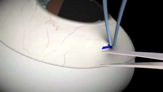
Glaucoma Surgery 3D Animation

A ventricular septal defect (VSD) is an opening or hole in the wall that separates the two lower chambers of the heart. This wall is called the ventricular septum. The hole causes oxygen-rich blood to leak from the left side of the heart to the right side. This causes extra work for the right side of the heart, since more blood than necessary is flowing through the right ventricle to the lungs. The hole is usually closed with surgery. However, in certain situations, your child's cardiologist and surgeon may think it is best to close the hole with a special device. This procedure is done in the heart catheterization lab.

Womens Issues and Blood Clotting

Care for Your Knee After Knee Replacement Surgery
In this video, Dr. Mark Hammerberg, provides details on two important activities to help during recovery from knee replacement surgery.
Denver Health's Orthopedics department offers many different types of treatments to help you, including surgical and non-surgical options. To find out if surgery is right for you, visit DenverHealth.org/Orthopedics or call 303-602-1590 to make an appointment.


Septoplasty (SEP-toe-plas-tee) is a surgical procedure to correct a deviated septum — a displacement of the bone and cartilage that divides your two nostrils. During septoplasty, your nasal septum is straightened and repositioned in the middle of your nose.

Watch that video to learn How to Know If You Are Pregnant

Sprains and Strains
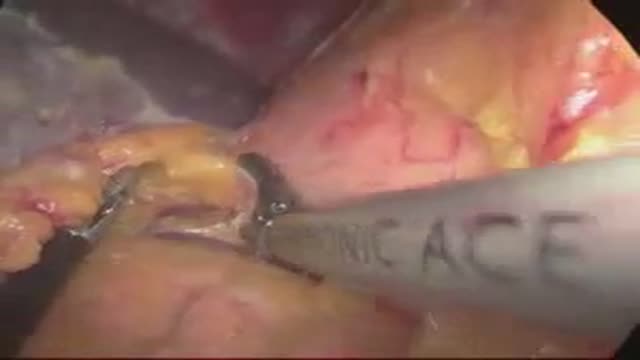
laparoscopic left adrenalectomy in 150kg patient with Cushings

Gastric bypass, also called Roux-en-Y gastric bypass surgery, is considered a “metabolic” procedure because it changes how your body absorbs fat, calories and nutrients. This metabolic change occurs because your gastrointestinal tract is altered when your gastric bypass surgeon attaches the smaller section of your stomach directly to your small intestine. As a result, your appetite changes and you feel full faster.