- Physical Examination
- Surgical Examination
- Ophthalmology
- Clinical Skills
- Orthopedics
- Surgery Videos
- Laparoscopy
- Pediatrics
- Funny Videos
- Cardiothoracic Surgery
- Nursing Videos
- Plastic Surgery
- Otorhinolaryngology
- Histology and Histopathology
- Neurosurgery
- Dermatology
- Pediatric Surgery
- Urology
- Dentistry
- Oncology and Cancers
- Anatomy Videos
- Health and Fitness
- Radiology
- Anaesthesia
- Physical Therapy
- Pharmacology
- Interventional Radiology
- Cardiology
- Endocrinology
- Gynecology
- Emergency Medicine
- Psychiatry and Psychology
- Childbirth Videos
- General Medical Videos
- Nephrology
- Physiology
- Diet and Food Health
- Diabetes Mellitus
- Neurology
- Women Health
- Osteoporosis
- Gastroenterology
- Pulmonology
- Hematology
- Rheumatology
- Toxicology
- Nuclear Medicine
- Infectious Diseases
- Vascular Disease
- Reproductive Health
- Burns and Wound Healing
- Other
Top videos
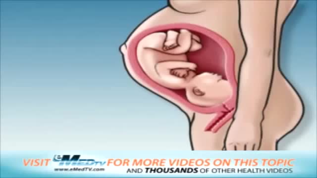
Your baby is still tiny, but already your body is changing. Your breasts start to swell and may feel tender. Tiredness, nausea, and a frequent need to pee are common pregnancy symptoms. In your second trimester, your growing uterus gradually rises up out of your pelvis.
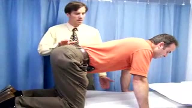
This is an educational medical video for Medical Students showing how to examine a hernia swelling

There are 3 major parts of the respiratory system: the airway, the lungs, and the muscles of respiration. The airway, which includes the nose, mouth, pharynx, larynx, trachea, bronchi, and bronchioles, carries air between the lungs and the body's exterior.
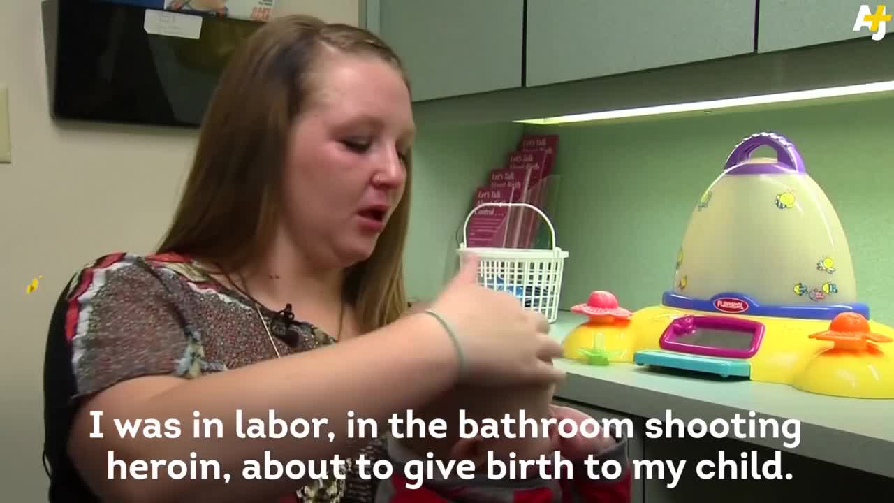
Each year, thousands of babies in the U.S. are born addicted to opiates. And the problem is getting worse.
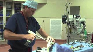
Dr. Eric Janssen of SportsMED Orthopaedic Surgery & Spine Center in Huntsville, Alabama demonstrates a total knee replacement using dry bones model. In this demonstration he uses the Wright Medical Evolution Knee implant. This demonstrations does not include soft tissue.
Terrifying Sinus Infection - Disturbing - Must Watch
An arthroscopic meniscectomy is a procedure to remove some or all of a meniscus from the tibio-femoral joint of the knee using arthroscopic (aka 'keyhole') surgery. In a complete meniscectomy the meniscus including the meniscal rim is removed. A partial meniscectomy involves partial removal of the meniscus. This may vary from minor trimming of a frayed edge to anything short of removing the rim. This is a minimally invasive procedure often done as day suas an outpatient in a one-day clinic [1] This procedure is performed when a meniscal tear is too large to be corrected by a surgical meniscal repair.[1] When non-operative therapy provides some degree of symptom relief over the long-term, these benefits may wane with continued meniscal degeneration. In such patients, arthroscopic partial meniscectomy can be effective in improving patient quality of life.

DermaClinix, The world's leading hair Transplantation and aesthetic dermatology clinic that offers the best hair transplant in Chennai. The clinic is fully equipped with infrastructure and the ultra-modern devices in the field of cosmetology and hair restoration. At DermaClinx Chennai offers a number of treatments including chemical peels for acne, pigmentation treatment, glow facials, hair growth therapies and many more. Hair transplant Doctors at DermaClinix Nungambakkam, Chennai have more than 12 years of experience in this field & holds the International accreditations and member of ISHRS (USA). Visit us today to know more about the hair growth treatments. https://www.dermatologistchennai.in/hair-transplant-surgeon-in-chennai.php Address: No:19 1st Floor TMA Tower Dr.Thirumurthy Nagar main road, Nungambakkam, Chennai, Tamil Nadu 600034 Call us at - +918939636222, +91 89398 81919 For more: Website - https://www.hairtransplantchennai.org/ Email - enquiry@hairtransplantchennai.org
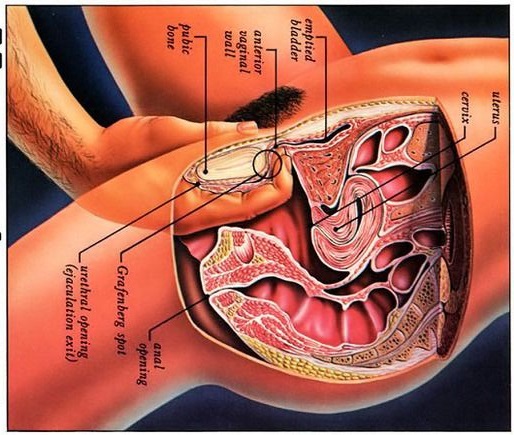
Watch that video to know what G spot is
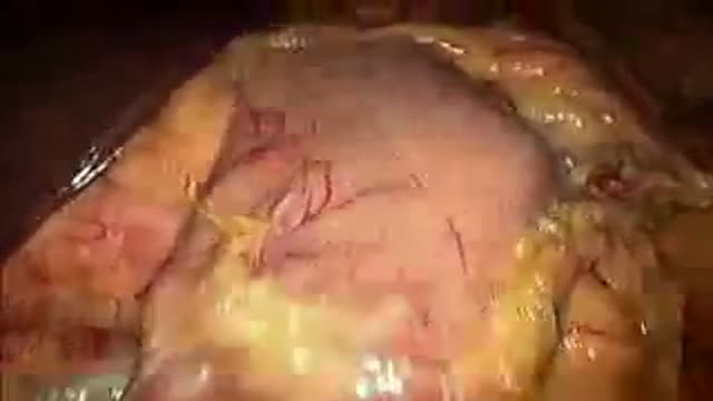
Revision of Mini Gastric ByPass
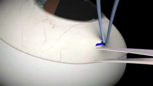
Glaucoma Surgery 3D Animation
The Spirotome belongs to the Direct & Frontal type of biopsy systems for taking large core biopsy from virtually every soft tissue in the body. The FDA has approved 13 applications. This video shows how easy it is to take a large core from a thoracic wall tumor mass. The size and quality of the sample allows quantitative molecular biology.
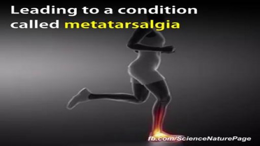
While it is unclear whether high heel shoes may or may not cause back pain, it is common for high heels to exacerbate an already present spinal condition. ... This pain in the back may also result from foot or leg fatigue that results from wearing these shoes and this can affect whole body mechanics.
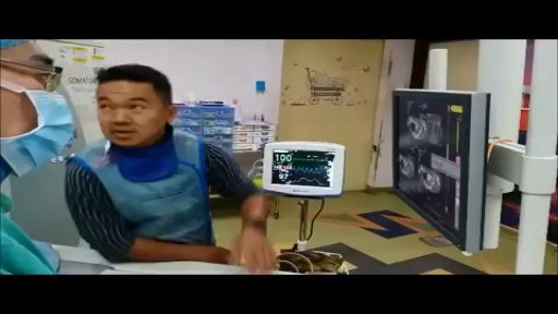
A computed tomography (CT) scan uses a special X-ray machine to take detailed pictures of the body’s organs and tissues. In a biopsy, a small piece of tissue is removed from your body. This tissue sample is then examined in the lab. A needle biopsy is the safest and easiest way to remove this tissue safely from the body. To do a needle biopsy, the radiologist will insert a needle through your skin and into your tissue. A syringe or an automated needle may be used to take the tissue sample.
Removal of Infected Hernia Mesh
Video demonstrates the action of the isolated lumbar multifidis muscle

Ultrasound Guided Sclerotherapy for Varicose Veins

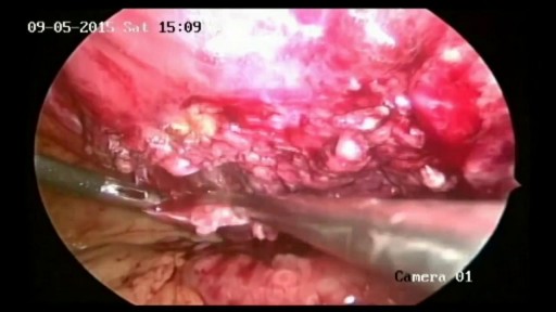
The video demonstrates complete excision of endometrosis in a variety of challenging situations.
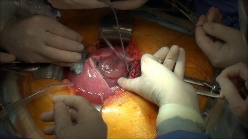
Prenatal repair of myelomeningocele (MMC), the most common and severe form of spina bifida, is a delicate surgical procedure where fetal surgeons open the uterus and close the opening in the baby's back while they are still in the womb.
