- Physical Examination
- Surgical Examination
- Ophthalmology
- Clinical Skills
- Orthopedics
- Surgery Videos
- Laparoscopy
- Pediatrics
- Funny Videos
- Cardiothoracic Surgery
- Nursing Videos
- Plastic Surgery
- Otorhinolaryngology
- Histology and Histopathology
- Neurosurgery
- Dermatology
- Pediatric Surgery
- Urology
- Dentistry
- Oncology and Cancers
- Anatomy Videos
- Health and Fitness
- Radiology
- Anaesthesia
- Physical Therapy
- Pharmacology
- Interventional Radiology
- Cardiology
- Endocrinology
- Gynecology
- Emergency Medicine
- Psychiatry and Psychology
- Childbirth Videos
- General Medical Videos
- Nephrology
- Physiology
- Diet and Food Health
- Diabetes Mellitus
- Neurology
- Women Health
- Osteoporosis
- Gastroenterology
- Pulmonology
- Hematology
- Rheumatology
- Toxicology
- Nuclear Medicine
- Infectious Diseases
- Vascular Disease
- Reproductive Health
- Burns and Wound Healing
- Other
Top videos
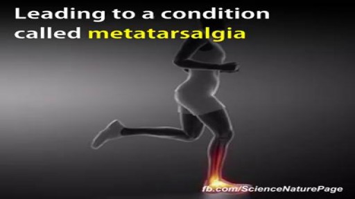
While it is unclear whether high heel shoes may or may not cause back pain, it is common for high heels to exacerbate an already present spinal condition. ... This pain in the back may also result from foot or leg fatigue that results from wearing these shoes and this can affect whole body mechanics.
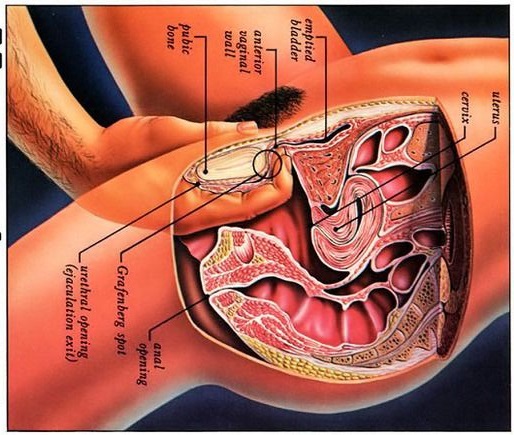
Watch that video to know what G spot is
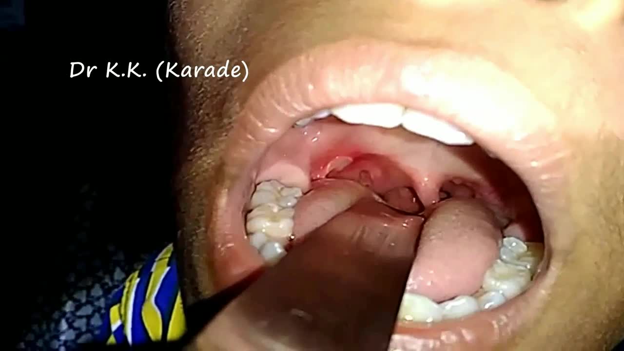
Canker sores (Aphthous ulcer) are small, painful ulcers on the inside of the mouth, tongue, lips, or throat.Canker sores are white or yellow and surrounded by a bright red area. They are not cancerous.
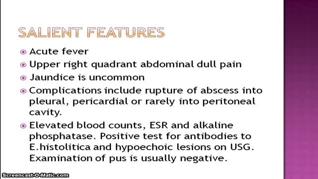
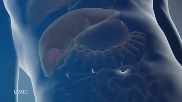
Bacterial abscess of the liver is relatively rare; however, it has been described since the time of Hippocrates (400 BCE), with the first published review by Bright appearing in 1936. In 1938, Ochsner's classic review heralded surgical drainage as the definitive therapy; however, despite the more aggressive approach to treatment, the mortality remained at 60-80%.[1] The development of new radiologic techniques, the improvement in microbiologic identification, and the advancement of drainage techniques, as well as improved supportive care, have reduced mortality to 5-30%; yet, the prevalence of liver abscess has remained relatively unchanged. Untreated, this infection remains uniformly fatal. The three major forms of liver abscess, classified by etiology, are as follows: Pyogenic abscess, which is most often polymicrobial, accounts for 80% of hepatic abscess cases in the United States Amebic abscess due to Entamoeba histolytica accounts for 10% of cases [2] Fungal abscess, most often due to Candida species, accounts for fewer than 10% of cases
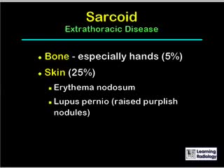
A diagnosis of sarcoidosis is established on the basis of compatible clinical and radiologic findings and histologic evidence of the presence of noncaseous epithelioid cell granulomas in one or more organs and the absence of causative organisms or particulates (16). Granulomas of known causes and local sarcoidlike reactions must be excluded. Granulomatous lesions may result from many conditions, including tuberculosis, berylliosis, leprosy, hypersensitivity pneumonitis, Crohn disease, primary biliary cirrhosis, and fungal disease. Moreover, local sarcoidlike reactions may be seen in lymph nodes that drain a neoplasm or a site of chronic inflammation (19). Such reactions also have been seen in patients who have undergone chemotherapy and radiation therapy (23). If biopsy of lymph nodes or pulmonary or pleural tissue is necessary for diagnosis, one of three techniques may be used: transbronchial biopsy, CT-guided biopsy, or surgical biopsy (24). The use of a surgical technique may be warranted when the results of biopsy with another procedure are not definitive and biopsy of mediastinal lymph nodes, lung, or both is required. This can generally be done with minimally invasive procedures, such as cervical mediastinoscopy, the Chamberlain procedure (a parasternal minithoracotomy for biopsy of the aortopulmonary window or para-aortic nodes), or video-assisted thoracoscopic surgical biopsy (25).
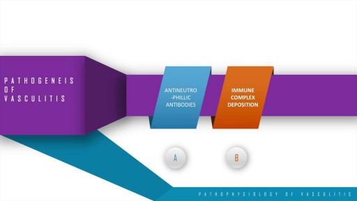
A step wise approach to the pathogenesis, types, disease entities and diagnosis of vasculitis. This discussion also includes the management options of vasculitis and their adverse drug reactions. In essence, vasculitis is a clfinicopathologic process characterised by inflammation and damage of blood vessels. This may be mainly due to three pathological processes which include immune complex deposition, anti-neutrophillic antibody formation and pathological T lymphocyte response and granuloma formation. The disease entities include Wegner's granulomatosis, Churg Strauss and many others. These present with palpable purpura, unexplained renal dysfunction etc which can be diagnosed based on biopsy and angiogram.

There are 3 major parts of the respiratory system: the airway, the lungs, and the muscles of respiration. The airway, which includes the nose, mouth, pharynx, larynx, trachea, bronchi, and bronchioles, carries air between the lungs and the body's exterior.
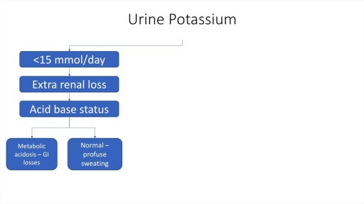
A step by step approach to Hypokalaemia including causes, diagnosis and management.
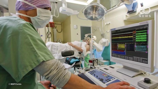
Depression is a very serious mental illness that affects millions worldwide. Could a small brain implant cure it?
The most frequent incision utilized to open the abdomen for liver surgery is called a chevron incision. In this incision a cut is made on the abdomen below the rib cage. The cut starts under the armpit below the ribs on the right side of the abdomen and continues all the way across the abdomen to the opposite arm pit thereby the whole width of the abdomen is cut to provide access to the liver. The average length of the incision is approximately 24 to 30 inches. This is one of the longest incisions is utilized in abdominal surgery. The incision is frequently associated with significant discomfort after the surgery and in some patients the discomfort can continue for many months, particularly when some of the nerves in the abdominal wall have been cut during the surgery. Laparoscopic surgery provides advantages over open surgery for the liver since the chevron incision is completely avoided and the surgery is performed through tiny incisions. As a consequence the duration of stay in hospital, the amount and duration of post operative discomfort, and the length of recovery is much shorter after the laparoscopic procedure compared to open surgery
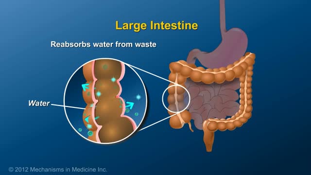
An ileostomy is an opening in the belly (abdominal wall) that’s made during surgery. The end of the ileum (the lowest part of the small intestine) is brought through this opening to form a stoma, usually on the lower right side of the abdomen. A Wound Ostomy Continence nurse (WOCN or WOC nurse) or the surgeon will figure out the best location for your stoma. (A WOC nurse is a specially trained registered nurse who takes care of and teaches ostomy patients. This nurse may also be called an ostomy nurse.)

Gastric bypass, also called Roux-en-Y gastric bypass surgery, is considered a “metabolic” procedure because it changes how your body absorbs fat, calories and nutrients. This metabolic change occurs because your gastrointestinal tract is altered when your gastric bypass surgeon attaches the smaller section of your stomach directly to your small intestine. As a result, your appetite changes and you feel full faster.
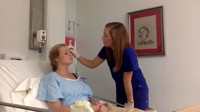
Physical assessment is taking an educated, systematic look at all aspects of an individual’s health status utilizing knowledge, skills and tools of health history and physical exam. To collect data- information about the client’s health, including physiological, psychological, sociocultural and spiritual aspects To establish actual and potential problems To establish the nurse-client relationship Method: The history is done first, then the physical examination focuses on finding data associated with the history. Health History- obtained through interview and record review. Physical exam- accomplished by tools and techniques ** A complete assessment is not necessarily carried out each time. A comprehensive assessment is part of a health screening examination. On admission, you will do an admission assessment (not necessarily including everything presented here) and document it on the admission form. You will do a daily shift assessment (patient systems review). And, if client has a specific problem, you may assess only that part of the body (focused). Data Collection: Information is organized into objective and subjective data: Subjective: Apparent only to person affected; includes client’s perceptions, feelings, thoughts, and expectations. It cannot be directly observed and can be discovered only asking questions. Objective: Detectable by an observer or can be tested against an acceptable standard; tangible, observable facts; includes observation of client behavior, medical records, lab and diagnostic tests, data collected by physical exam. ** To obtain data for the nursing health history, you must utilize good interview techniques and communications skills. Record accurately. DO NOT ASSUME. D. Frameworks for Health Assessment There are two main frameworks utilized in health assessment: Head to Toe- systematic collection of data starting with the head and working downward. Functional Health Assessment- Gordon’s 11 functional health patterns that address the behaviors a person uses to maintain health. PERSON is the ACC-ADN framework for assessment. It is similar to Gordon's functional health patterns.
Video demonstrates the action of the isolated lumbar multifidis muscle

Ultrasound Guided Sclerotherapy for Varicose Veins

Examination of Neck Swelling

Capsaicin binds to pain receptors on our nerves called TRPV1. Normally, it reacts to heat by sending warning signals to the brain. Capsaicin causes TRPV1 to send those same signals. So, you react as if there's something hot in your mouth
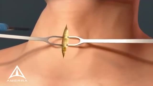
Tracheotomy is a surgical procedure which consists of making an incision on the anterior aspect of the neck and opening a direct airway through an incision in the trachea (windpipe). The resulting stoma (hole), or tracheostomy, can serve independently as an airway or as a site for a tracheostomy tube to be inserted; this tube allows a person to breathe without the use of his or her nose or mouth. Both surgical and percutaneous techniques are widely used in current surgical practice. It is among the oldest described procedures.
