- Physical Examination
- Surgical Examination
- Ophthalmology
- Clinical Skills
- Orthopedics
- Surgery Videos
- Laparoscopy
- Pediatrics
- Funny Videos
- Cardiothoracic Surgery
- Nursing Videos
- Plastic Surgery
- Otorhinolaryngology
- Histology and Histopathology
- Neurosurgery
- Dermatology
- Pediatric Surgery
- Urology
- Dentistry
- Oncology and Cancers
- Anatomy Videos
- Health and Fitness
- Radiology
- Anaesthesia
- Physical Therapy
- Pharmacology
- Interventional Radiology
- Cardiology
- Endocrinology
- Gynecology
- Emergency Medicine
- Psychiatry and Psychology
- Childbirth Videos
- General Medical Videos
- Nephrology
- Physiology
- Diet and Food Health
- Diabetes Mellitus
- Neurology
- Women Health
- Osteoporosis
- Gastroenterology
- Pulmonology
- Hematology
- Rheumatology
- Toxicology
- Nuclear Medicine
- Infectious Diseases
- Vascular Disease
- Reproductive Health
- Burns and Wound Healing
- Other
Top videos
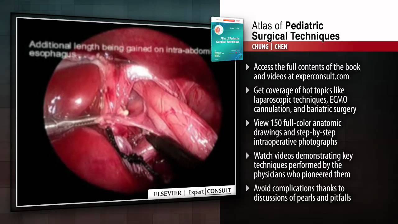
This title in the new Surgical Techniques Atlas series presents state-of-the-art updates on the full range of pediatric surgical techniques performed today. Expand your repertoire and hone your clinical skills thanks to the expert advice, procedural videos, and online access at expertconsult.com. For more information, please visit http://www.us.elsevierhealth.com/product.jsp?sid=EHS_US_BS-SPE-59&isbn=9781416046899&dmnum=null&elsca1=CriticalCare&elsca2=soc_med&elsca3=null&elsca4=youtube_ELSpromovideos

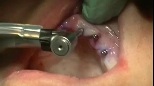
A Beautiful Smile at Lake Pointe is Sugar Land premier dentistry practice. Dr. Lance Jue has been serving patients' preventive, restorative and cosmetic dental needs here in Sugar Land for over 19 years. Book an appointment online now with Dr. Lance Jue

Hemorrhoidectomy Ligasure
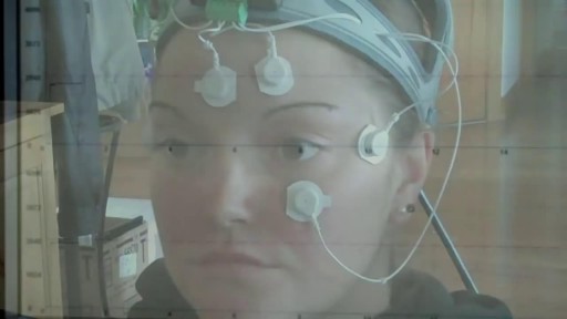
Electronystagmography (ENG) is a diagnostic test to record involuntary movements of the eye caused by a condition known as nystagmus. It can also be used to diagnose the cause of vertigo, dizziness or balance dysfunction by testing the vestibular system.
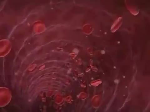
Sickle cell anemia (sickle cell disease) is a disorder of the blood caused by an inherited abnormal hemoglobin (the oxygen-carrying protein within the red blood cells). The abnormal hemoglobin causes distorted (sickled) red blood cells.
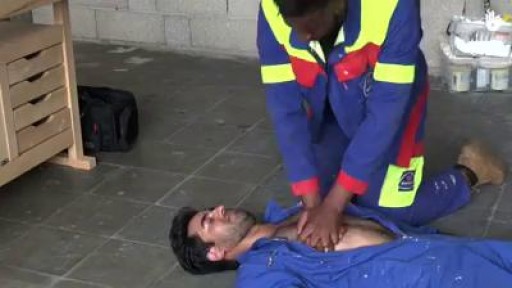
Choking occurs when a foreign object becomes lodged in the throat or windpipe, blocking the flow of air. In adults, a piece of food often is the culprit. Young children often swallow small objects. Because choking cuts off oxygen to the brain, administer first aid as quickly as possible. The universal sign for choking is hands clutched to the throat. If the person doesn't give the signal, look for these indications: Inability to talk Difficulty breathing or noisy breathing Inability to cough forcefully Skin, lips and nails turning blue or dusky Loss of consciousness
Arm Replantation of a Child By Dr. Omid Liaghat : https:drliaghatclinic.com
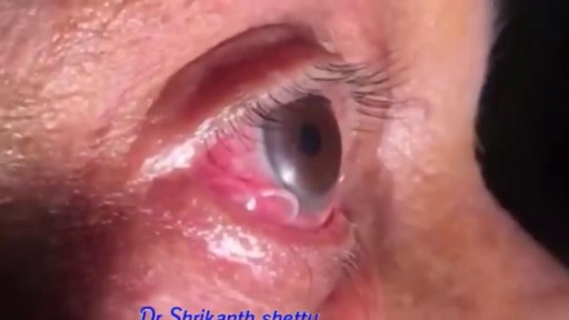
Parasitic Worm removed from Man's EYE
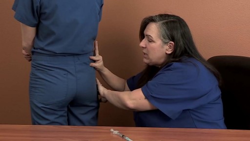
This video demonstrates the process for preparing and administering an intramuscular injection (IM)
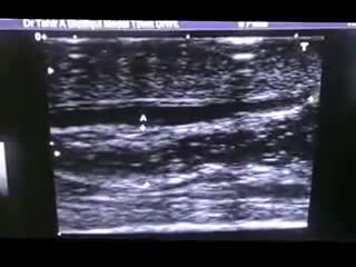
The epididymis is a long coiled tube that lies above and behind each testicle. The epididymis collects and transports sperm from the testis to the vas deferens (tubes that transport sperm to the urethra). An epididymal cyst is a cyst-like mass in the epididymis that contains clear fluid. Typically, epididymal cysts and spermatoceles do not cause symptoms. When discovered, the epididymal cyst is usually about the size of a pea and feels separate from the top of the testis. Spermatoceles typically arise from the head of the epididymis, and are felt on the top portion of the testicle. Epididymal cysts and spermatoceles are often incidental findings on testicular self-examination or routine physical examination. It is important that any mass noted in the scrotum be examined by a urologist in order to obtain an accurate diagnosis, especially a mass on the testicle itself. Our team in the Division of Urology will typically be able to confirm the diagnosis on physical exam. However, a scrotal ultrasound may also be used in order to rule out other conditions.
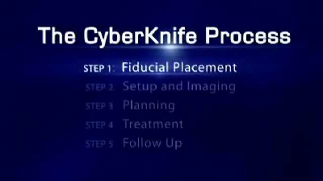
Radiosurgery: Radiosurgery devices, such as the CyberKnife Robotic Radiosurgery System, offer patients a new option for the treatment of lung cancer. The CyberKnife® System is used to treat lung cancer patients who cannot tolerate surgery, have an inoperable tumor, or are seeking an alternative to surgery.

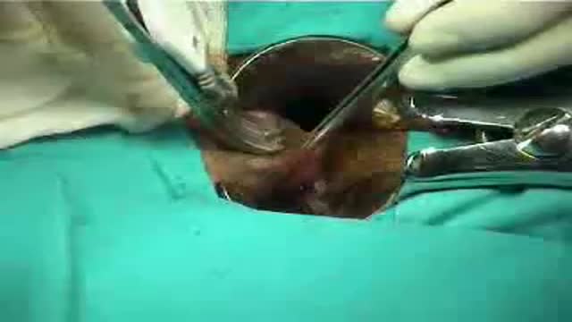
Tracheostomy

Integrative Physical Examination Lecture
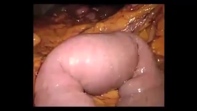
Gallstone ileus is an important, though infrequent, cause of mechanical bowel obstruction, affecting older adult patients who often have other significant medical conditions. It is caused by impaction of a gallstone in the ileum after being passed through a biliary-enteric fistula. The diagnosis is often delayed since symptoms may be intermittent and investigations fail to identify the cause of the obstruction. The mainstay of treatment is removal of the obstructing stone after resuscitating the patient. Gallstone ileus continues to be associated with relatively high rates of morbidity and mortality.


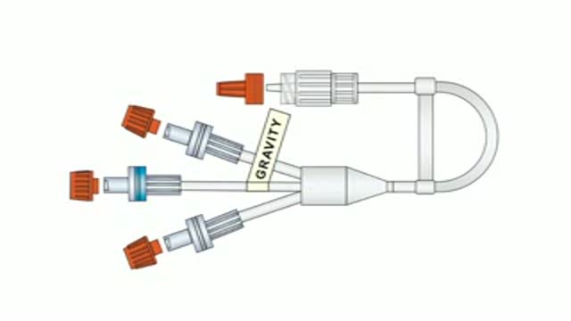
To facilitate the delivery of fluids during labour, obstetric anaesthetists from Coventry designed a triple IV peripheral connector with a central high-flow anti-reflux valve. This connector, now rightly known as the Coventry valve, can be used for all theatre settings and is especially useful in obstetrics, ICU, HDU and orthopaedics. More details on www.mediplus.co.uk
-Tibial stress fractures are common in athletes and nonathletes who suddenly increase their physical activity. Clinical features include pain, localized tenderness, and swelling. Plain x-ray is <50% sensitive for stress fractures, especially in the first 2-3 weeks after the onset of symptoms. MRI is preferred over bone scan or ultrasound as it can show the fracture line that extends through the cortex into the medullary line. MRI can also identify ligament, muscle, and cartilage injuries. However, MRI findings may be persistently abnormal for up to 1 year after the stress fracture has healed.
