- Physical Examination
- Surgical Examination
- Ophthalmology
- Clinical Skills
- Orthopedics
- Surgery Videos
- Laparoscopy
- Pediatrics
- Funny Videos
- Cardiothoracic Surgery
- Nursing Videos
- Plastic Surgery
- Otorhinolaryngology
- Histology and Histopathology
- Neurosurgery
- Dermatology
- Pediatric Surgery
- Urology
- Dentistry
- Oncology and Cancers
- Anatomy Videos
- Health and Fitness
- Radiology
- Anaesthesia
- Physical Therapy
- Pharmacology
- Interventional Radiology
- Cardiology
- Endocrinology
- Gynecology
- Emergency Medicine
- Psychiatry and Psychology
- Childbirth Videos
- General Medical Videos
- Nephrology
- Physiology
- Diet and Food Health
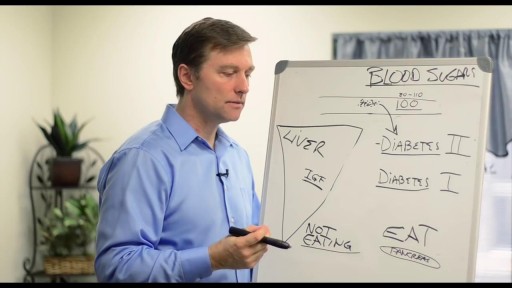
- Diabetes Mellitus
- Neurology
- Women Health
- Osteoporosis
- Gastroenterology
- Pulmonology
- Hematology
- Rheumatology
- Toxicology
- Nuclear Medicine
- Infectious Diseases
- Vascular Disease
- Reproductive Health
- Burns and Wound Healing
- Other
Top videos
In this podcast, CDC's Dr. Barbara Reynolds discusses best practices in crisis and emergency risk communication. She characterizes the initial phase of the crisis communication lifecycle and describes the five most common mistakes made in emergency communication to the public and how to counter them.

Revision of Mini Gastric ByPass

Classical PKU is an autosomal recessive disorder, caused by mutations in both alleles of the gene for phenylalanine hydroxylase (PAH), found on chromosome 12. In the body, phenylalanine hydroxylase converts the amino acid phenylalanine to tyrosine, another amino acid.

Cervicitis is an inflammation of the cervix, the lower, narrow end of the uterus that opens into the vagina. Possible symptoms of cervicitis include bleeding between menstrual periods, pain with intercourse or during a cervical exam, and abnormal vaginal discharge. However, it's also possible to have cervicitis and not experience any signs or symptoms. Often, cervicitis results from a sexually transmitted infection, such as chlamydia or gonorrhea. Cervicitis can develop from noninfectious causes, too. Successful treatment of cervicitis involves treating the underlying cause of the inflammation.
Problems that affect ovulation, and the hormones involved with ovulation, are the most common cause of female infertility. They include: Polycystic Ovarian Syndrome (PCOS). Women with PCOS do not ovulate regularly and they experience infrequent or absent menstrual cycles.

Hemophilia B is a hereditary bleeding disorder caused by a lack of blood clotting factor IX. Without enough factor IX, the blood cannot clot properly to control bleeding.

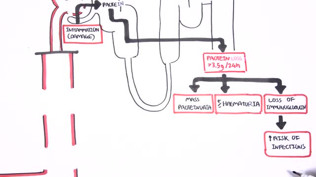
Nephrotic syndrome is a kidney disorder that causes your body to excrete too much protein in your urine. Nephrotic syndrome is usually caused by damage to the clusters of small blood vessels in your kidneys that filter waste and excess water from your blood. Nephrotic syndrome causes swelling (edema), particularly in your feet and ankles, and increases the risk of other health problems. Treatment for nephrotic syndrome includes treating the underlying condition that's causing it and taking medications. Nephrotic syndrome can increase your risk of infections and blood clots. Your doctor may recommend medications and dietary changes to prevent these and other complications of nephrotic syndrome.
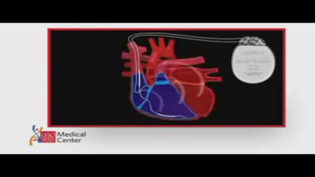
ICDs are useful in preventing sudden death in patients with known, sustained ventricular tachycardia or fibrillation. Studies have shown ICDs to have a role in preventing cardiac arrest in high-risk patients who haven't had, but are at risk for, life-threatening ventricular arrhythmias. View an animation of an ICD. Newer-generation ICDs may have a dual function which includes the ability to serve as a pacemaker. The pacemaker feature would stimulate the heart to beat if the heart rate is detected to be too slow. What is an Implantable Cardioverter Defibrillator (ICD)? An ICD is a battery-powered device placed under the skin that keeps track of your heart rate. Thin wires connect the ICD to your heart. If an abnormal heart rhythm is detected the device will deliver an electric shock to restore a normal heartbeat if your heart is beating chaotically and much too fast. ICDs have been very useful in preventing sudden death in patients with known, sustained ventricular tachycardia or fibrillation. Studies have shown that they may have a role in preventing cardiac arrest in high-risk patients who haven't had, but are at risk for, life-threatening ventricular arrhythmias.

New device claims to stimulate brain for depression treatment
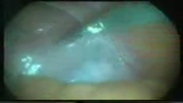
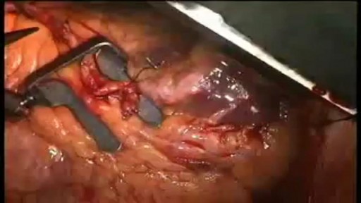
Laparoscopic varicocellectomy Surgery
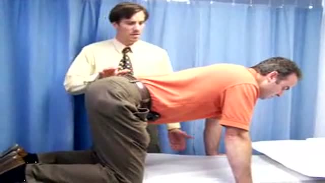
Video demonstrates the action of the isolated lumbar multifidis muscle

Ultrasound Guided Sclerotherapy for Varicose Veins

How do you make a working human heart? Scientists can turn stem cells into beating heart cells, but getting them to organize into a 3D heart requires a scaffold. At the Massachusetts General Hospital in Boston, Harald Ott and his team are reusing the scaffold that nature provides. They’re stripping away all the living cells from dead hearts, before filling in the leftover matrix with healthy new cells. In this video, Brendan Maher finds out how the technique could be used to develop parts of the heart, like the aortic root and valve, for transplant.
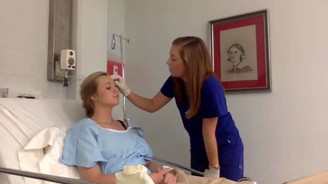
Physical assessment is taking an educated, systematic look at all aspects of an individual’s health status utilizing knowledge, skills and tools of health history and physical exam. To collect data- information about the client’s health, including physiological, psychological, sociocultural and spiritual aspects To establish actual and potential problems To establish the nurse-client relationship Method: The history is done first, then the physical examination focuses on finding data associated with the history. Health History- obtained through interview and record review. Physical exam- accomplished by tools and techniques ** A complete assessment is not necessarily carried out each time. A comprehensive assessment is part of a health screening examination. On admission, you will do an admission assessment (not necessarily including everything presented here) and document it on the admission form. You will do a daily shift assessment (patient systems review). And, if client has a specific problem, you may assess only that part of the body (focused). Data Collection: Information is organized into objective and subjective data: Subjective: Apparent only to person affected; includes client’s perceptions, feelings, thoughts, and expectations. It cannot be directly observed and can be discovered only asking questions. Objective: Detectable by an observer or can be tested against an acceptable standard; tangible, observable facts; includes observation of client behavior, medical records, lab and diagnostic tests, data collected by physical exam. ** To obtain data for the nursing health history, you must utilize good interview techniques and communications skills. Record accurately. DO NOT ASSUME. D. Frameworks for Health Assessment There are two main frameworks utilized in health assessment: Head to Toe- systematic collection of data starting with the head and working downward. Functional Health Assessment- Gordon’s 11 functional health patterns that address the behaviors a person uses to maintain health. PERSON is the ACC-ADN framework for assessment. It is similar to Gordon's functional health patterns.
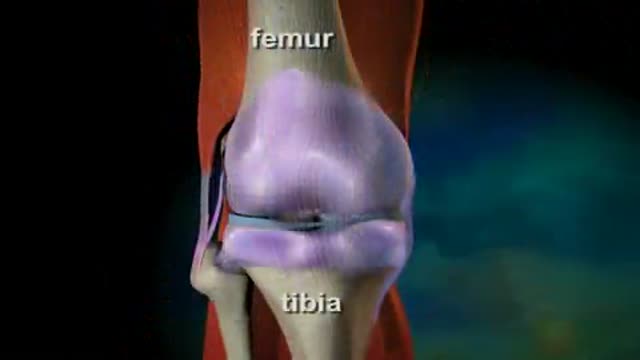
Osteoarthritis is a degenerative joint disease that is caused by the chronic breakdown and eventual loss of cartilage within the joints. As the cartilage wears away, the bones that meet at the joint begin to rub against each other. This can cause extreme pain and can severely reduce movement and flexibility of the joint. Growths of bone, called bone spurs, can also form around the edges of the joint and cause pain. Joint swelling can also occur if the synovial membrane lining the joint becomes irritated, producing excess fluid that collects inside the joint. What Causes Osteoarthritis? More than half of the population age 65 or older have osteoarthritis in at least one joint. Osteoarthritis usually results from injury to a joint or from wear and tear over time. Heredity, lack of use, and being overweight also contribute to the development of osteoarthritis. Treating Osteoarthritis Treatment can include weight loss, physiotherapy, and medication. If the condition becomes severe and mobility is greatly reduced, hip replacement surgery may be necessary.
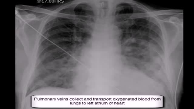
Expand Section. Pulmonary edema is often caused by congestive heart failure. When the heart is not able to pump efficiently, blood can back up into the veins that take blood through the lungs. As the pressure in these blood vessels increases, fluid is pushed into the air spaces (alveoli) in the lungs.
When your arteries cannot supply enough blood to your heart, your doctor may recommend coronary artery bypass graft (CABG) surgery. One of the most common heart surgeries in the United States, CABG surgery restores blood flow to your heart. Approximately every 10 minutes, someone has beating heart or "off-pump" bypass surgery1. Beating heart bypass surgery is — in simple terms — bypass surgery that is performed on your heart while it is beating. Your heart will not be stopped during surgery. You will not need a heart-lung machine. Your heart and lungs will continue to perform during your surgery. Surgeons use a tissue stabilization system to immobilize the area of the heart where they need to work. Beating heart bypass surgery is also called Off Pump Coronary Artery Bypass Surgery (OPCAB). Both OPCAB and conventional on-pump surgery restore blood flow to the heart. However, off-pump bypass surgery has proven to reduce side effects in certain types of patients.
