- Physical Examination
- Surgical Examination
- Ophthalmology
- Clinical Skills
- Orthopedics
- Surgery Videos
- Laparoscopy
- Pediatrics
- Funny Videos
- Cardiothoracic Surgery
- Nursing Videos
- Plastic Surgery
- Otorhinolaryngology
- Histology and Histopathology
- Neurosurgery
- Dermatology
- Pediatric Surgery
- Urology
- Dentistry
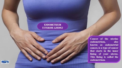
- Oncology and Cancers
- Anatomy Videos
- Health and Fitness
- Radiology
- Anaesthesia
- Physical Therapy
- Pharmacology
- Interventional Radiology
- Cardiology
- Endocrinology
- Gynecology
- Emergency Medicine
- Psychiatry and Psychology
- Childbirth Videos
- General Medical Videos
- Nephrology
- Physiology
- Diet and Food Health
- Diabetes Mellitus
- Neurology
- Women Health
- Osteoporosis
- Gastroenterology
- Pulmonology
- Hematology
- Rheumatology
- Toxicology
- Nuclear Medicine
- Infectious Diseases
- Vascular Disease
- Reproductive Health
- Burns and Wound Healing
- Other
Top videos
An omentectomy is a surgical procedure designed to remove the omentum, which is a thin fold of abdominal tissue that encases the stomach, large intestine and other abdominal organs. This fatty lining contains lymph nodes, lymph vessels, nerves and blood vessels.
Revision of Mini Gastric ByPass

Generic minoxidil is known to treat hair-fall issues in men and women, it is best for hair growth, hair re-development, etc. it is available in the strength of 5mg and easily available at online pharmacy store. For more information visit to http://www.medstorerx.com/generic-minoxidil.aspx
Spermatogenesis is the process in which spermatozoa are produced from spermatogonial stem cells by way of mitosis and meiosis. The initial cells in this pathway are called spermatogonia, which yield primary spermatocytes by mitosis.

Gastric bypass, also called Roux-en-Y gastric bypass surgery, is considered a “metabolic” procedure because it changes how your body absorbs fat, calories and nutrients. This metabolic change occurs because your gastrointestinal tract is altered when your gastric bypass surgeon attaches the smaller section of your stomach directly to your small intestine. As a result, your appetite changes and you feel full faster.
Obesity is the abnormal condition that causes a person to put on excessive amounts of weight due to accumulation of fat in their body. This extreme weight causes a variety of other disorders and diseases as complications associated with it. https://goo.gl/GgSAoY

Ascites, the collection of fluid within the peritoneal space is caused due to a variety of causes including cirrhosis, cardiac causes, sinusoidal obstruction syndrome, tubercular peritonitis and pancreatitis, amongst others. Most commonly, the cause of ascots may be cirrhosis , which in turn, is most frequently causes by alcohol use, hepatitis C and non-alcoholic steatohepatitis. At the heart of the ascitic fluid analysis is the serum albumin ascitic gradient, the differential diagnosis of which has been discussed in detail in this presentation. Both low SAAG and high SAAG ascites have been dealt with in some depth, with a brief overview of the management of these conditions

Generalized Anxiety Disorder, Symptoms Of Anxiety Attack, Shortness Of Breath Anxiety --- http://panic-attacks-anxiety.good-info.co --- Newly Discovered Panic "Off Switch" Gives You Anxiety Relief Without Pills or Therapy Here's an interesting fact about anxiety and panic attacks: Did you know that just like the hiccups, doctors still can't agree exactly why they happen to you? And did you also know there's a 60-second solution to panic and anxiety that you can do anywhere? Yes, it takes you just one minute and I'm going to share it with you today. Until one day about a year ago, I thought I might be doomed to let panic attacks rule my life. And I made this free online presentation to tell you about the one discovery about panic and general anxiety that finally cut through the confusion and changed everything. Pay very close attention, because whether you've only had one or two "attacks" so far… or even if you've been having them for years and it seems like a life sentence you'll never escape from… You're about to discover one weird thing that panic, anxiety and the hiccups – yes, the hiccups – have in common that goes right back to the stone age. Discover How To Begin Eliminating Panic And Anxiety From Your Life Forever Click Here: http://panic-attacks-anxiety.good-info.co
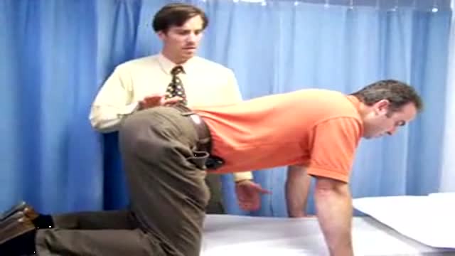
Video demonstrates the action of the isolated lumbar multifidis muscle
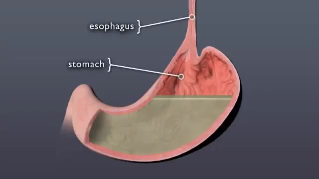
Discover what happens to pill when it swallowed

Ultrasound Guided Sclerotherapy for Varicose Veins

People with celiac disease may lose weight because their bodies are not able to absorb enough nutrients from food. Over time, a range of problems may develop as a result of the body's reaction to gluten — from skin rashes and lactose intolerance to infertility, bone weakness and nerve damage.

ROTIGS medical device by Honolulu inventor Dr. Brad NaPier makes airway intubations easier for medical professionals.

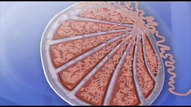
It is a specialized bodily fluid that supplies essential substances and nutrients, such as sugar, oxygen, and hormones to our cells, and carries waste away from those cells, this waste is eventually flushed out of the body in urine, feces, sweat, and lungs (carbon dioxide). Blood also contains clotting agents.

Classical PKU is an autosomal recessive disorder, caused by mutations in both alleles of the gene for phenylalanine hydroxylase (PAH), found on chromosome 12. In the body, phenylalanine hydroxylase converts the amino acid phenylalanine to tyrosine, another amino acid.
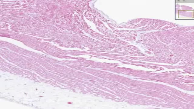
Histology of Heart Cardiac Muscle
The spleen is one of the most overlooked organs. Rarely does it get attention unless there is some kind of accident or trauma. However, I find spleen dysfunction to be very prevalent. This video talks about some of the symptoms.

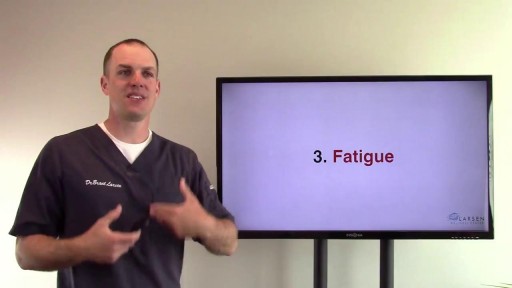
You might not notice signs or symptoms of Hashimoto's disease at first, or you may notice a swelling at the front of your throat (goiter). Hashimoto's disease typically progresses slowly over years and causes chronic thyroid damage, leading to a drop in thyroid hormone levels in your blood. The signs and symptoms are mainly those of an underactive thyroid gland (hypothyroidism). Signs and symptoms of hypothyroidism include: Fatigue and sluggishness Increased sensitivity to cold Constipation Pale, dry skin A puffy face Hoarse voice Unexplained weight gain — occurring infrequently and rarely exceeding 10 to 20 pounds, most of which is fluid Muscle aches, tenderness and stiffness, especially in your shoulders and hips Pain and stiffness in your joints and swelling in your knees or the small joints in your hands and feet Muscle weakness, especially in your lower extremities Excessive or prolonged menstrual bleeding (menorrhagia) Depression
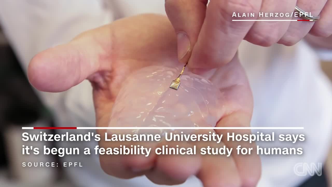
Scientists have developed a wireless brain implant that enabled a paralyzed monkey to walk again.
