- Physical Examination
- Surgical Examination
- Ophthalmology
- Clinical Skills
- Orthopedics
- Surgery Videos
- Laparoscopy
- Pediatrics
- Funny Videos
- Cardiothoracic Surgery
- Nursing Videos
- Plastic Surgery
- Otorhinolaryngology
- Histology and Histopathology
- Neurosurgery
- Dermatology
- Pediatric Surgery
- Urology
- Dentistry
- Oncology and Cancers
- Anatomy Videos
- Health and Fitness
- Radiology
- Anaesthesia
- Physical Therapy
- Pharmacology
- Interventional Radiology
- Cardiology
- Endocrinology
- Gynecology
- Emergency Medicine
- Psychiatry and Psychology
- Childbirth Videos
- General Medical Videos
- Nephrology
- Physiology
- Diet and Food Health
- Diabetes Mellitus
- Neurology
- Women Health
- Osteoporosis
- Gastroenterology
- Pulmonology
- Hematology
- Rheumatology
- Toxicology
- Nuclear Medicine
- Infectious Diseases
- Vascular Disease
- Reproductive Health
- Burns and Wound Healing
- Other
Top videos
A talus fracture is a break in one of the bones that forms the ankle. This type of fracture often occurs during a high-energy event, such as a car collision or a high-velocity fall. Because the talus is important for ankle movement, a fracture often results in significant loss of motion and function. In addition, a talus fracture that does not heal properly can lead to serious complications, including chronic pain. For this reason, many talus fractures require surgery.

Female Condom Demonstration
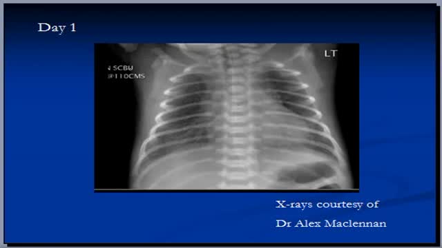
The video will describe RDS in premature babies. Please see website for disclaimer

Male and female Foley catheter insertion into bladder. Kearn how to
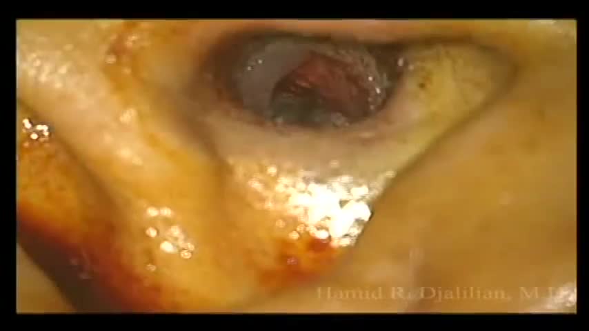
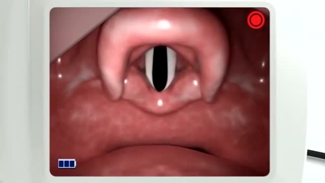
This is a surgery showing the removal of a large exostosis. Exostoses are bony growths in the ear canal from chronic exposure to cold water/air, most commonly from surfing. This patient had growths in both ears, which were completely obstructing the ear canals. This patient had a single exostosis that was blocking this side (the right side).

This particular video is intended as a demonstration of Neurologic Examination. This demonstration is intended as an example of a neurologic exam which may be used as part of the initial evaluation of patients with complaints that may have an underlying neurologic origin. This video is solely for educational purposes and intended for use to prepare for OSCEs incorporating standardized patient encounters. It is not intended as a demonstration of a comprehensive neurologic examination and is not intended as medical advice or medical guidelines.
It is not intended as a complete instructional video and should not be considered a source of complete physical examination instruction.
Instead, it should be treated as a supplement to independent learning using primary Osteopathic Clinical Skills instructional resources. Clinical skills are best learned and developed with support from faculty in the context of a complete Osteopathic Medical School Curriculum.
Osteopathic Clinical Skills is a channel dedicated to discussing and exploring Osteopathic Clinical Skills concepts for medical students, residents, and clinicians and presenting them in an easy to understand manner.
Attributions:
Many thanks to the University of North Texas Health Science Center Texas College of Osteopathic Medicine (UNTHSC - TCOM) for permitting use of the Simulation facilities and equipment during the production of this video.
Additional thanks to the UNTHSC-TCOM standardized patient and faculty volunteers who participated in this production and provided permission for the use of their image in this video.

Each month inside your ovaries, a group of eggs starts to grow in small, fluid-filled sacs called follicles. Eventually, one of the eggs erupts from the follicle (ovulation). It usually happens about 2 weeks before your next period. Hormones Rise After the egg leaves the follicle, the follicle develops into something called the corpus luteum. The corpus luteum releases a hormone that helps thicken the lining of your uterus, getting it ready for the egg. The Egg Travels to the Fallopian Tube After the egg is released, it moves into the Fallopian tube. It stays there for about 24 hours, waiting for a single sperm to fertilize it. All this happens, on average, about 2 weeks after your last period.
Pediatric 4-Step Basic Technique
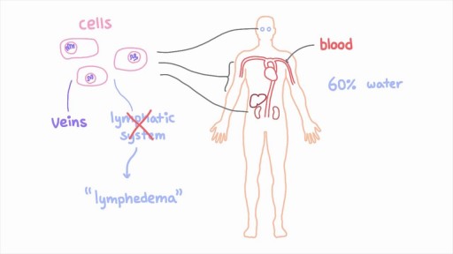
The lymphatic system is a network of specialized vessels (lymph vessels) throughout the body whose purpose is to collect excess lymph fluid with proteins, lipids, and waste products from the tissues. This fluid is then carried to the lymph nodes, which filter waste products and contain infection-fighting cells called lymphocytes. The excess fluid in the lymph vessels is eventually returned to the bloodstream. When the lymph vessels are blocked or unable to carry lymph fluid away from the tissues, localized swelling (lymphedema) is the result.
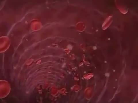
Sickle cell anemia (sickle cell disease) is a disorder of the blood caused by an inherited abnormal hemoglobin (the oxygen-carrying protein within the red blood cells). The abnormal hemoglobin causes distorted (sickled) red blood cells.
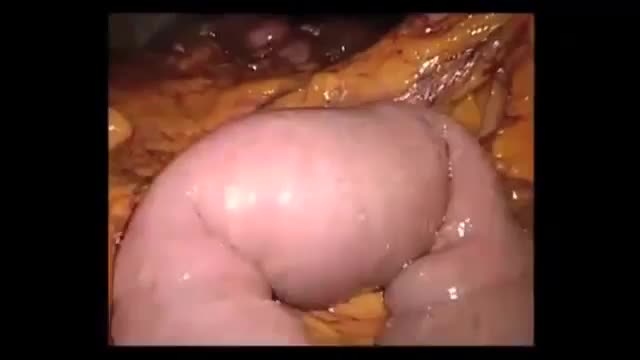
Gallstone ileus is an important, though infrequent, cause of mechanical bowel obstruction, affecting older adult patients who often have other significant medical conditions. It is caused by impaction of a gallstone in the ileum after being passed through a biliary-enteric fistula. The diagnosis is often delayed since symptoms may be intermittent and investigations fail to identify the cause of the obstruction. The mainstay of treatment is removal of the obstructing stone after resuscitating the patient. Gallstone ileus continues to be associated with relatively high rates of morbidity and mortality.

What is vascular access? What are the different types of accesses for hemodialysis? Does vascular access require surgery? Adina Voiculescu, M.D., FASDIN, General and Interventional Nephrologist at Brigham and Women's Hospital and Assistant Professor at Harvard Medical School, discusses the different types of vascular access, such as AV fistulas and AV grafts, and how to stay healthy while on hemodialysis.
Subscribe Link: https://www.youtube.com/channe....l/UCYrLjATd88gPwIKnt
0:00 - Intro
0:29 - Peritoneal dialysis & Hemodialysis
0:44 - Types of access to perform dialysis
1:48 - Recommendations
About Mass General Brigham:
Mass General Brigham combines the strength of two world-class academic medical centers, five nationally ranked specialty hospitals, 11 community hospitals, and dozens of health centers. Our doctors and researchers accelerate medical breakthroughs and drive innovations in patient care. They are leaders in medical education, serving as Harvard Medical School faculty and training the next generation of physicians. Mass General Brigham’s mission is to deliver the best, affordable health care to patients everywhere. Together, we transform the health of our communities and beyond.
#MassGeneralBrigham #MGB #Hemodialysis
Visit Mass General Brigham: https://www.massgeneralbrigham.org/
Find us on social:
Twitter: https://twitter.com/MassGenBrigham
Instagram: https://www.instagram.com/massgeneralbrigham/
Facebook: https://www.facebook.com/MassGeneralBrigham/
LinkedIn: https://www.linkedin.com/compa....ny/mass-general-brig
Mass General Brigham:
https://www.youtube.com/massgeneralbrigham
Hemodialysis: Types of Accesses for Kidney Dialysis and How to Stay Healthy | Mass General Brigham
https://youtu.be/_bxLpudpqnc

There's a small area called the Grafenberg spot, or G-spot, inside the vagina. It's located about an inch or so inside the vaginal opening on the upper vaginal wall — closest to the bellybutton. The G-spot is sexually sensitive and swells slightly during arousal and feels raised or bumpy
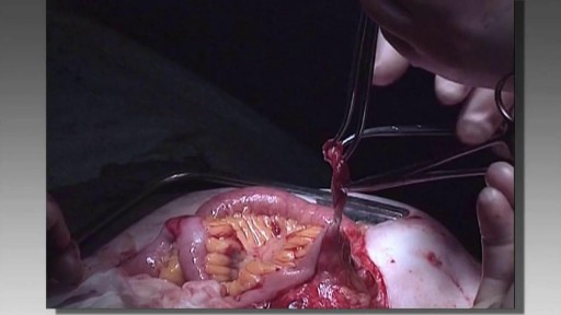
Totally Stapled Bowel Resection and Anastomosis

.

Ever wanted to see an open heart surgery? Dr. Sandwith, the only open-heart surgeon in the tri-county area, takes you into the OR to improve the life of a gentlemen with congenital heart disease.
#HCA_FL #FortWaltonDestinHospital
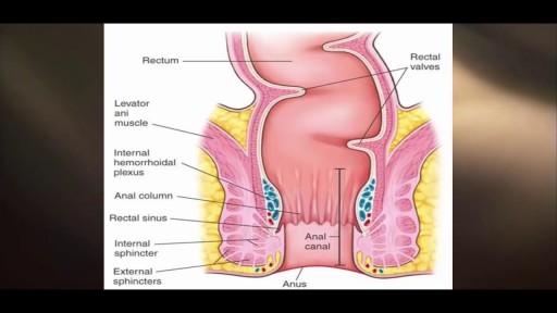
The only way to completely avoid anal sex risks is to abstain from anal sex. If you engage in anal sex, it is always important to use a condom to protect against the spread of infections and diseases.

Rare condition disorder known as Diprosopus, also known as craniofacial duplication. Diprosopus is a congenital defect also known as craniofacial duplication. The exact description of diprosopus refers to a fetus with a single trunk, normal limbs, and facial features that are duplicated to a certain degree. A less severe instance is when the fetus has a duplicated nose and the eyes are spaced far apart. In the most extreme instances, the entire face is duplicated, hence the name diprosopus, which is Greek for two-faced. Fetuses with diprosopus often also lack brains (anencephaly), have neural tube defects, or heart malformations. In some cases, if the brain is formed, it may have duplicated structures. Most infants with diprosopus are stillborn and there are fewer than fifty cases documented since 1864.
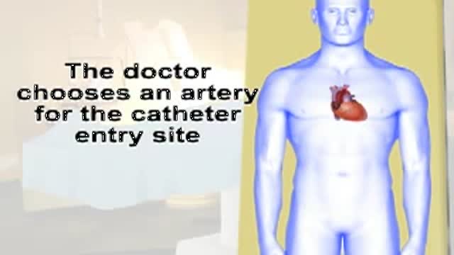
This video gives you an overview of how a cardiac catheterization is performed.
