- Physical Examination
- Surgical Examination
- Ophthalmology
- Clinical Skills
- Orthopedics
- Surgery Videos
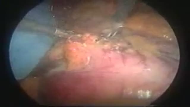
- Laparoscopy
- Pediatrics
- Funny Videos
- Cardiothoracic Surgery
- Nursing Videos
- Plastic Surgery
- Otorhinolaryngology
- Histology and Histopathology
- Neurosurgery
- Dermatology
- Pediatric Surgery
- Urology
- Dentistry
- Oncology and Cancers
- Anatomy Videos
- Health and Fitness
- Radiology
- Anaesthesia
- Physical Therapy
- Pharmacology
- Interventional Radiology
- Cardiology
- Endocrinology
- Gynecology
- Emergency Medicine
- Psychiatry and Psychology
- Childbirth Videos
- General Medical Videos
- Nephrology
- Physiology
- Diet and Food Health
- Diabetes Mellitus
- Neurology
- Women Health
- Osteoporosis
- Gastroenterology
- Pulmonology
- Hematology
- Rheumatology
- Toxicology
- Nuclear Medicine
- Infectious Diseases
- Vascular Disease
- Reproductive Health
- Burns and Wound Healing
- Other
Top videos

The menstrual cycle is the regular natural change that occurs in the female reproductive system that makes pregnancy possible. The cycle is required for the production of oocytes, and for the preparation of the uterus for pregnancy.
Watch that video of a Snake bite causes girl’s leg to rot away with necrosis
The spleen is one of the most overlooked organs. Rarely does it get attention unless there is some kind of accident or trauma. However, I find spleen dysfunction to be very prevalent. This video talks about some of the symptoms.

Bone pain: Pain is the most common sign of bone cancer, and may become more noticeable as the tumor grows. Bone pain can cause a dull or deep ache in a bone or bone region (e.g., back, pelvis, legs, ribs, arms). Early on, the pain may only occur at night, or when you are active.
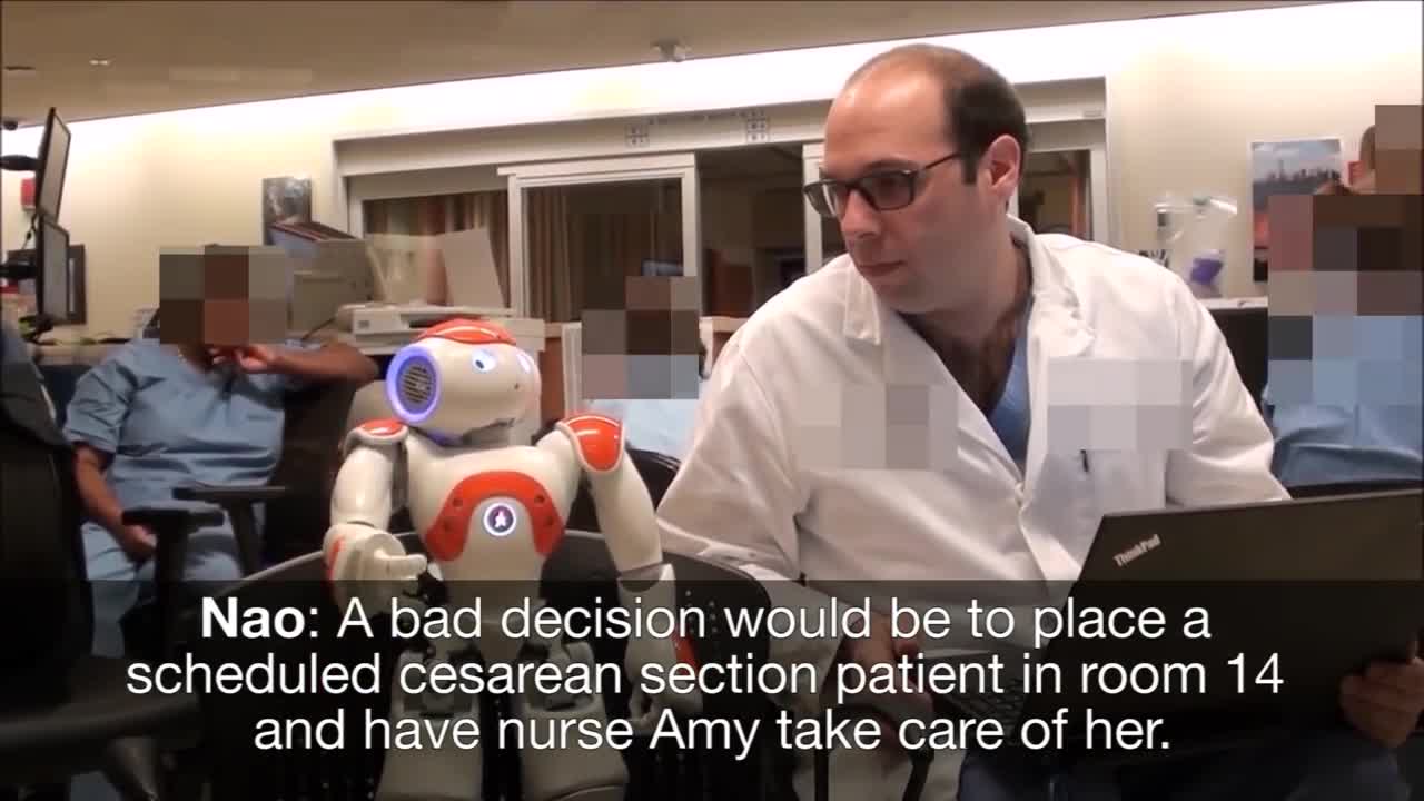
Medical Robot Assistants, new technology
Mini Gastric Bypass surgery Operation
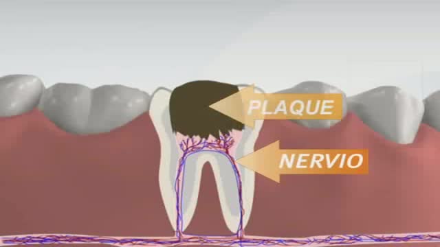
Root canals are common procedures and can help save your tooth from extraction. Dentists at Aspen Dental practices have been safely and expertly performing root canal procedures for over two decades.
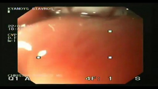
A 76 year-old, female, presented with a three day history of melena without any abdominal pain. She had one episode of hematemesis (about 100 ml blood) in the emergency room, patient has a strong alcoholic drink abuse.
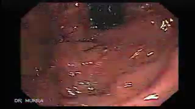
An upper endoscopy with magnification was performed.
multiple ulcers were detected across of the gastric camera,
esophageal varices was also detected
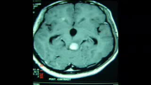
Complete Pineal Tumor excision by using Supracerebellar Infratentorial approach in sitting position was performed. The young adult male is up and about after surgery.
Hysterectomy is the surgical removal of the uterus. It ends menstruation and the ability to become pregnant. Depending on the reason for the surgery, a hysterectomy may also involve the removal of other organs and tissues such as the ovaries and/or fallopian tubes.
Barrett's esophagus is a complication of chronic (long lasting) and usually severe gastrointestinal reflux disease (GERD), but occurs in only a small percentage of patients with GERD. Criteria are needed for screening patients with GERD for Barrett's esophagus. Until validated criteria are available, it seems reasonable to do screening endoscopies in GERD patients who cannot be taken off acid suppression therapy after two to three years. The diagnosis of Barrett's esophagus rests upon seeing (at endoscopy) a pink esophageal lining that extends a short distance (usually less than 2.5 inches) up the esophagus from the gastroesophageal junction and finding intestinal type cells (goblet cells) on biopsy of the lining. There is a small but definite increased risk of cancer of the esophagus (adenocarcinoma) in patients with Barrett's esophagus.

Thoracic outlet syndrome is a group of disorders that occur when blood vessels or nerves in the space between your collarbone and your first rib (thoracic outlet) are compressed. This can cause pain in your shoulders and neck and numbness in your fingers. Common causes of thoracic outlet syndrome include physical trauma from a car accident, repetitive injuries from job- or sports-related activities, certain anatomical defects (such as having an extra rib), and pregnancy. Sometimes doctors can't determine the cause of thoracic outlet syndrome. Treatment for thoracic outlet syndrome usually involves physical therapy and pain relief measures. Most people improve with these approaches. In some cases, however, your doctor may recommend surgery.

Generic minoxidil is known to treat hair-fall issues in men and women, it is best for hair growth, hair re-development, etc. it is available in the strength of 5mg and easily available at online pharmacy store. For more information visit to http://www.medstorerx.com/generic-minoxidil.aspx
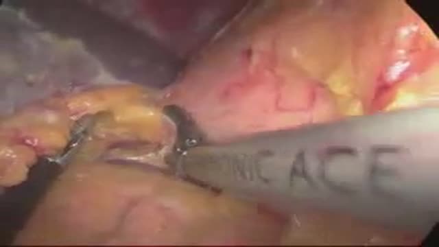
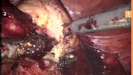
laparoscopic left adrenalectomy in 150kg patient with Cushings

Retinoblastoma is an eye cancer that begins in the retina — the sensitive lining on the inside of your eye. Retinoblastoma most commonly affects young children, but can rarely occur in adults. Your retina is made up of nerve tissue that senses light as it comes through the front of your eye. The retina sends signals through your optic nerve to your brain, where these signals are interpreted as images. A rare form of eye cancer, retinoblastoma is the most common form of cancer affecting the eye in children. Retinoblastoma may occur in one or both eyes.
TPE removes large-molecular-weight substances such as harmful antibodies from the plasma. It is usually carried out using an automated blood cell separator to ensure fluid balance and maintain a normal plasma volume. This may require the insertion of a femoral or jugular line to allow adequate blood flow. Typically, 30–40 mL/kg of plasma (1–1.5 plasma volumes) are removed at each procedure and replaced with isotonic 4.5 or 5.0% human albumin solution (some services substitute 25–50% of replacement volume with 0.9% saline). Exchange with fresh frozen plasma (FFP) is reserved for the replacement of ADAMTS13 in thrombotic thrombocytopenic purpura (see below) or to replace clotting factors. A one plasma volume exchange removes about 66% of an intravascular constituent and a two plasma volume exchange approximately 85%. TPE is normally combined with disease modifying treatment, such as immunosuppressive drugs, for the underlying condition.

How a simple conversation about colon cancer screening can save your life.

It is a specialized bodily fluid that supplies essential substances and nutrients, such as sugar, oxygen, and hormones to our cells, and carries waste away from those cells, this waste is eventually flushed out of the body in urine, feces, sweat, and lungs (carbon dioxide). Blood also contains clotting agents.

People with Extremely Large Body Parts

Rhinoplasty Nose Surgery. one of the most common cosmetic plastic surgery done world wide. Central part of face including forehead , nose and peri oral area ( lips and adjacent area) gives our face a unique feature. Nose, Being the most projected part of face is noticed first when one sees our face. #nosejob #nosereshaping #nosetip #nosesurgery #rhinoplasty #rhinoplastysurgery #cost #price Get more http://www.themedspa.us/cosmetic-surgery/nose-surgery.html Get more http://www.bestfacesurgeryindia.com
