- Physical Examination
- Surgical Examination
- Ophthalmology
- Clinical Skills
- Orthopedics
- Surgery Videos
- Laparoscopy
- Pediatrics
- Funny Videos
- Cardiothoracic Surgery
- Nursing Videos
- Plastic Surgery
- Otorhinolaryngology
- Histology and Histopathology
- Neurosurgery
- Dermatology
- Pediatric Surgery
- Urology
- Dentistry
- Oncology and Cancers
- Anatomy Videos
- Health and Fitness
- Radiology
- Anaesthesia
- Physical Therapy
- Pharmacology
- Interventional Radiology
- Cardiology
- Endocrinology
- Gynecology
- Emergency Medicine
- Psychiatry and Psychology
- Childbirth Videos
- General Medical Videos
- Nephrology
- Physiology
- Diet and Food Health
- Diabetes Mellitus
- Neurology
- Women Health
- Osteoporosis
- Gastroenterology
- Pulmonology
- Hematology
- Rheumatology
- Toxicology
- Nuclear Medicine
- Infectious Diseases
- Vascular Disease
- Reproductive Health
- Burns and Wound Healing
- Other
Top videos
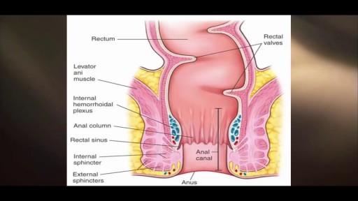
The only way to completely avoid anal sex risks is to abstain from anal sex. If you engage in anal sex, it is always important to use a condom to protect against the spread of infections and diseases.

Pediatric Urine Samples Collection
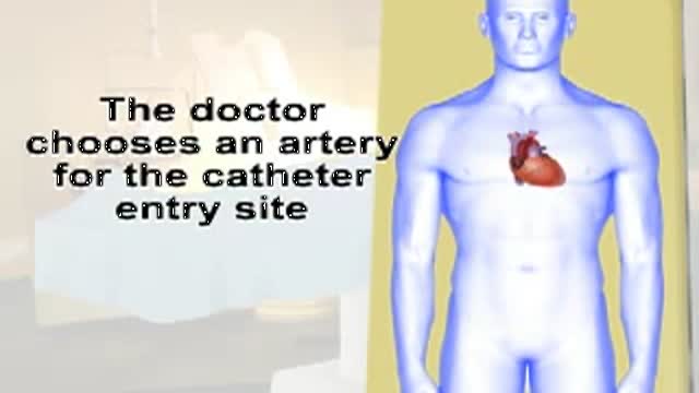
This video gives you an overview of how a cardiac catheterization is performed.

What is vascular access? What are the different types of accesses for hemodialysis? Does vascular access require surgery? Adina Voiculescu, M.D., FASDIN, General and Interventional Nephrologist at Brigham and Women's Hospital and Assistant Professor at Harvard Medical School, discusses the different types of vascular access, such as AV fistulas and AV grafts, and how to stay healthy while on hemodialysis.
Subscribe Link: https://www.youtube.com/channe....l/UCYrLjATd88gPwIKnt
0:00 - Intro
0:29 - Peritoneal dialysis & Hemodialysis
0:44 - Types of access to perform dialysis
1:48 - Recommendations
About Mass General Brigham:
Mass General Brigham combines the strength of two world-class academic medical centers, five nationally ranked specialty hospitals, 11 community hospitals, and dozens of health centers. Our doctors and researchers accelerate medical breakthroughs and drive innovations in patient care. They are leaders in medical education, serving as Harvard Medical School faculty and training the next generation of physicians. Mass General Brigham’s mission is to deliver the best, affordable health care to patients everywhere. Together, we transform the health of our communities and beyond.
#MassGeneralBrigham #MGB #Hemodialysis
Visit Mass General Brigham: https://www.massgeneralbrigham.org/
Find us on social:
Twitter: https://twitter.com/MassGenBrigham
Instagram: https://www.instagram.com/massgeneralbrigham/
Facebook: https://www.facebook.com/MassGeneralBrigham/
LinkedIn: https://www.linkedin.com/compa....ny/mass-general-brig
Mass General Brigham:
https://www.youtube.com/massgeneralbrigham
Hemodialysis: Types of Accesses for Kidney Dialysis and How to Stay Healthy | Mass General Brigham
https://youtu.be/_bxLpudpqnc
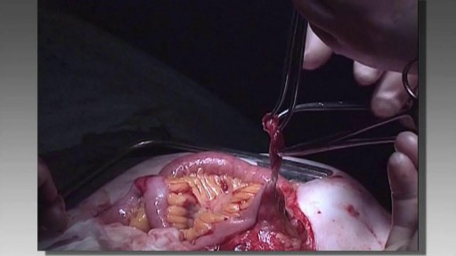
Totally Stapled Bowel Resection and Anastomosis

Rare condition disorder known as Diprosopus, also known as craniofacial duplication. Diprosopus is a congenital defect also known as craniofacial duplication. The exact description of diprosopus refers to a fetus with a single trunk, normal limbs, and facial features that are duplicated to a certain degree. A less severe instance is when the fetus has a duplicated nose and the eyes are spaced far apart. In the most extreme instances, the entire face is duplicated, hence the name diprosopus, which is Greek for two-faced. Fetuses with diprosopus often also lack brains (anencephaly), have neural tube defects, or heart malformations. In some cases, if the brain is formed, it may have duplicated structures. Most infants with diprosopus are stillborn and there are fewer than fifty cases documented since 1864.

Introduction to the Brachial Plexus Examination, 4 of 5 videos demonstrating the physical exam for evaluation of Brachial Plexus conditions.
Brachial plexus injury - Care at Mayo Clinic:
https://www.mayoclinic.org/dis....eases-conditions/bra
Watch all the videos in this series on this playlist:
https://www.youtube.com/playli....st?list=PLSWR1ylG_6J

Menorrhagia is the medical term for menstrual periods with abnormally heavy or prolonged bleeding. Although heavy menstrual bleeding is a common concern, most women don't experience blood loss severe enough to be defined as menorrhagia. With menorrhagia, you can't maintain your usual activities when you have your period because you have so much blood loss and cramping. If you dread your period because you have such heavy menstrual bleeding, talk with your doctor. There are many effective treatments for menorrhagia.
A central venous catheter, also called a central line, is a long, thin, flexible tube used to give medicines, fluids, nutrients, or blood products over a long period of time, usually several weeks or more. A catheter is often inserted in the arm or chest through the skin into a large vein.
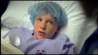
Our surgeons take a compassionate, family-centered approach to both inpatient and outpatient care. We’re committed to making sure both you and your child understand our process. Told through a kid's eyes, this video tour reveals our caring approach.
To learn more about pediatric surgery at Stamford Hospital, visit: https://www.stamfordhealth.org..../care-treatment/pedi
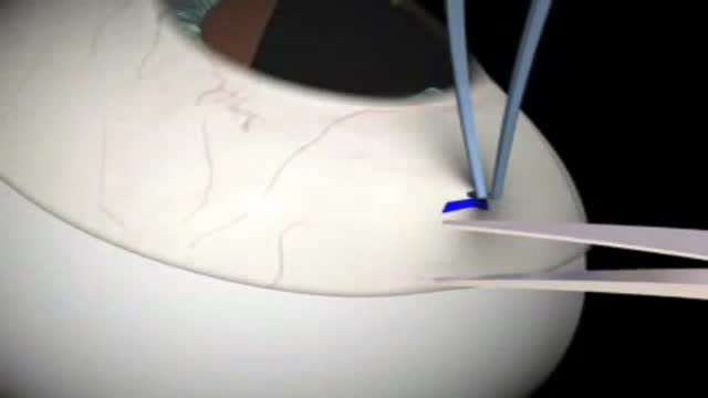
Glaucoma Surgery 3D Animation

Bunions can be very painful. ... Bunion removal is a surgical procedure that corrects a deformed area of the foot near the big toe. Bunion removal is sometimes called a bunionectomy, bunion surgery, or hallux valgus correction. Hallux valgus is a Latin phrase that means “foot deformity

To understand congenital heart defects, it's helpful to know how a healthy heart works. Your child's heart is a muscle about the size of his or her fist. The heart works like a pump and beats 100,000 times a day. The heart has two sides, separated by an inner wall called the septum. The right side of the heart pumps blood to the lungs to pick up oxygen. The left side of the heart receives the oxygen-rich blood from the lungs and pumps it to the body. The heart has four chambers and four valves and is connected to various blood vessels. Veins are blood vessels that carry blood from the body to the heart. Arteries are blood vessels that carry blood away from the heart to the body.

Add Me on
Instagram-https://instagram.com/_dialysi....s_therapist?igshid=Y
Telegram-https://t.me/dialysistherapist
Dialysis
Dialysis technician
Dialysis nurse
Kidney dialysis
Hemolysis
Dialysis technology
Dialysis therapist
Dialysis technician vacancy
Dialysis technician salary
Dialysis scope
Dialysis course scope
B.sc dialysis scope
Salary of dialysis technician
Role of dialysis technician
Salary of dialysis technician in india

According to a recent study, most people's sexual romps last about 1.5–7 minutes. But, as Dr. Harry Fisch writes in his new book The New Naked: The Ultimate Sex Education for Grownups, 45 percent of men come in two minutes or less, leaving their female partners orgasmless. Here are some ways to extend your man's sexual stamina, and more likely have an orgasm yourself in the process.
What is a brain aneurysm? A brain (cerebral) aneurysm is a bulging, weak area in the wall of an artery that supplies blood to the brain. In most cases, a brain aneurysm causes no symptoms and goes unnoticed. In rare cases, the brain aneurysm ruptures, releasing blood into the skull and causing a stroke. When a brain aneurysm ruptures, the result is called a subarachnoid hemorrhage. Depending on the severity of the hemorrhage, brain damage or death may result.

Endoscopic fenestration of suprasellar cyst in a 4 years old girl
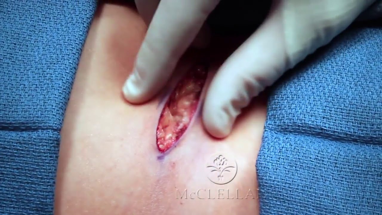
Watch that video to know How to Perform Invisible Skin Sutures Technique

To avoid pregnancy and STDs, always remember to use a condom every time you have sex — including oral, vaginal, or anal sex. Whenever oral sex is being performed on a girl, a dental dam should be used. A guy receiving oral sex should wear a latex condom — or, if he or his partner is allergic to latex, a polyurethane condom.
