- Physical Examination
- Surgical Examination
- Ophthalmology
- Clinical Skills
- Orthopedics
- Surgery Videos
- Laparoscopy
- Pediatrics
- Funny Videos
- Cardiothoracic Surgery
- Nursing Videos
- Plastic Surgery
- Otorhinolaryngology
- Histology and Histopathology
- Neurosurgery
- Dermatology
- Pediatric Surgery
- Urology
- Dentistry
- Oncology and Cancers
- Anatomy Videos
- Health and Fitness
- Radiology
- Anaesthesia
- Physical Therapy
- Pharmacology
- Interventional Radiology
- Cardiology
- Endocrinology
- Gynecology
- Emergency Medicine
- Psychiatry and Psychology
- Childbirth Videos
- General Medical Videos
- Nephrology
- Physiology
- Diet and Food Health
- Diabetes Mellitus
- Neurology
- Women Health
- Osteoporosis
- Gastroenterology
- Pulmonology
- Hematology
- Rheumatology
- Toxicology
- Nuclear Medicine
- Infectious Diseases
- Vascular Disease
- Reproductive Health
- Burns and Wound Healing
- Other
Top videos
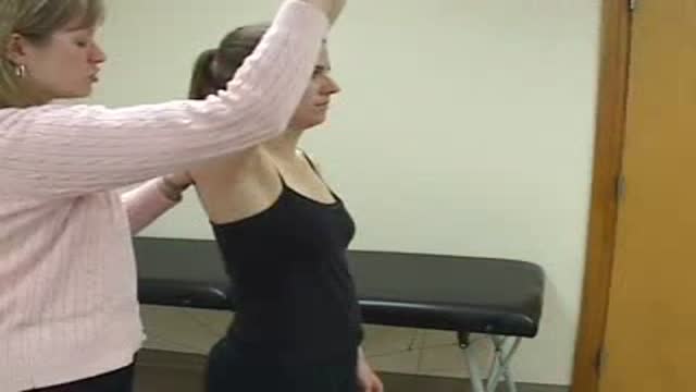
Neer's Sign

a video of abdominal physical examination including all the required items:
-Inspection
-Palpation
-Percussion
-Auscultation

This video demonstrates why ears become clogged and why ear popping helps. The video also explains why ear popping may become difficult resulting in a persistent clogged or muffled ear especially after an ear infection.
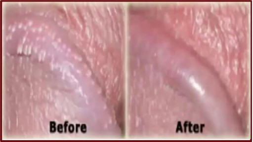
http://penilepapules.plus101.com/ ----- White Spots On Shaft, Pearly Penile Papules Treatment Cream, Single Red Bump On Shaft, Ppp Surgery. Common Home Made Remedies for Pearly Penile Papules. When it comes to treating pearly penile papules many people find it very difficult to reach one of the medical treatments. This is mainly because they are highly expensive and not many people can afford spending large amounts of money on surgery and recovery. In addition to that, these procedures have been reported as being quite risky, which make the men suffering from pearly penile papules think twice before going for one of the available surgeries. This is why, along the time, many homemade, natural treatments have been experienced, so that a cheaper and less risky way of curing pearly penile papules would be found. Some of the methods which have been tried proved to be very less effective, while some did not have any effect at all. Yet, there have also been methods which not only proved to be effective, but they were also considered to be much better than the medical treatment. Most of those who have tried the tea tree oil treatment reported significant diminish of the number of the papules from their penises. In addition to the clearing of the skin, they have also noticed that there were no side effects and the skin remained soft after the papules were removed. As the method was quite simple to put in practice (it requires the application of tea tree oil on the affected area with a cotton swab for three or four times per day), many men decided this was indeed a great solution to their problem.

Traditional Liposuction VS Vaser Liposuction
A side-by-side comparison of traditional liposuction and a #Vaser liposuction. Both of these were performed by our skilled surgeons at Divine Cosmetic Surgery.
#vaserliposuction #liposuction #liposuctionDelhi #liposuctionresults #shorts #vaserliposuctionDelhi
Know more about liposuction
https://www.divinecosmeticsurg....ery.com/liposuction-
Traditional Liposuction vs 360 High Def Vaser Liposuction - https://www.youtube.com/watch?v=r_bBI2p9fVI&t=14s
-------------------------------------------------------------------------------
Why Vaser Is Best For Thigh Liposuction - https://youtu.be/dlzpdDEZcS4
-------------------------------------------------------------------------------
Abdomen Vaser Liposuction - Live - https://www.youtube.com/watch?v=_Cvl2Txn8LQ
-------------------------------------------------------------------------------
Back Vaser Liposuction In Female - https://youtu.be/OC60UdgtIWU
-------------------------------------------------------------------------------
For more details about Liposuction Visit - https://www.divinecosmeticsurgery.com/
-------------------------------------------------------------------------------
Dr. Amit Gupta
MBBS, M.S., DNB (Plastic & Cosmetic Surgery)
Divine Cosmetic Surgery | +91 9811994417
info@divinecosmeticsurgery.com | 01141828787
Delhi | Mumbai | Gurgaon
𝗦𝗼𝗰𝗶𝗮𝗹 𝗠𝗲𝗱𝗶𝗮 𝗮𝗻𝗱 𝗬𝗼𝘂𝘁𝘂𝗯𝗲 𝘃𝗶𝗱𝗲𝗼 𝗰𝗵𝗮𝗻𝗻𝗲𝗹 : -
🎦 https://www.youtube.com/c/DrAm....itGuptaBestPlasticCo
👍🏻 https://www.facebook.com/dramitguptaplasticsurgeon
📷 https://www.instagram.com/divineaesthetics_delhi/
🐥 https://twitter.com/dramitguptajee
🖇️ https://www.linkedin.com/compa....ny/divinecosmeticsur
📌 https://pinterest.com/divinesurgery
#Liposuction #vaserliposuction #liposuctioncostinindia #liposuctiondelhi #liposuction #liposuctioncost #liposuctioncostfactors #liposuctioncostindelhi #DrAmitGuptaPlasticSurgeon #DivineCosmeticSurgery #dramitgupta
Disclaimer: The information on our videos & social media is provided for informational purposes only and is not meant for the advice provided by your surgeon.
We are not responsible for any harm if anyone misguides you from our name. Our all-social media official handles are linked up on our website. All images & content used on our videos & social media are for illustrative concerns only, original results and processes may vary.
“People need to realize this is imminently preventable,” he said. Lyme disease develops following an infection with the bacteria Borrelia burgdorferi. It's transmitted to humans through the bite of infected blacklegged ticks. The tick must be attached to its host for 36 to 48 hours to transmit the bacteria.
A video showing the process of childbirth via vaginal delivery.
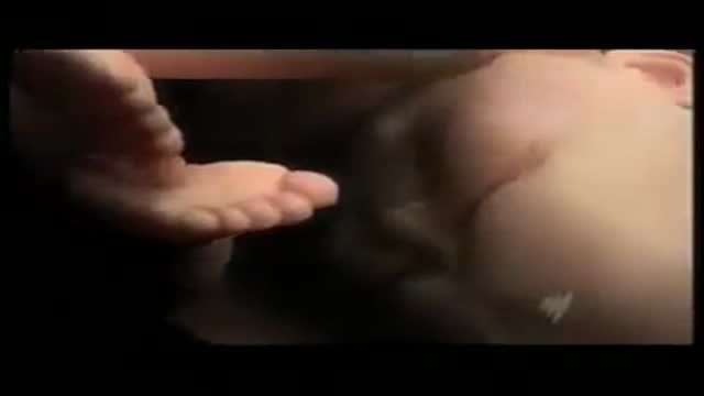
Penile implant surgery for dysfunctional erection of the penis

Histology of Medium Artery and Vein

Robotics is the engineering science and technology of robots, and their design, manufacture, application, and structural disposition.
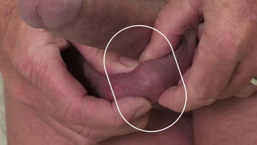
Penile implants are devices placed inside the penis to allow men with erectile dysfunction (ED) to get an erection. Penile implants are typically recommended after other treatments for ED fail. There are two main types of penile implants, semirigid and inflatable.
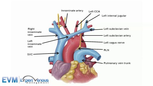
The superior vena cava (SVC, also known as the cava or cva) is a short, but large diameter vein located in the anterior right superior mediastinum.

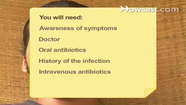
Clean hands can help prevent the spread of infectious diseases, such as flu. This podcast explains the proper way to wash your hands.
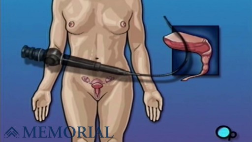
Cystoscopy (sis-TOS-kuh-pee) is a procedure that allows your doctor to examine the lining of your bladder and the tube that carries urine out of your body (urethra). A hollow tube (cystoscope) equipped with a lens is inserted into your urethra and slowly advanced into your bladder.

Childbirth (also called labour, birth, partus or parturition) is the culmination of a human pregnancy or gestation period with the birth of one or more newborn infants from a woman's uterus. The process of normal human childbirth is categorized in three stages of labour: the shortening and dilation of the cervix, descent and birth of the infant, and birth of the placenta. In many cases, with increasing frequency, childbirth is achieved through caesarean section, the removal of the neonate through a surgical incision in the abdomen, rather than through vaginal birth. In the U.S. and Canada it represents nearly 1 in 3 (31.8%) and 1 in 4 (22.5%) of all childbirths, respectively.

Pompe disease is a rare multisystem genetic disorder that is characterized by absence or deficiency of the lysosomal enzyme alpha-glucosidase (GAA). This enzyme is required to breakdown (metabolize) the complex carbohydrate glycogen and convert it into the simple sugar glucose.
Do you suffer with depression? Maybe you’ve recently been diagnosed or are a caregiver to someone with depression. Learn more about this common mood disorder, including depression causes, risk factors, and prevention. We’ll help you take control of your depression and live an active, healthy life.

some knowledge

Oral sex is a commonly performed act of foreplay involving the kissing or licking of the genital area to pleasure a partner. However, it is sometimes stated that the act alone can increase the risk of throat cancer. Is this really the case?

This system treats type 2 diabetes by promoting weight loss.
