- Physical Examination
- Surgical Examination
- Ophthalmology
- Clinical Skills
- Orthopedics
- Surgery Videos
- Laparoscopy
- Pediatrics
- Funny Videos
- Cardiothoracic Surgery
- Nursing Videos
- Plastic Surgery
- Otorhinolaryngology
- Histology and Histopathology
- Neurosurgery
- Dermatology
- Pediatric Surgery
- Urology
- Dentistry
- Oncology and Cancers
- Anatomy Videos
- Health and Fitness
- Radiology
- Anaesthesia
- Physical Therapy
- Pharmacology
- Interventional Radiology
- Cardiology
- Endocrinology
- Gynecology
- Emergency Medicine
- Psychiatry and Psychology
- Childbirth Videos
- General Medical Videos
- Nephrology
- Physiology
- Diet and Food Health
- Diabetes Mellitus
- Neurology
- Women Health
- Osteoporosis
- Gastroenterology
- Pulmonology
- Hematology
- Rheumatology
- Toxicology
- Nuclear Medicine
- Infectious Diseases
- Vascular Disease
- Reproductive Health
- Burns and Wound Healing
- Other
Top videos
Dr. Linder is removing a patients breast implants after having five breast augmentations from three previous surgeons. She has baker 4 capsular contracture and is look forward to having them removed. The most common reasons for removing a breast implant include; heath reasons such as back pain, reoccurring complications and the desire for a different shape or size. For implant removal surgery, Dr. Linder makes an inframammary incision (along the breast crease). The implant can be removed intact, or it may need to be punctured before removal. An antibiotic solution is used to irrigate the breast pocket after implant removal. For more information about breast implant removal go to www.implantremoval.net or call Dr. Linder's office at 310-275-4513

Egg Freezing Oocyte Cryopreservation
The G-SHOT® (clinical description: G-Spot Amplification™ or GSA™), is a simple, nonsurgical, physician-administered treatment that can temporarily augment the Grafenburg spot (G-Spot) in sexually active women with normal sexual function.
Robotic-assisted endoscopic thyroid surgery using the daVinci® Surgical System can safely and effectively offer those needing thyroid surgery relief without neck incisions. Dr. Ron Kuppersmith and Dr. Andrew deJong are now performing this procedure at the College Station Medical Center in Texas.
To avoid pregnancy and STDs, always remember to use a condom every time you have sex — including oral, vaginal, or anal sex. Whenever oral sex is being performed on a girl, a dental dam should be used. A guy receiving oral sex should wear a latex condom — or, if he or his partner is allergic to latex, a polyurethane condom.
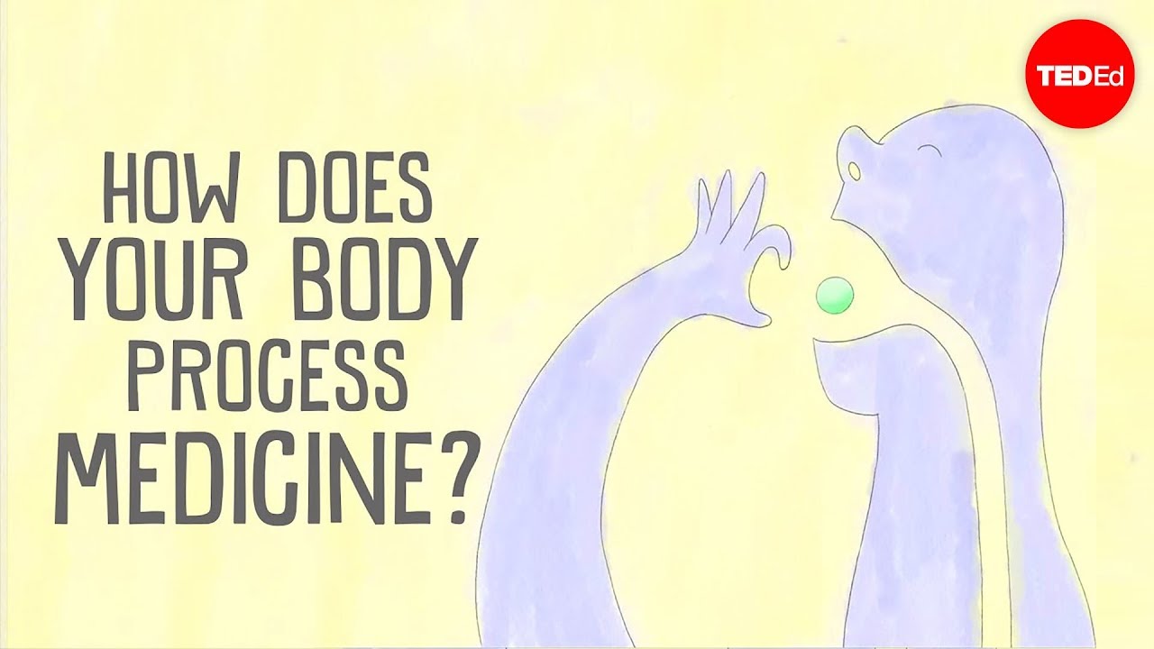
View full lesson: http://ed.ted.com/lessons/how-....does-your-body-proce
Have you ever wondered what happens to a painkiller, like ibuprofen, after you swallow it? Medicine that slides down your throat can help treat a headache, a sore back, or a throbbing sprained ankle. But how does it get where it needs to go in the first place? Céline Valéry explains how your body processes medicine.
Lesson by Céline Valéry, animation by Daniel Gray.
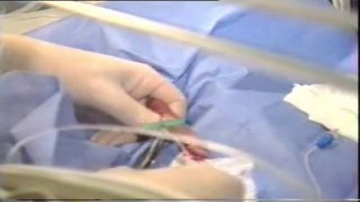
Catheters and Long Lines are introduced in Neonates to administer fluid and Total Parentral Nutrition. The proceedure is not easy to perform and is prone to get infections.
Strict Aseptic technique is mandatory

The thyroid gland lies in the midline of the anterior neck, just caudal to the thyroid cartilage. To inspect the thyroid gland, the examiner stands in front of the patient. The examiner asks the seated patient to dorsiflex (extend) the neck and swallow a sip of water. Minor enlargement of the gland may only become apparent on inspection in this position. Palpation of the thyroid gland is typically performed with the examiner standing behind the patient. Both lobes and the isthmus of the thyroid gland should be palpated for any nodules or diffuse enlargement. Mobility of the thyroid gland with swallowing should be assessed with palpation. Nodules arising from the thyroid gland typically move with swallowing. A hard, fixed thyroid gland could indicate malignancy. If a central nodule is identified, the patient is asked to protrude the tongue. Upward movement of the central nodule on protrusion of the tongue indicates a thyroglossal cyst. Auscultation is performed at the superior poles of bilateral lobes as this is where the superior thyroid artery is most superficial and bifurcates into its terminal branches. A bilateral bruit over the superior poles suggests Graves disease. Examination of the thyroid gland is completed by palpating the regional cervical lymph nodes for any enlargement.
Subscribe to AMBOSS YouTube for the latest clinical examination videos, medical student interviews, study tips and tricks, and live webinars!
Free 5 Day Trial: https://go.amboss.com/amboss-YT
Instagram: https://www.instagram.com/amboss_med/
Facebook: https://www.facebook.com/AMBOSS.Med/
Twitter: https://twitter.com/ambossmed
Blog: https://blog.amboss.com/us
#AMBOSSMed #ClinicalExamination #USMLE
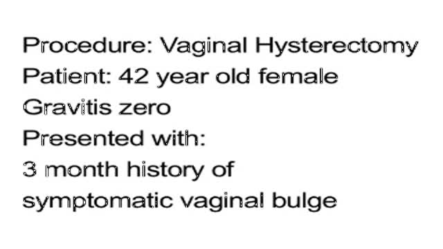
Vaginal Hysterectomy Procedure of a 42 years old female patient with a 3 months history of symptomatic vaginal bulge
For more information about Mohs surgery, please visit https://cle.clinic/3x7CRTy
Mohs surgery is a highly effective skin cancer removal procedure that takes just a few hours. It is most often used to treat basal cell and squamous cell carcinomas, the two most common skin cancers.
Chapters:
0:00 How effective is Mohs Surgery?
0:23 When is Mohs Surgery used?
0:50 How does Mohs Surgery work?
1:55 Does Mohs Surgery cure skin cancer?
2:06 How long is the recovery period after Mohs Surgery?
Resources:
Skins Cancer: https://cle.clinic/3G2MMM8
How Skin Cancer Is Found and Removed — At the Same Appointment: https://cle.clinic/3r9Wzu6
The Best Strategies To Reduce Your Risk of Skin Cancer: https://cle.clinic/38Bazqn
The information in this video was accurate as of 4.8.2022 and is for information purposes only. Consult your local medical authority or your healthcare practitioner for advice.
▶Share this video with others: https://youtu.be/aCV1UZ0Yj-o
▶Subscribe to learn more about Cleveland Clinic:
https://www.youtube.com/user/C....levelandClinic?sub_c
#ClevelandClinic #MohsSurgery #SkinCancer
AUTO-HEMOTHERAPY IN HERPES CASES. THE STORY OF A DOCTOR IN FERME-NEUVE. CBC NEWS 1977.
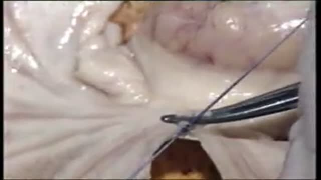
Mesenteric Vessel Ligation
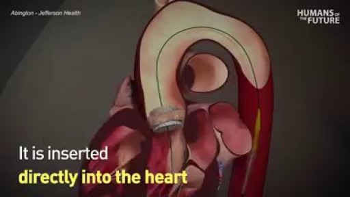
Could this be a viable alternative to open heart surgery?

Business Insider's Michelle Yan has been nearsighted since she was 9 years old. After laser eye surgery, she has 20/20. She walks us through the pre-surgery steps, the actual surgery, as well as the recovery process.
MORE MEDICAL TECH:
8 Medical Procedures That Are Improving Lives
https://www.youtube.com/watch?v=kTMMrAP6DNI
13 Medical Procedures Changing The Health World
https://www.youtube.com/watch?v=VAR44vnxWis
Lifelike Medical Robot Actually Bleeds
https://www.youtube.com/watch?v=IjnhmcCQLsc
------------------------------------------------------
#Lasik #Surgery #TechInsider
Tech Insider tells you all you need to know about tech: gadgets, how-to's, gaming, science, digital culture, and more.
Visit us at: https://www.businessinsider.com
TI on Facebook: https://www.facebook.com/techinsider
TI on Instagram: https://www.instagram.com/tech_insider/
TI on Twitter: https://twitter.com/techinsider
TI on Amazon Prime: http://read.bi/PrimeVideo
INSIDER on Snapchat: https://insder.co/2KJLtVo
------------------------------------------------------
What It's Like To Get Laser Eye Surgery
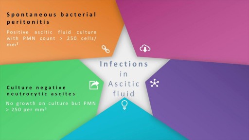
Ascites, the collection of fluid within the peritoneal space is caused due to a variety of causes including cirrhosis, cardiac causes, sinusoidal obstruction syndrome, tubercular peritonitis and pancreatitis, amongst others. Most commonly, the cause of ascots may be cirrhosis , which in turn, is most frequently causes by alcohol use, hepatitis C and non-alcoholic steatohepatitis. At the heart of the ascitic fluid analysis is the serum albumin ascitic gradient, the differential diagnosis of which has been discussed in detail in this presentation. Both low SAAG and high SAAG ascites have been dealt with in some depth, with a brief overview of the management of these conditions

A Lecture Presented by Dr. Mostafa Yakoot to Vascular Surgery Congress. TITLE: SAFETY & EFFICACY OF A NEW HONEY OINTMENT (PEDYPHAR) FOR DIABETIC FOOT ULCERS. Based on the original article in JWC by: Yakoot M, Abdelatif M, Etman M.
